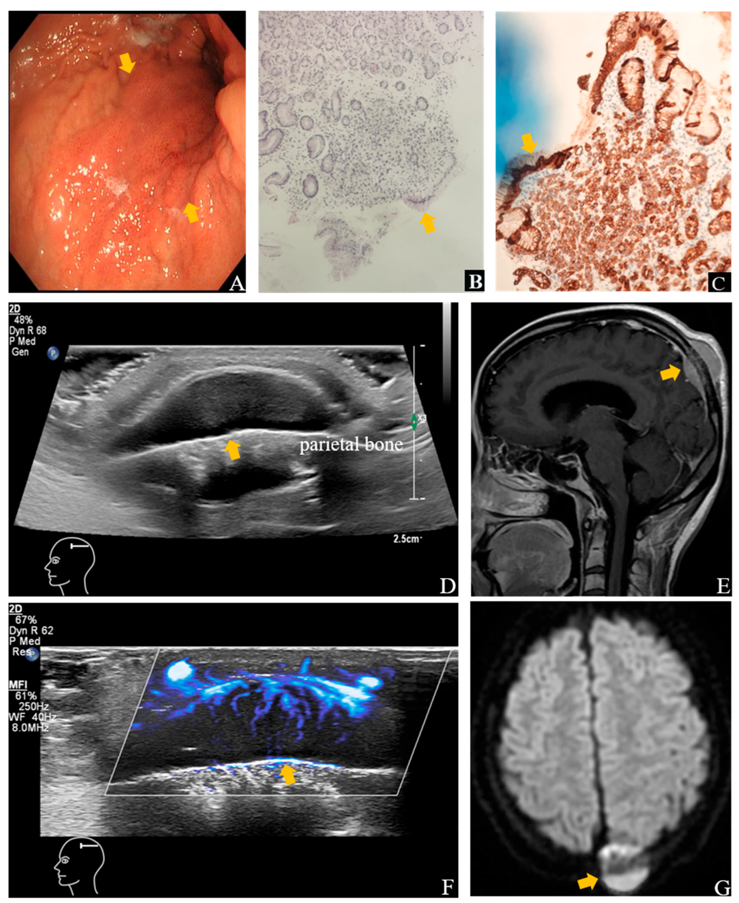
Figure 1.
Ultrasound (US) and contrast-enhanced MRI images show the lesion in the left parietal bone. An endoscopic biopsy ((A), arrow) performed four months earlier in a 26-year-old woman with epigastric discomfort confirmed SRCC on histopathological examination ((B), arrow), Hematoxylin and Eosin staining (H&E), ×10 and (C), arrow), Immunohistochemical Staining (IHC), AE1/AE3 (+)). She complained of right intercostal pain and palpable, firm, immobile masses over the scalp, left neck, and lower abdomen. Her maternal grandmother had a history of cardia cancer. Gray-scale image showed a 3.1 × 2.9 × 0.8 cm solid mass in the subgaleal layer over the left parietal region adjacent to the skull (arrow), with ill-defined margins and abundant vascularity (arrow) on SMI (D,F). Disruption of the adjacent calvarial cortex was noted. Contrast-enhanced MRI of the head revealed a hyperintense lesion in the left parietal bone (arrow) with osteolytic destruction (E,G), consistent with osseous metastasis. One month later, follow-up ultrasound showed enlargement of the scalp lesion to 3.7 × 3.6 × 1.5 cm, and after two months, further progression to 5.7 × 5.1 × 2.0 cm.
![Diagnostics 15 02177 g001 Diagnostics 15 02177 g001]()
Figure 2.
US images show metastatic lesions involving the cervical lymph nodes and intercostal soft tissues. Gray-scale image showed confluent lymph node clusters in the left cervical and supraclavicular regions (arrow) with increased vascular flow (arrow) on SMI (A,B). Fine-needle aspiration of the supraclavicular lymph node revealed infiltrating atypical epithelial cells ((C), H&E, ×10). Immunohistochemistry was consistent with metastatic gastric adenocarcinoma. Gray-scale images showed several solid nodules in the subcutaneous and muscular layers of the right intercostal region, the largest measuring 2.0 × 0.7 cm (D), arrow), accompanied by rich vascularity (E), arrow) and cortical disruption of adjacent ribs (F), arrow). Contrast-enhanced CT of the chest revealed bilateral pleural thickening suggestive of metastases.
Figure 3.
US and Contrast-enhanced CT images show metastatic lesions involving the pelvic cavity, pancreatic-splenic hilum region, peritoneum and omentum. Pelvic ultrasonography disclosed an 11.0 × 7.6 × 9.5 cm infiltrative mass involving uterus and bladder (
arrow), with prominent vascular signals (
arrow) (
A,
B). Contrast-enhanced CT showed an ill-defined heterogeneous mass in the bilateral adnexal areas (
arrow) (
C,
F). Additional findings included a 1.9 × 1.6 cm vascularized nodule on the bladder wall (
D),
arrow), an 8.7 × 6.2 × 6.5 cm poorly demarcated mass in the pancreatic-splenic hilum region on ultrasonography (
E),
arrow) and contrast-enhanced CT (
I),
arrow), and diffuse peritoneal and omental thickening with multiple nodules (
arrow), the largest measuring 3.8 × 2.7 cm (
arrow) (
G,
H). The pelvic mass was consistent with a Krukenberg tumor, typically appearing as a heterogeneous solid lesion with potential cystic areas [
1]. The irregular peritoneal thickening and omental cake appearance indicated metastatic spread [
2]. The patient received seven cycles of a modified BEMA regimen combined with nivolumab. Initial treatment response was stable disease; however, follow-up showed progression at these sites, and the patient ultimately succumbed to acute obstructive renal failure due to tumor progression, which aligns with the aggressive nature and poor prognosis of advanced SRCC [
3,
4]. Systemic ultrasonography, combining high-frequency imaging and microvascular techniques, plays a crucial role in detecting rare metastatic sites and monitoring therapeutic response in real-time, offering an early, sensitive, radiation-free, and repeatable assessment for patients with poor general condition [
5,
6].
![Diagnostics 15 02177 g003 Diagnostics 15 02177 g003]()
Author Contributions
The manuscript writing and images collection were completed by X.D. and L.Z. The image analysis was completed by X.L. The methodology was investigated by L.G. The project administration was handled by J.L. All authors have read and agreed to the published version of the manuscript.
Funding
This work was supported by National High Level Hospital Clinical Research Funding (Grant number: 2022-PUMCH-B-064) to Jianchu Li.
Institutional Review Board Statement
Ethical review and approval were waived for this study due to the nature of a retrospective case report, which did not impact the management of the patient.
Informed Consent Statement
Written informed consent has been obtained from the patient’s family to publish this paper.
Acknowledgments
We sincerely thank the departments of Ultrasound, Gastroenterology, and Pathology of Beijing Sixth Hospital for the endoscopic and gastric pathology images, and the department of Pathology of Peking Union Medical College Hospital for the lymph node pathology images.
Conflicts of Interest
The authors declare no conflicts of interest.
References
- Liu, J.; Chang, C.; Zhang, H. Grayscale ultrasound feature typing of metastatic ovarian tumors, particularly signet-ring cell carcinoma. Quant. Imaging Med. Surg. 2023, 13, 49–57. [Google Scholar] [CrossRef]
- Rui, L.; Min, Q.; Xin, M.; Yujun, C.; Yihong, G.; Ruilong, Y.; Bo, W.; Tengfei, Y. Advances and innovations in ultrasound-based tumor management: Current applications and emerging directions. Ultrasound J. 2025, 17, 40. [Google Scholar] [CrossRef]
- Kao, Y.C.; Fang, W.L.; Wang, R.F.; Li, A.F.; Yang, M.H.; Wu, C.W.; Shyr, Y.M.; Huang, K.H. Clinicopathological differences in signet ring cell adenocarcinoma between early and advanced gastric cancer. Gastric Cancer 2019, 22, 255–263. [Google Scholar] [CrossRef] [PubMed]
- Kwon, K.J.; Shim, K.N.; Song, E.M.; Choi, J.Y.; Kim, S.E.; Jung, H.K.; Jung, S.A. Clinicopathological characteristics and prognosis of signet ring cell carcinoma of the stomach. Gastric Cancer 2014, 17, 43–53. [Google Scholar] [CrossRef]
- Cooper, N.; Meehan, H.; Linton-Reid, K.; Barcroft, J.; Danin, J.; Seah, M.; Sadigh, S.; Bharwani, N.; Sur, S.; Fotopoulou, C.; et al. Clinical utility of ultrasonography in pediatric and adolescent gynecology: Retrospective review of 1313 ultrasound examinations. Ultrasound Obstet. Gynecol. 2025, 65, 226–234. [Google Scholar] [CrossRef] [PubMed]
- Komatsu, M.; Teraya, N.; Natsume, T.; Harada, N.; Takeda, K.; Hamamoto, R. Clinical Application of Artificial Intelligence in Ultrasound Imaging for Oncology. JMA J. 2025, 8, 18–25. [Google Scholar] [CrossRef] [PubMed]
| Disclaimer/Publisher’s Note: The statements, opinions and data contained in all publications are solely those of the individual author(s) and contributor(s) and not of MDPI and/or the editor(s). MDPI and/or the editor(s) disclaim responsibility for any injury to people or property resulting from any ideas, methods, instructions or products referred to in the content. |
© 2025 by the authors. Licensee MDPI, Basel, Switzerland. This article is an open access article distributed under the terms and conditions of the Creative Commons Attribution (CC BY) license (https://creativecommons.org/licenses/by/4.0/).


