Article

Ocular Structural and Vascular Changes in Patients with Severe Asymptomatic Carotid Disease After Undergoing Carotid Endarterectomy (CEA) and Carotid Artery Stenting (CAS)

by
Foteini Xanthou
1,*,†,
Anna Dastiridou
1,†,
Athanasios Giannoukas
2,
Miltiadis Matsagkas
2,
Chara Tzavara
3,
Athanasios Chaidoulis
2,
Sofia Androudi
1 and
Evangelia E. Tsironi
1
1
Ophthalmology Department, Faculty of Medicine, School of Health Sciences, University of Thessaly, University Hospital of Larissa, 41110 Larissa, Greece
2
Vascular Surgery Department, Faculty of Medicine, School of Health Sciences, University of Thessaly, University Hospital of Larissa, 41110 Larissa, Greece
3
Biostatistics Department, National and Kapodistrian University of Athens, 15784 Athens, Greece
*
Author to whom correspondence should be addressed.
These authors contributed equally to this work.
Diagnostics 2025, 15(14), 1826; https://doi.org/10.3390/diagnostics15141826
Submission received: 16 May 2025 / Revised: 15 July 2025 / Accepted: 18 July 2025 / Published: 21 July 2025
(This article belongs to the Section Clinical Diagnosis and Prognosis)

Abstract

Background/Objectives: This study aimed to prospectively assess the incidence of retinal embolization and to evaluate the vascular and structural changes in the retina and choroid in 52 patients with asymptomatic severe carotid artery disease who underwent carotid artery revascularization. Methods: In our study, 35 patients underwent carotid endarterectomy (CEA) and 17 underwent carotid artery stenting (CAS). Biomicroscopy, fundoscopy, optical coherence tomography (OCT), and OCT-angiography (OCTA) were performed at baseline and 1 month after revascularization. Results: The subfoveal choroidal thickness (SFCT), peripapillary choroidal thickness inferior to the optic nerve head (ppCTi), total overall average retinal vascular density (rVDtot), and total overall average choriocapillaris vascular density (ccVDtot) of the eyes ipsilateral to the operated carotid artery increased significantly after revascularization, whereas a statistically significant increase was also found in the SFCT, rVDtot, and ccVDtot of the contralateral eyes in the overall cohort. Comparing the two study groups, we found that the SFCT, superior and inferior peripapillary choroidal thicknesses (ppCTs, ppCTi), rVDtot, and ccVDtot increased in both groups after revascularization, but significantly only in the CEA group. Furthermore, the temporal choriocapillaris vascular density (ccVDt) increased significantly after revascularization in both groups to a similar degree. Conclusions: Carotid artery revascularization led to a statistically significant increase in retinal and choroidal vascular densities, which indicates significantly improved ocular perfusion. The analysis of the findings of the two study groups suggests the superiority of CEA in terms of improving ocular perfusion in asymptomatic severe carotid artery disease. The rate of retinal embolization was similar in both surgical groups.

1. Introduction

Carotid artery disease, as a manifestation of atherosclerosis, is a major risk factor for vascular events in multiple locations, including ipsilateral ischemic stroke and ocular ischemic syndrome, and therefore represents a major cause of morbidity and mortality. Carotid artery stenosis has become a prominent public health concern. The estimated rate of ipsilateral carotid-related acute ischemic stroke in asymptomatic patients with severe (70–99%) carotid artery stenosis is 4.7% over 5 years [1]. Overall, carotid artery stenosis is the cause of 10–20% of ischemic strokes, which are the second most common cause of death worldwide [2]. It is important to distinguish between asymptomatic and symptomatic carotid artery stenosis, as they are associated with different stroke risks [3]. A symptomatic carotid lesion is referred to as a symptomatic lesion after a stroke, transient ischemic attack, or amaurosis fugax. Moreover, ocular signs and symptoms may be the first manifestations of carotid artery disease [4,5], as the blood supply to the eye is mostly provided by branches of the ophthalmic artery, which is the first branch of the internal carotid artery (ICA). Notably, retinal embolization is associated with an increased risk of stroke [6,7].
Furthermore, asymptomatic carotid stenosis has been associated with neurocognitive decline, possibly as the result of silent microinfarctions, which have been found in 17–33% of patients with asymptomatic carotid disease [8]. However, the mechanisms contributing to the development of vascular cognitive impairment associated with carotid artery stenosis are multifaceted and not fully understood.
The objective of carotid artery revascularization is to prevent primary or secondary strokes. The methods available for carotid artery revascularization include carotid endarterectomy (CEA), intraluminal restoration with carotid artery stenting (CAS), and transcarotid artery revascularization (TCAR). Carotid endarterectomy (CEA) was the first method that was developed; it was first performed in 1954 and remained the only method for about 40 years. Currently, CEA is the standard treatment. CEA involves exposure of the carotid artery and mechanical removal of plaque, most typically from the carotid bulb and the proximal internal carotid artery, via a neck incision. CAS, which is an endovascular, minimally invasive procedure, involves femoral (less often radial) puncture, catheter introduction to the carotid artery proximal to the site of stenosis, neuroprotection system (distal or proximal) activation, balloon dilatation of the stenotic area, and finally, stent implantation for endovascular lumen reconstruction. If performed with the routine use of neuroprotection and second-generation stent (mesh-covered, antiembolic) implantation, CAS is safe and at least as effective as CEA in stroke prevention. The third method is the hybrid procedure of transcarotid artery revascularization (TCAR), which has emerged as a valid alternative to both open CEA and transfemoral/transradial CAS. In TCAR, a direct puncture approach involves accessing the carotid artery through a small incision in the neck. Subsequent procedural steps are similar to CAS. This technique enables a more direct and less invasive approach to revascularization, potentially reducing complications compared with traditional methods. These procedures effectively reduce the risk of first and recurrent ischemic stroke. Choosing between the aforementioned carotid revascularization procedures depends on several factors, including the patient’s anatomy, age, gender, and procedural risk.
It is currently not known whether CEA, CAS, or both lead to a change in ocular perfusion. This could be observed using OCT-angiography, a new noninvasive imaging modality that can provide qualitative and quantitative information about retinal and choroidal microcirculation [9]. Therefore, the objective of this study was to examine whether there are any differences in the ipsilateral choroidal and retinal perfusion or retinal embolization after CEA vs. CAS, and to investigate the effect of revascularization on the contralateral eye.

2. Materials and Methods

This single-center prospective study was conducted at the University Hospital of Larissa, Greece, from October 2018 to December 2022. Patients were eligible to participate in this study if they had asymptomatic, severe (>70–99%) stenosis of the internal carotid artery (ICA) and were scheduled for carotid artery revascularization. Asymptomatic was defined as the absence of symptoms (stroke, TIA, and amaurosis fugax) for at least 6 months. The percentage of the carotid stenosis was evaluated by both ultrasound and OCT-angiography. They were divided into two groups, one undergoing carotid endarterectomy (CEA) and the second undergoing carotid artery stenting (CAS). The vascular surgeons selected the surgical procedure. This study’s protocol adheres to the Declaration of Helsinki and was approved by the University Hospital of Larissa Review Board. Before enrolling, eligible participants were required to sign an informed consent form.
Patients who met any of the following criteria were excluded: symptomatic, severe carotid artery stenosis; uncontrolled arterial hypertension; poor glycemic control; best-corrected visual acuity (BCVA) < 0.7; ocular hypertension; glaucoma; severe non-proliferative or proliferative diabetic retinopathy; prior pan-retinal photocoagulation; history of retinal vein or artery occlusion; history of posterior uveitis; ocular ischemic syndrome; prior or current treatment with intravitreal anti-VEGF or steroid injections; refractive error with spherical equivalent > 3 diopters; corneal opacification; dense cataract; prior ocular surgeries other than uneventful cataract extraction.
The patients were examined 1–3 days before and 1 month after revascularization. Preoperative and postoperative ophthalmological examinations were performed at the same time of day (within a 3 h span) to avoid significant diurnal variations, and all patients had normal blood pressure at the time of examination. BCVA, tonometry, biomicroscopy, and fundoscopy were performed to exclude ocular pathology. Additionally, macular OCT, OCT of the optic nerve head, and OCT-angiography (OCTA) of the retina and choriocapillaris were included in each ophthalmological examination. OCT and OCTA are suitable for the quantitative estimation of indicators such as the choroidal thickness, retinal structure, and retinal and choroidal vascular densities, which are currently used as biomarkers for ischemic ophthalmopathy [10]. In addition, fundus color images were obtained to further document the absence of ocular diseases.
OCT and OCTA imaging were performed using the Triton Plus swept source OCT (Topcon Inc., Tokyo, Japan) with its 100 kHz scanning speed and wavelength of 1050 nm, and the coupled SMARTTrackTM real-time eye-tracking system. The scanning protocols included a 6 × 6 mm 3D OCT-angiography scan centered on the fovea with a resolution of 320 × 320 pixels and a 3D OCT 7 × 7 mm scan with a resolution of 512 × 256 pixels (Scheme 1a,b). OCT retinal macular thickness (MT) and choroidal thickness (CT) were quantified using the instrument’s built-in software version IMAGEnet 6 1.36. For the macular retinal and choroidal thicknesses, seven areas were analyzed: central (c), superior (s), inferior (i), superonasal (sn), inferonasal (in), superotemporal (st), and inferotemporal (it); the peripapillary choroidal thickness (ppCT) was quantified in four areas: superior, inferior, temporal, and nasal. The automatic segmentation algorithms of the OCT-angiography device were used. The choriocapillaris default segmentation was from Bruch’s membrane down to 20.8 μm. For the macular vascular density (VD), six areas (overall/total, fovea, superior, inferior, nasal, and temporal) were analyzed. The overall macular vascular density of the retina (rVDtot) and choriocapillaris (ccVDtot) was recorded as the mean of the foveal, superior, inferior, nasal, and temporal vascular density in each segment. The qualitative evaluation was performed by two investigators.
In the CEA group, the same procedure using bovine patch angioplasty was performed on all patients. CAS was performed through a femoral approach using filtering cerebral protection devices.
The quantitative variables are expressed as mean values (SD), while qualitative variables are expressed as absolute and relative frequencies. Paired t-tests or Wilcoxon signed-rank tests were used for time comparisons for the whole sample. For the comparison of proportions, the chi-square and Fisher’s exact tests were used. Student’s t-tests were computed for comparisons between the two groups. Time comparisons were analyzed using the Wilcoxon signed-rank test. Repeated measures analysis of variance (ANOVA) was adopted to evaluate the changes in the indexes of the two types of revascularization over the follow-up period. Log transformations were used in the repeated measures analysis of variance. All reported p-values are two-tailed. Statistical significance was set at p < 0.05, and the analyses were conducted using the SPSS statistical software (version 26.0).

3. Results

We initially examined 81 patients. Twenty-one of them were excluded because of an underlying, previously undiagnosed ocular disease, and eight of them were excluded because of their inability to cooperate in order to acquire sufficient quality OCT exams. Fifty-two patients (82.7% males) with a mean age of 68.4 years (SD = 7.9 years) were analyzed. There were 35 patients (67.3%) in the CEA group and 17 patients (32.7%) in the CAS group. Table 1 provides the participant demographic information as well as information regarding their medical histories. The patient characteristics of the two groups were similar.
The postoperative changes in the ipsilateral eyes are presented in Table 2. The ppCTi, MTs, SFCT, rVDn, rVDtot, ccVDt, ccVDn, and ccVDtot increased significantly after revascularization, while MTst and MTit decreased significantly after revascularization (CEA and CAS).
Table 3 presents the postoperative changes in the contralateral eyes. The MTsn, subfoveal choroidal thickness (SFCT), superior retinal vascular density (rVDs), rVDi, rVDt, rVDtot, ccVDs, ccVDt, and ccVDn increased significantly after revascularization (CEA and CAS).
The changes in the nerve fiber layer thickness, choroidal thickness around the optic nerve, and topographic measurements of the macula of the ipsilateral eye after each type of revascularization are presented in Table 4. The preoperative and postoperative values in the two groups were similar (p > 0.05). Regarding changes over time, it was found that in the CEA group, the ppCTs, ppCTi, and ppCTn increased significantly while MTst and MTit decreased significantly after revascularization.
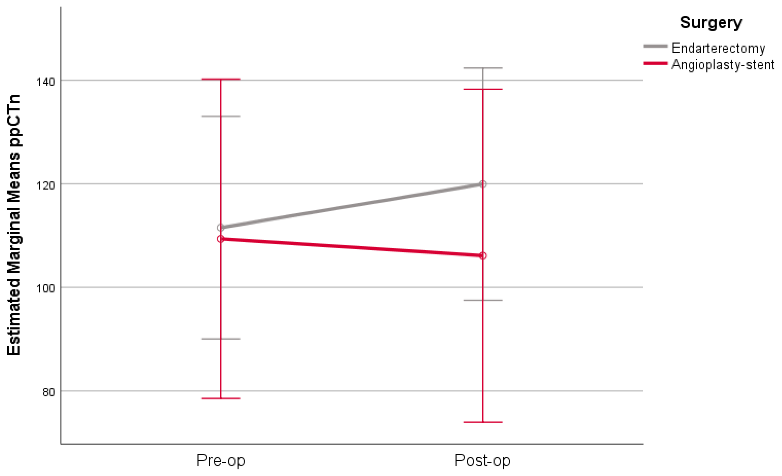
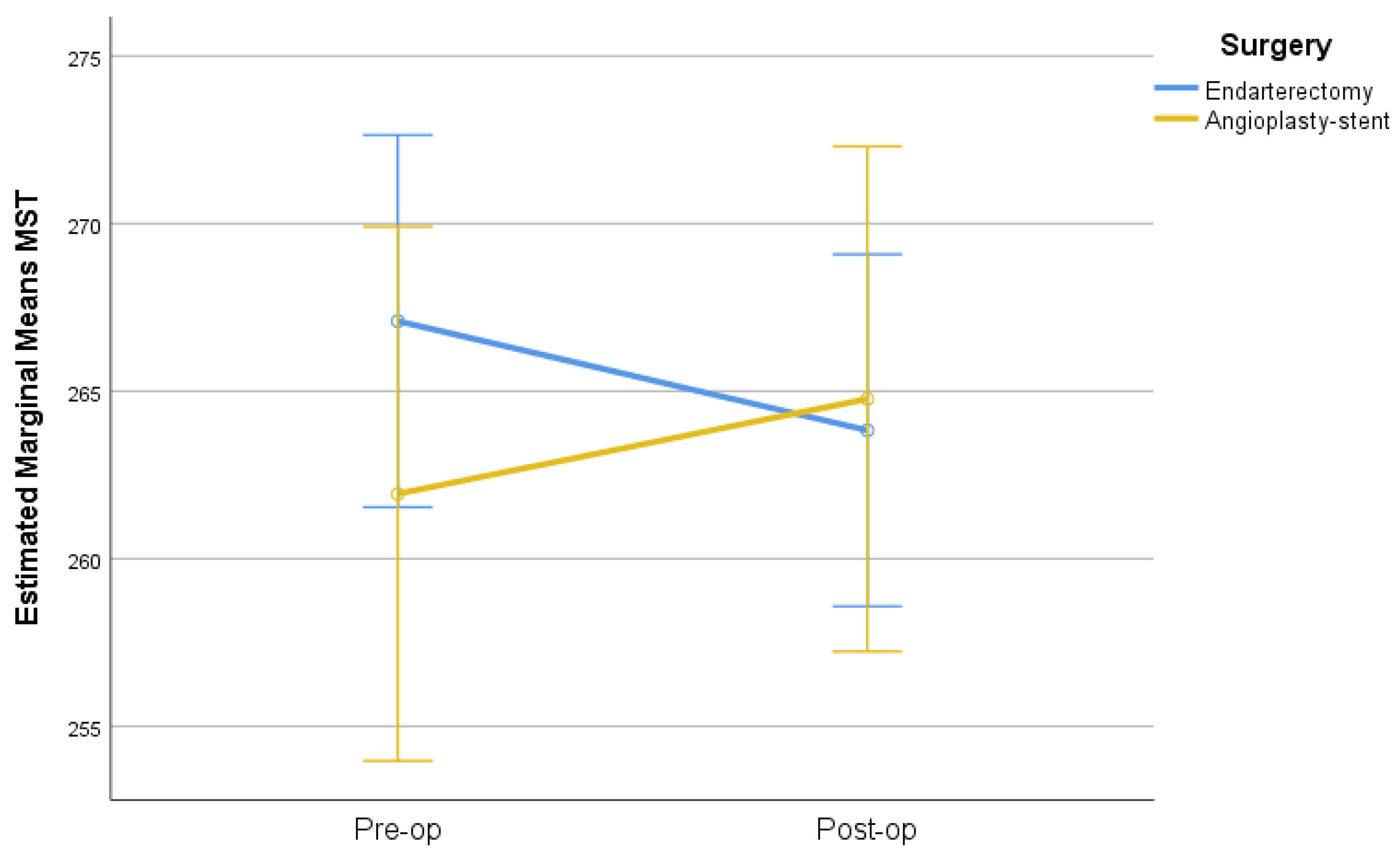
In the CAS group, the central (foveal) MT (CMT) decreased significantly after revascularization. The degree of change in the ppCTn and MTst differed significantly between the two revascularization groups (p = 0.038 and p = 0.047, respectively). In the CEA group, the ppCTn values increased significantly, while they remained similar in the CAS group (Figure 1).
The MTst values also decreased significantly in the CEA group but did not change in the CAS group (Figure 2).
The changes in the macular choroidal thickness, retinal vascular density, and vascular density of the choriocapillaris of the ipsilateral eyes in the two revascularization groups are presented in Table 5.
Interestingly, the SFCT, inferior choroidal thickness (CTi), inferonasal choroidal thickness (CTin), rVDs, rVDtot, and ccVDtot increased significantly after revascularization but only in the CEA group. Furthermore, ccVDt increased significantly after revascularization in both groups to a similar degree. Only the degree of change in the rVDs differed significantly between the two revascularization procedures (p = 0.007): it increased significantly in the CEA group and did not change in the CAS group (Figure 3).

4. Discussion

In the present study, we found a postoperative increase in choroidal and retinal thicknesses and vascular densities in the ipsilateral eye as a result of carotid artery revascularization. We also found an improvement in ocular perfusion, more specifically, a postoperative increase in SFCT and vascular density in the retina and choriocapillaris, in the contralateral eye. This is not surprising considering the anatomy of collateral orbital vessels. The development of the ophthalmic artery involves multiple anastomoses with both the internal and external carotid arteries. Multiple anastomoses are evident in the maxillary artery branch, an external carotid artery branch, and the distal branch of the ophthalmic artery [11,12], which often create a retrograde blood flow stemming from the external carotid artery in advanced ICA stenosis and ICA occlusion. The results of previous studies also support the hypothesis that revascularization to treat unilateral carotid stenosis can increase hemispheric cerebral blood flow on both sides [13]. It is also important to note that in our study, no significant inflammation was detected in either the CEA or CAS groups. Inflammation is a common concern following CAS and CEA, both aimed at treating carotid artery disease, but with distinct profiles of post-procedural responses. After CAS, inflammation often results from the physical disruption of plaques, injury to the artery lining, and the body’s immune response to the stent as a foreign object. This inflammatory process can lead to complications such as periprocedural ischemic lesions, restenosis, or in-stent restenosis, which involves the proliferation of cells narrowing the artery. Late inflammatory reactions may also occur because of prosthetic materials used during surgery. In our study, no significant inflammation was detected in either the CEA or CAS groups. In our patients, the typical inflammation at the incision site subsided within a few days without the use of antibiotics or steroids. Follow-up carotid artery ultrasound exams conducted one month after revascularization for both groups revealed a healthy, unobstructed carotid artery with no abnormalities.
Our study adds to the existing body of literature studying OCTA changes in patients with carotid artery stenosis undergoing treatment. Lahme et al. reported significantly improved flow density in the optic nerve head after carotid endarterectomy [14]. In their study, Cao et al. found that carotid artery stenting led to a significant increase in macular VD and choroidal vascular volume in a cohort of both asymptomatic and symptomatic patients (Cao et al.). Lee et al. were able to detect an increase in macular vessel densities in both eyes following unilateral CAS for severe carotid stenosis (Lee et al., Sci Rep). Our study is, in fact, the first to investigate the vascular changes in the retina and choriocapillaris after carotid revascularization in a cohort of asymptomatic patients with severe carotid artery stenosis undergoing CEA and CAS.
Comparing the two study groups, those undergoing CEA and those in the CAS group. There was a statistically significant ipsilateral increase in the ppCTs, ppCTi, ppCTn, SFCT, CTi, CTin, rVDs, rVDtot, and ccVDtot in the CEA group, suggesting improved ocular perfusion and indicating that CEA is inferior in terms of improving ocular perfusion in asymptomatic severe carotid artery disease. Interestingly, until now, this increase in postoperative choroidal thickness was only noted in cases where the extent of the stenosis was 50–70% but not in patients with a higher degree of stenosis [15]. In our study, we found a statistically significant postoperative increase in the peripapillary and macular choroidal thickness in patients with severe (70–80%) asymptomatic carotid disease.
Remarkably, the superotemporal and inferotemporal retinal macular thickness (MTst and MTit) decreased significantly after surgery in the CEA group. A possible explanation for this segmental decrease in macular retinal thickness could be the regression of preoperative functional hyperemia because of retinal vascular autoregulation in order to maintain an adequate oxygen supply. Interestingly, the retinal microcirculation has end arteries without anastomoses, which play barrier and autoregulatory roles, and is a relatively low-flow/high-oxygen extraction system [16]. The retina has small local oxygen and energy reserves and therefore, in conditions of hypoperfusion, functional hyperemia could counter the metabolic demands. This theory is also supported by the findings of Akcay et al., who investigated alterations in the choroid in patients with severe carotid disease and found a compensatory SFCT increase in ipsilateral internal carotid stenoses greater than 70% [17].
Furthermore, the significant rise in the aforementioned parameters suggests that carotid revascularization improves the ocular microcirculation, reduces the risk of developing an ocular ischemic syndrome, and indicates a positive prognosis for the patients’ visual function.
Previous studies explored the changes in CT as well as retinal and choroidal vascular changes, using the choroidal vascular index, before and after carotid artery revascularization (CEA or CAS). Initially, those studies measured the flow in the ophthalmic artery and later the macular and choroidal thickness. In 2015, Sayin et al. were the first to show that the macular and choroidal thicknesses were reduced in the eyes ipsilateral to the carotid stenosis, and later, a weak negative correlation between the mean macular thickness and the duration and percentage of the ICA stenosis was found [18,19]. CEA led to a significant increase in the SFCT, which was more noticeable in the ipsilateral eye [20]. It was shown that CAS also has the potential to improve the SFCT [21] as well as the vascular density of the deep vascular retinal complex in both eyes [22]. Severe and long-lasting ICA stenosis can potentially alter choroidal vessels so that no change in SFCT will occur [23]. Reversed blood flow in the ophthalmic artery, which can be observed in ophthalmic artery Doppler waveforms, is a predictor for advanced ICA stenosis with poor functional outcomes [24]. Although retrograde blood flow in the ophthalmic artery can fully recover after carotid revascularization [25], ocular function remains poor. This observation is supported by other studies that showed that chronic high-grade carotid artery stenosis in combination with retrograde ophthalmic artery blood flow leads to a significant and chronic reduction in blood flow in the ipsilateral central retinal artery [26,27]. The clinical consequence of the chronic reduction in blood flow is hypoperfusion retinopathy, progressing to ocular ischemic syndrome with devastating ocular results, such as ocular ischemia, retinal neovascularization, and neovascular glaucoma with secondary angle closure. OCTA and the evaluation of ocular microvasculature could be essential in the assessment of therapy success and could be used as noninvasive indicators to monitor asymptomatic carotid artery stenosis [28,29].
Although OCTA segmentation algorithms worked well, we had to manually correct the sclerochoroidal segmentation in three patients. This was counterbalanced by the fact that two graders showed complete agreement, and there was high intraindividual repeatability. Furthermore, OCTA is prone to certain imaging artifacts [30]. Nevertheless, only high-quality images were used in the analysis.
A small sample size is more affected by random fluctuations and outliers and is prone to the risk of type I and type II errors. Therefore, the small number of participants in our study constitutes a limitation. Despite the short follow-up period, our findings are supported by recent studies that suggested that improvements in choroidal thickness and vascular flow can be seen in the early postsurgical period [31]. Another limitation of our study is that, despite only including eyes with a refractive status of ±3D and measuring the CT at the same time of day to avoid diurnal effects on the CT, specific ocular biometric parameters such as axial length, anterior chamber depth or lens thickness that could modify the CT [32,33] and the accuracy of the OCTA findings were not measured. Moreover, a further limitation is that the vasculature of the retina and choriocapillaris was only analyzed in two dimensions from the OCTA scans, whereas the vasculature exhibits a three-dimensional configuration. Larger studies could allow for not only intrasubject but also intersubject comparisons and the development of software capable of examining the volume of the retinal and choriocapillaris vasculature. A complementary evaluation of the flow direction in the ophthalmic artery would also provide a more accurate answer to the question of which surgical procedure is more promising in terms of improving ocular perfusion.
Since the optimal treatment of asymptomatic carotid artery disease remains under debate, the examination of the retinal and choroidal microcirculation could be utilized as an additional biomarker to evaluate the clinical consequences of each surgical procedure and inform decision-making regarding selecting a surgical procedure. Determining the differences in ocular vascular structures is therefore essential for predicting long-term visual prognosis, preventing complications such as ocular ischemic syndrome, and preserving ocular function and also patients’ survival, since the retinal and choroidal microvasculature can be considered a window into the cerebral microvascular systems. Studying retinal microvasculature provides a unique opportunity for studying cerebral small vessel disease since cerebral microcirculation and retinal microvessels share similar anatomy, physiology, and embryology [34]. The direct visualization of retinal vessels to study retinal microvascular health as a proxy for brain microvascular health in patients with carotid artery disease could have many benefits [35]. Therefore, the retinal and choroidal microcirculation could serve as biomarkers for the severity of underlying cardiovascular, neurodegenerative, and microvascular diseases [36].

Author Contributions

Conceptualization, F.X. and E.E.T.; methodology, F.X., E.E.T., A.D. and S.A.; formal analysis, F.X., A.D. and C.T.; investigation, F.X. and A.D.; resources, A.G., M.M. and A.C.; data curation, F.X.; writing—original draft preparation, F.X. and A.D.; writing—review, F.X., A.D. and E.E.T.; editing, F.X.; supervision, E.E.T. All authors have read and agreed to the published version of the manuscript.

Funding

This research received no external funding.

Institutional Review Board Statement

This study was conducted in accordance with the Declaration of Helsinki and approved by the Institutional Review Board of the University Hospital of Larissa, Greece (protocol code: 3359, date: 22 July 2016).

Informed Consent Statement

Informed consent was obtained from all subjects involved in the study.

Data Availability Statement

Data are available upon reasonable request.

Conflicts of Interest

The authors declare no conflicts of interest.

References

  1. Chang, R.W.; Tucker, L.Y.; Rothenberg, K.A.; Lancaster, E.; Faruqi, R.M.; Kuang, H.C.; Flint, A.C.; Avins, A.L.; Nguyen-Huynh, M.N. Incidence of Ischemic Stroke in Patients With Asymptomatic Severe Carotid Stenosis Without Surgical Intervention. JAMA 2022, 327, 1974–1982. [Google Scholar] [CrossRef] [PubMed]
  2. Wabnitz, A.M.; Turan, T.N. Symptomatic Carotid Artery Stenosis: Surgery, Stenting, or Medical Therapy? Curr. Treat Options Cardiovasc. Med. 2017, 19, 62. [Google Scholar] [CrossRef] [PubMed]
  3. Halliday, A.; Harrison, M.; Hayter, E.; Kong, X.; Mansfield, A.; Marro, J.; Pan, H.; Peto, R.; Potter, J.; Rahimi, K.; et al. Asymptomatic Carotid Surgery Trial (ACST) Collaborative Group. 10-year stroke prevention after successful carotid endarterectomy for asymptomatic stenosis (ACST-1): A multicentre randomised trial. Lancet 2010, 376, 1074–1084. [Google Scholar] [CrossRef] [PubMed]
  4. Biousse, V. Carotid disease and the eye. Curr. Opin. Ophthalmol. 1997, 8, 16–26. [Google Scholar] [CrossRef] [PubMed]
  5. Biousse, V.; Newman, N.J. Eye syndromes and the neuro-ophthalmology of stroke. In Handbook of Clinical Neurology; Elsevier: Amsterdam, The Netherlands, 2008; pp. 595–611. [Google Scholar]
  6. Wong, T.Y.; Klein, R. Retinal arteriolar emboli: Epidemiology and risk of stroke. Curr. Opin. Ophthalmol. 2002, 13, 142–146. [Google Scholar] [CrossRef] [PubMed]
  7. Song, G.; Sun, R.; Chen, Y.F.; Ma, Y.; Wang, Y.B.; Jiao, L.Q.; Ling, F. Retinal embolization after carotid endarterectomy and stenting for carotid artery stenosis. J. Clin. Neurosci. 2015, 22, 1298–1302. [Google Scholar] [CrossRef] [PubMed]
  8. Rudolph, C.; Eldrup, N. Asymptomatic carotid stenosis and concomitant silent brain infarctions. Vascular 2020, 28, 7–15. [Google Scholar] [CrossRef] [PubMed]
  9. Laíns, I.; Wang, J.C.; Cui, Y.; Katz, R.; Vingopoulos, F.; Staurenghi, G.; Vavvas, D.G.; Miller, J.W.; Miller, J.B. Retinal applications of swept source optical coherence tomography (OCT) and optical coherence tomography angiography (OCTA). Prog. Retin. Eye Res. 2021, 84, 100951. [Google Scholar] [CrossRef] [PubMed]
  10. Phuljhele, S.; Sharma, S.; Chawla, R.; Saxena, R.; Sharma, P. Evaluation of optical coherence tomography angiography changes in nonarteritic anterior ischemic optic neuropathy. Indian. J. Ophthalmol. 2023, 71, 2020–2026. [Google Scholar] [CrossRef] [PubMed]
  11. Alvernia, J.E.; Hidalgo, J.; Sindou, M.P.; Washington, C.; Luzardo, G.; Perkins, E.; Nader, R.; Mertens, P. The maxillary artery and its variants: An anatomical study with neurosurgical applications. Acta Neurochir. 2017, 159, 655–664. [Google Scholar] [CrossRef] [PubMed]
  12. Michalinos, A.; Zogana, S.; Kotsiomitis, E.; Mazarakis, A.; Troupis, T. Anatomy of the Ophthalmic Artery: A Review concerning Its Modern Surgical and Clinical Applications. Anat. Res. Int. 2015, 2015, 591961. [Google Scholar] [CrossRef] [PubMed]
  13. Sadato, A.; Maeda, S.; Hayakawa, M.; Adachi, K.; Toyama, H.; Nakahara, I.; Hirose, Y. Carotid stenting for unilateral stenosis can increase contralateral hemispheric cerebral blood flow. J. Neurointerv Surg. 2018, 10, 351–354. [Google Scholar] [CrossRef] [PubMed]
  14. Lahme, L.; Marchiori, E.; Panuccio, G.; Nelis, P.; Schubert, F.; Mihailovic, N.; Torsello, G.; Eter, N.; Alnawaiseh, M. Changes in retinal flow density measured by optical coherence tomography angiography in patients with carotid artery stenosis after carotid endarterectomy. Sci. Rep. 2018, 8, 17161. [Google Scholar] [CrossRef] [PubMed]
  15. Akca Bayar, S.; Kayaarası Öztürker, Z.; Pınarcı, E.Y.; Ercan, Z.E.; Akay, H.T.; Yılmaz, G. Structural Analysis of the Retina and Choroid before and after Carotid Artery Surgery. Curr. Eye Res. 2020, 45, 496–503. [Google Scholar] [CrossRef] [PubMed]
  16. Patton, N.; Aslam, T.; Macgillivray, T.; Pattie, A.; Deary, I.J.; Dhillon, B. Retinal vascular image analysis as a potential screening tool for cerebrovascular disease: A rationale based on homology between cerebral and retinal microvasculatures. J. Anat. 2005, 206, 319–348. [Google Scholar] [CrossRef] [PubMed]
  17. Akçay, B.İ.; Kardeş, E.; Maçin, S.; Ünlü, C.; Özgürhan, E.B.; Maçin, A.; Bozkurt, T.K.; Ergin, A.; Surmeli, R. Evaluation of Subfoveal Choroidal Thickness in Internal Carotid Artery Stenosis. J. Ophthalmol. 2016, 2016, 5296048. [Google Scholar] [CrossRef] [PubMed]
  18. Sayin, N.; Kara, N.; Uzun, F.; Akturk, I.F. A quantitative evaluation of the posterior segment of the eye using spectral-domain optical coherence tomography in carotid artery stenosis: A pilot study. Ophthalmic Surg. Lasers Imaging Retina 2015, 46, 180–185. [Google Scholar] [CrossRef] [PubMed]
  19. Dagdelen, K.; Muz, O.E. Investigation of macular and optic nerve head structural changes using spectral domain optical coherence tomography in internal carotid artery stenosis. Int. Ophthalmol. 2021, 41, 875–882. [Google Scholar] [CrossRef] [PubMed]
  20. Lareyre, F.; Nguyen, E.; Raffort, J.; Carboni, J.; Doyen, J.; Hassen-Khodja, R.; Gastaud, P.; Chofflet, J.; Jean-Baptiste, E. Changes in Ocular Subfoveal Choroidal Thickness After Carotid Endarterectomy Using Enhanced Depth Imaging Optical Coherence Tomography: A Pilot Study. Angiology 2018, 69, 574–581. [Google Scholar] [CrossRef] [PubMed]
  21. Biberoglu, E.; Eraslan, M.; Midi, I.; Baltacioglu, F.; Bitargil, M. Ocular blood flow and choroidal thickness changes after carotid artery stenting. Arq. Bras. Oftalmol. 2020, 83, 417–423. [Google Scholar] [CrossRef] [PubMed]
  22. Lee, C.W.; Cheng, H.C.; Chang, F.C.; Wang, A.G. Optical Coherence Tomography Angiography Evaluation of Retinal Microvasculature Before and After Carotid Angioplasty and Stenting. Sci. Rep. 2019, 9, 14755. [Google Scholar] [CrossRef] [PubMed]
  23. Ala-Kauhaluoma, M.; Koskinen, S.M.; Silvennoinen, H.; Vikatmaa, P.; Nuotio, K.; Ijäs, P.; Relander, K.; Lindsberg, P.J.; Soinne, L.; Summanen, P.A. Subfoveal choroidal thickness in ipsi- and contralateral eyes of patients with carotid stenosis before and after carotid endarterectomy: A prospective study. Acta Ophthalmol. 2021, 99, 545–552. [Google Scholar] [CrossRef] [PubMed]
  24. Tsai, C.L.; Lee, J.T.; Cheng, C.A.; Liu, M.T.; Chen, C.Y.; Hu, H.H.; Peng, G.S. Reversal of ophthalmic artery flow as a predictor of intracranial hemodynamic compromise: Implication for prognosis of severe carotid stenosis. Eur. J. Neurol. 2013, 20, 564–570. [Google Scholar] [CrossRef] [PubMed]
  25. Ishii, M.; Hayashi, M.; Yagi, F.; Sato, K.; Tomita, G.; Iwabuchi, S. Relationship between the Direction of Ophthalmic Artery Blood Flow and Ocular Microcirculation before and after Carotid Artery Stenting. J. Ophthalmol. 2016, 2016, 2530914. [Google Scholar] [CrossRef] [PubMed]
  26. Heßler, H.; Zimmermann, H.; Oberwahrenbrock, T.; Kadas, E.M.; Mikolajczak, J.; Brandt, A.U.; Kauert, A.; Paul, F.; Schreiber, S.J. No Evidence for Retinal Damage Evolving from Reduced Retinal Blood Flow in Carotid Artery Disease. Biomed. Res. Int. 2015, 2015, 604028. [Google Scholar] [CrossRef] [PubMed]
  27. Schappe, L.; Klein, C.; Stögbauer, J.; Federspiel, J.; Lochner, P. Die Rolle der Duplexsonographie der Arteria ophthalmica bei Stenosen der Arteria carotis interna [Role of duplex ultrasonography of the ophthalmic artery in internal carotid artery stenosis]. Radiologie 2024, 64, 687–693. [Google Scholar] [CrossRef] [PubMed]
  28. Xu, Q.; Sun, H.; Yi, Q. Association Between Retinal Microvascular Metrics Using Optical Coherence Tomography Angiography and Carotid Artery Stenosis in a Chinese Cohort. Front. Physiol. 2022, 13, 824646. [Google Scholar] [CrossRef] [PubMed]
  29. Cao, L.; Wu, J.; Wang, H.; Kwapong, W.R.; Yan, Y.; Wan, J.; Wang, P.; Liu, G.; Wang, R.; Hu, F.; et al. Influence of Carotid Artery Stenting on the Retina and Choroid. Transl Vis Sci Technol. 2024, 13, 5. [Google Scholar] [CrossRef] [PubMed]
  30. Spaide, R.F.; Fujimoto, J.G.; Waheed, N.K.; Sadda, S.R.; Staurenghi, G. Optical coherence tomography angiography. Prog. Retin. Eye Res. 2018, 64, 1–55. [Google Scholar] [CrossRef] [PubMed]
  31. Durusoy, G.K.; Gumus, G.; Onay, M.; Altay, C.M.; Binboga, A.B. Early choroidal structure and choroidal vascularity index change after carotid stenting. Photodiagnosis Photodyn. Ther. 2022, 38, 102748. [Google Scholar] [CrossRef] [PubMed]
  32. Li, X.Q.; Larsen, M.; Munch, I.C. Subfoveal choroidal thickness in relation to sex and axial length in 93 Danish university students. Invest. Ophthalmol. Vis. Sci. 2011, 52, 8438–8441. [Google Scholar] [CrossRef] [PubMed]
  33. Wei, W.B.; Xu, L.; Jonas, J.B.; Shao, L.; Du, K.F.; Wang, S.; Chen, C.X.; Xu, J.; Wang, Y.X.; Zhou, J.Q.; et al. Subfoveal choroidal thickness: The Beijing Eye Study. Ophthalmology 2013, 120, 175–180. [Google Scholar] [CrossRef] [PubMed]
  34. István, L.; Czakó, C.; Élő, Á.; Mihály, Z.; Sótonyi, P.; Varga, A.; Ungvári, Z.; Csiszár, A.; Yabluchanskiy, A.; Conley, S.; et al. Imaging retinal microvascular manifestations of carotid artery disease in older adults: From diagnosis of ocular complications to understanding microvascular contributions to cognitive impairment. Geroscience 2021, 43, 1703–1723. [Google Scholar] [CrossRef] [PubMed]
  35. Lamb, T.D.; Collin, S.P.; Pugh, E.N., Jr. Evolution of the vertebrate eye: Opsins, photoreceptors, retina and eye cup. Nat. Rev. Neurosci. 2007, 8, 960–976. [Google Scholar] [CrossRef] [PubMed]
  36. Frost, S.; Kanagasingam, Y.; Sohrabi, H.; Vignarajan, J.; Bourgeat, P.; Salvado, O.; Villemagne, V.; Rowe, C.C.; Macaulay, S.L.; Szoeke, C.; et al. Retinal vascular biomarkers for early detection and monitoring of Alzheimer’s disease. Transl. Psychiatry 2013, 3, 233. [Google Scholar] [CrossRef] [PubMed]
Scheme 1. (a): pre-procedural vascular density map of superficial retinal vascular plexus; (b): post-procedural vascular density map of superficial retinal vascular plexus.
Diagnostics 15 01826 sch001
Figure 1. Changes in nasal peripapillary choroidal thickness (ppCTn) in ipsilateral eyes in the two revascularization groups; data are reported as the mean +/− SD.
Diagnostics 15 01826 g001
Figure 2. Changes in superotemporal macular thickness (MTst) in ipsilateral eyes in the two revascularization groups; data are reported as the mean +/− SD.
Diagnostics 15 01826 g002
Figure 3. Changes in superior retinal vascular density (rVDs) in ipsilateral eyes in the two revascularization groups; data are reported as the mean +/− SD.
Diagnostics 15 01826 g003
Table 1. Cohort characteristics.
Total Sample (N = 52; 100%)Revascularizationp
CEA
(N = 35; 67.3%)
CAS
(N = 17; 32.7%)
Ν (%)Ν (%)Ν (%)
Gender
Male43 (82.7)28 (80.0)15 (88.2)0.700 ++
Female9 (17.3)7 (20.0)2 (11.8)
Age, mean (SD)68.4 (7.9)67.9 (8.2)69.3 (7.3)0.560 ‡
Coronary heart disease22 (42.3)13 (37.1)9 (52.9)0.279 +
Hypertension47 (90.4)30 (85.7)17 (100)0.159 ++
Hypercholesterolemia52 (100)35 (100)17 (100)-
Diabetes17 (32.7)13 (37.1)4 (23.5)0.326 +
Peripheral artery disease3 (5.9)2 (5.9)1 (5.9)>0.999 ++
Nicotine use29 (56.9)20 (58.8)9 (52.9)0.689 +
Bilateral carotid stenosis26 (50)17 (48.6)9 (52.9)0.768 +
Side
Left22 (42.3)12 (34.3)10 (58.8)0.093 +
Right30 (57.7)23 (65.7)7 (41.2)
+ Pearson’s chi-square test; ++ Fisher’s exact test; ‡ Student’s t-test.
Table 2. Changes in eyes ipsilateral to carotid artery revascularization, showing significant values in bold.
Pre-RevascularizationPost-RevascularizationChangep (Wilcoxon Signed-Rank Test)
Mean (SD)Median (IQR)Mean (SD)Median (IQR)Mean (SD)
ppCTs119.7 (70.9)89.5 (67–160)123.3 (69.3)97 (75.5–154)3.6 (15.2)0.100
ppCTi98.6 (62.5)79.5 (57–128.5)104.6 (62.3)86 (65.5–131)6 (16.6)0.004
ppCTt117.1 (64.7)103 (62.5–163.5)117 (63.7)102 (67.5–159.5)−0.1 (27)0.694
ppCTn110.8 (62.7)89 (62–129)115.4 (65.7)95 (65.5–132)4.6 (21.6)0.117
CMT247.8 (23.7)248.1 (232.5–259)246.9 (24.5)247.5 (232.5–258)−1 (16.3)0.070
MTs276.8 (18.4)277.3 (267.5–289)278.1 (24.3)276.5 (265–287)1.3 (18)0.049
MTi274.4 (17.4)274.5 (263.5–284)271.4 (16.1)271 (260–283)−3 (9.5)0.066
MTst265.4 (16.4)267.5 (257.5–275.5)264.1 (15.3)265.5 (255.5–274)−1.3 (10.6)0.021
MTit268.1 (14.8)269 (258–279.5)265.9 (14.2)265.5 (255.5–277)−2.2 (7.4)0.013
MTsn288.3 (21.3)287.5 (276–300.5)286.6 (18.1)285.5 (275–298.5)−1.7 (11.1)0.068
MTin286.2 (17.8)286 (276.5–296.5)286 (19.4)285.5 (275.5–296)−0.2 (7.3)0.783
SFCT212 (93.7)201.4 (131–281.5)220.9 (92.2)204 (138.5–292.5)8.9 (30.2)0.011
CTs225.8 (68.5)230.7 (190.5–240.5)220.4 (66.7)228.6 (176–235.5)−5.4 (27.2)0.383
CTi227.8 (89.6)234.6 (178.5–254)226.7 (93.1)237.2 (164–256)−1.1 (24.3)0.201
CTst216.5 (60.3)215.2 (200.5–239.5)209.8 (60.5)210.8 (176–230)−6.7 (31.6)0.062
Ctit219 (75.6)221.5 (181.5–268.5)219.9 (81)221.6 (160.5–283.5)0.9 (30.3)0.474
CTsn187.3 (78.5)193 (146.5–198.1)187 (77.2)197.5 (135–204.5)−0.3 (24.7)0.498
CTin186.6 (84.1)191.4 (139–196.2)186.2 (84.6)192.6 (130–205)−0.4 (19.2)0.206
rVDc18.3 (5)18 (15.4–21.5)18.4 (5.1)17.6 (15.1–21.2)0.1 (4.6)0.848
rVDs42.2 (5.1)42.1 (41–44.1)43.2 (4.6)43.6 (41.5–44.7)1 (4.6)0.140
rVDi42 (4.7)42.3 (40.8–44.6)42.5 (4.4)42.7 (40.6–44.4)0.5 (4.6)0.358
rVDt42 (3.7)42 (40.9–43.5)42.6 (3.5)42.7 (40–43.3)0.6 (3.6)0.343
rVDn40.6 (5.9)40.4 (39–41.8)41.7 (6)41.7 (39.7–43.4)1.2 (5.2)0.033
rVDtot37 (3.6)36.8 (35.8–38.3)37.7 (3.3)37.8 (35.8–39.2)0.7 (2.7)0.018
ccVDc50.2 (6.8)49.6 (46.3–52.5)49.8 (5.7)49.8 (48.2–52.7)−0.4 (5.5)0.891
ccVDs49.3 (4.9)49.2 (48.7–51)50.1 (3.4)50.2 (49.2–51.3)0.8 (3)0.092
ccVDi49.8 (5.3)50.8 (49.2–52.2)50.2 (3.2)50.3 (48.6–51.8)0.3 (4.4)0.967
ccVDt51.1 (3.8)51.3 (50.4–52.5)52.2 (2.9)52.2 (51–53)1.1 (2.7)0.001
ccVDn51 (5.1)51.2 (50.7–53.1)52.1 (4)52.4 (50.9–53.6)1.1 (3.6)0.049
ccVDtot50.3 (3)50.4 (49.7–51.6)50.9 (2.1)50.7 (50.4–51.8)0.6 (1.7)0.015
SD: standard deviation; ppCTs: superior peripapillary choroidal thickness; ppCTi: inferior peripapillary choroidal thickness; ppCTt: temporal peripapillary choroidal thickness; ppCTn: nasal peripapillary choroidal thickness; CMT: central macular thickness; MTs: superior macular thickness; MTi: inferior macular thickness; MTst: superotemporal macular thickness; MTit: inferotemporal macular thickness; MTsn: superonasal macular thickness; MTin: inferonasal macular thickness; SFCT: subfoveal choroidal thickness; CTs: superior choroidal thickness; CTi: inferior choroidal thickness; CTst: superotemporal choroidal thickness; CTit: choroidal thickness inferotemporal; CTsn: choroidal thickness superonasal; CTin: choroidal thickness inferonasal; rVDc: central retinal vascular density; rVDs: superior retinal vascular density; rVDi: inferior retinal vascular density; rVDt: temporal retinal vascular density; rVDn: nasal retinal vascular density; rVDtot: total overall average retinal vascular density; ccVDc: central choriocapillaris vascular density; ccVDs: superior choriocapillaris vascular density; ccVDi: inferior choriocapillaris vascular density; ccVDt: temporal choriocapillaris vascular density; ccVDn: nasal choriocapillaris vascular density; ccVDtot: total overall average choriocapillaris vascular density.
Table 3. Changes in eyes contralateral to the carotid artery revascularization, showing significant values in bold.
Pre-RevascularizationPost-RevascularizationChangep (Wilcoxon Signed-Rank Test)
Mean (SD)Median (IQR)Mean (SD)Median (IQR)Mean (SD)
ppCTs123.1 (59.5)108 (80–152)123.1 (59.5)111 (75.5–155.5)0 (15.4)0.535
ppCTi93.4 (58.9)84.5 (54–106)93.9 (58.5)84 (54–105)0.5 (14.3)0.963
ppCTt111.5 (58.2)103.5 (70–132)115.4 (64.5)103.5 (70.5–127)3.9 (22.6)0.265
ppCTn115.8 (56.1)105.5 (70.5–146.5)112.5 (57.1)100.5 (69–138.5)−3.3 (26.2)0.622
CMT246.6 (25.2)249 (234.5–262.5)245.8 (25.2)247 (229.5–262.5)−0.8 (13)0.177
MTs271.8 (24.8)273.5 (267–284.5)277 (17.7)277.5 (268–286.5)5.2 (19.6)0.161
MTi271 (18.2)271.5 (260.5–280)269.4 (18.5)272 (258.5–279)−1.6 (8)0.136
MTst260.5 (23.1)263.5 (255.5–274.5)265.4 (15.7)265 (258.5–275.5)4.9 (18.5)0.152
MTit266.3 (15.8)270 (258.5–274.5)266.3 (16.5)269 (257.5–275)0 (4.9)0.895
MTsn285.4 (21)285 (276.5–296)286.6 (20.8)287.5 (276.5–297.2)1.2 (8.2)0.042
MTin283.6 (21.9)285.5 (272–294.5)283.4 (21.2)284 (272.5–292.8)−0.2 (6.6)0.975
SFCT223.8 (85)217 (160.5–265)230.3 (83.7)220 (166–272)6.5 (20)0.050
CTs221.2 (62.9)228.7 (197.5–237)224.4 (65.8)235.1 (193.5–238)3.2 (16.7)0.091
CTi216.9 (80.1)224.3 (181.5–249.5)212.9 (78.8)225.4 (169–248.5)−4 (20)0.359
CTst211.4 (63)218.3 (184.5–229.5)215 (65)221.4 (186.5–229.5)3.6 (18.7)0.076
CTit215.6 (68)219.1 (175.5–235.5)213.6 (68.4)219.8 (163.5–231)−2 (15.9)0.547
CTsn187.8 (76.7)202.7 (142.5–205.4)186.8 (77.1)200.1 (134–205)−1.1 (22.1)0.359
CTin178.8 (70.7)187.5 (139–195.2)181.1 (80.8)195.5 (124–196)2.3 (25.9)0.355
rVDc18.2 (4.3)17.4 (15.9–19.8)18.5 (4.5)17.7 (16.9–20.6)0.3 (2.8)0.268
rVDs40.3 (4.9)40.3 (39.5–42.6)41.8 (3.5)41.9 (41.1–44)1.6 (4.5)0.022
rVDi40.7 (4.3)40.7 (38.4–42.9)42.3 (8.9)42.2 (40.5–44)1.6 (9.8)0.003
rVDt40.7 (3.6)40.7 (39.5–43)41.7 (3.4)42.2 (40.2–43.8)1 (3.8)0.043
rVDn40 (4.7)40.3 (38.1–42.3)40.6 (3.5)41.1 (39.7–42.5)0.7 (4.1)0.133
rVDtot36 (2.8)36.2 (35.2–37.5)37 (2.6)37 (35.5–38.1)1 (2.2)0.001
ccVDc49.3 (4.8)49.4 (46.9–51.5)49.9 (4.3)49.5 (47.6–52.2)0.6 (4.7)0.530
ccVDs48.7 (3.2)48.9 (48.1–50.7)49.7 (3.7)50 (49.5–51.3)1 (3.6)0.005
ccVDi49.7 (3.3)50.3 (48.3–51.3)49.7 (3.4)50.2 (48.7–51.7)0.1 (3.8)0.330
ccVDt51.4 (2.5)51.6 (50.8–52.8)52 (2.7)52.2 (51.2–52.7)0.6 (2.3)0.018
ccVDn51.1 (3.7)51.4 (49–53.4)51.8 (3.6)51.8 (51–53.4)0.7 (2.7)0.038
ccVDtot50 (2.3)50.3 (49.4–51.3)50.6 (2)50.7 (49.8–51.5)0.6 (2.1)0.077
SD: standard deviation; ppCTs: superior peripapillary choroidal thickness; ppCTi: inferior peripapillary choroidal thickness; ppCTt: temporal peripapillary choroidal thickness; ppCTn: nasal peripapillary choroidal thickness; CMT: central macular thickness; MTs: superior macular thickness; MTi: inferior macular thickness; MTst: superotemporal macular thickness; MTit: inferotemporal macular thickness; MTsn: superonasal macular thickness; MTin: inferonasal macular thickness; SFCT: subfoveal choroidal thickness; CTs: superior choroidal thickness; CTi: inferior choroidal thickness; CTst: superotemporal choroidal thickness; CTit: choroidal thickness inferotemporal; CTsn: choroidal thickness superonasal; CTin: choroidal thickness inferonasal; rVDc: central retinal vascular density; rVDs: superior retinal vascular density; rVDi: inferior retinal vascular density; rVDt: temporal retinal vascular density; rVDn: nasal retinal vascular density; rVDtot: total overall average retinal vascular density; ccVDc: central choriocapillaris vascular density; ccVDs: superior choriocapillaris vascular density; ccVDi: inferior choriocapillaris vascular density; ccVDt: temporal choriocapillaris vascular density; ccVDn: nasal choriocapillaris vascular density; ccVDtot: total overall average choriocapillaris vascular density.
Table 4. Ipsilateral changes in nerve fiber layer thickness, peripapillary choroidal thickness, and topographic measurements of the macula after CEA and CAS, showing significant values in bold.
RevascularizationPre-RevascularizationPost-RevascularizationChangep 1p 2
Mean (SD)Median (IQR)Mean (SD)Median (IQR)Mean (SD)
RNFLtotCEA104.9 (15.6)104 (97–116)104.3 (14.6)104 (93–115)−0.6 (3.9)0.6470.356
CAS102.9 (8.9)103 (102–107)103.5 (9.6)103 (101–109)0.5 (3.6)0.698
RNFLsCEA130 (41.4)130 (106–144)124.6 (23.5)125 (106–142)−5.4 (29.9)0.5750.511
CAS124.7 (17.6)124 (112–140)124.2 (17.9)125 (110–134)−0.5 (6.3)0.637
RNFLiCEA140.4 (41.3)139 (121–149)135 (20.2)140 (121–152)−5.4 (35.3)0.9670.260
CAS129.1 (14.1)130 (128–135)132.2 (15)131 (128–135)3.1 (8.4)0.452
RNFLtCEA79.8 (42.7)72 (65–83)74.7 (17.3)72 (65–81)−5.1 (29.3)0.2120.497
CAS71.3 (8.6)71.3 (64–79)71.5 (9.6)72 (63–81)0.2 (3.6)0.469
RNFLnCEA90.3 (40)85 (76–93)84 (15.1)83 (73–94)−6.3 (31.3)0.1570.505
CAS87.4 (14.7)86 (81–96)86.5 (15.8)87 (76–93)−0.9 (4.5)0.420
ppCTsCEA119.9 (77.4)87 (67–161)125.1 (73.1)93 (77–155)5.2 (16)0.0300.090
CAS119.3 (57.1)106 (67–160)119.6 (62.8)98 (70–153)0.4 (13.1)0.925
ppCTiCEA100.7 (64.4)79 (61–133)109.4 (61.5)96 (70–139)8.8 (17)0.0020.127
CAS94.5 (60)81 (44–118)94.8 (64.6)75 (48–118)0.3 (14.6)0.460
ppCTtCEA115.3 (63.2)101 (57–165)116.9 (65.8)102 (68–165)1.6 (27.7)0.6740.526
CAS120.8 (69.4)120.8 (69–148)117.1 (61)109 (63–154)−3.7 (26)0.924
ppCTnCEA111.5 (63.9)104 (61–125)119.9 (65.2)112 (70–134)8.4 (22.3)0.0340.038
CAS109.4 (62)85 (63–133)106.1 (67.7)79 (63–113)−3.3 (18.3)0.649
CMTCEA247.6 (26.6)248 (228–261)247 (27.8)248 (232–261)−0.6 (19.9)0.3980.856
CAS248.3 (16.9)248.3 (240–254)246.5 (16.3)247 (239–250)−1.7 (3.1)0.037
MTsCEA276.5 (17)277 (265–290)278 (27.6)276 (260–288)1.4 (20.4)0.1110.892
CAS277.3 (21.5)277.3 (270–286)278.4 (16.2)279 (266–281)1 (12.5)0.255
MTiCEA275 (19.3)276 (264–284)272.5 (17)273 (260–284)−2.6 (9.8)0.2890.536
CAS273.2 (13)273.2 (263–283)269.2 (14.3)266 (257–274)−4 (8.9)0.100
MTstCEA267.1 (14.6)268 (258–277)263.8 (16.1)266 (252–275)−3.3 (8.7)0.0080.047
CAS261.9 (19.6)263 (255–274)264.8 (14.1)264 (256–272)2.8 (13.1)0.756
MTitCEA269.5 (15.1)269 (259–280)266.5 (15)267 (254–278)−3 (7.7)0.0100.280
CAS265.4 (14.1)267 (256–272)264.8 (12.7)265 (256–271)−0.6 (6.5)0.488
MTsnCEA287.2 (22.6)287 (275–302)285 (18.6)286 (272–300)−2.2 (11.4)0.1220.655
CAS290.6 (18.8)290.6 (282–300)289.9 (17.1)285 (279–297)−0.7 (10.7)0.343
MTinCEA284.5 (18.2)284 (276–295)285.6 (20.1)289 (275–296)1.1 (6.4)0.3290.075
CAS289.7 (16.9)289.7 (277–298)286.9 (18.2)283 (276–296)−2.8 (8.4)0.344
1 p-value for time effect (Wilcoxon signed-rank test). 2 Repeated measures ANOVA p-value (using logarithmic transformations) for the time × surgery effect. SD: standard deviation; RNFLtot: overall average thickness of retinal nerve fiber layer; RNFLs: thickness of superior retinal nerve fiber layer; RNFLi: thickness of inferior retinal nerve fiber layer; RNFLt: thickness of temporal retinal nerve fiber layer; RNFLn: thickness of nasal retinal nerve fiber layer; ppCTs: superior peripapillary choroidal thickness; ppCTi: inferior peripapillary choroidal thickness; ppCTt: temporal peripapillary choroidal thickness; ppCTn: nasal peripapillary choroidal thickness; CMT: central macular thickness; MTs: superior macular thickness; MTi: inferior macular thickness; MTst: superotemporal macular thickness; MTit: inferotemporal macular thickness; MTsn: superonasal macular thickness; MTin: inferonasal macular thickness.
Table 5. Changes in subfoveal and choroidal thickness, flow in retinal vessels, and choriocapillaris blood flow of the ipsilateral eye after CEA and CAS.
PrePostChangep 1p 2
Mean (SD)Median (IQR)Mean (SD)Median (IQR)Mean (SD)
SFCTCEA213.5 (95.8)194 (130–284)223.1 (95)206 (134–300)9.6 (28.7)0.0110.928
CAS208.9 (92.1)208.9 (134–254)216.5 (88.8)202 (163–266)7.6 (34.1)0.407
CTsCEA230.7 (62.6)230.7 (198–239)228.5 (60.9)228.6 (203–230)−2.2 (19.3)0.7230.168
CAS215.5 (80.3)215.5 (168–242)203.6 (76.6)203.6 (147–245)−11.9 (38.7)0.246
CtiCEA234.6 (82.2)234.6 (206–242)237.2 (82.6)237.2 (186–249)2.6 (17.2)0.0010.075
CAS213.7 (104.6)213.7 (143–262)205.1 (111.4)191 (135–261)−8.6 (34.1)0.276
CTstCEA215.2 (51.6)215.2 (203–228)210.8 (52.5)210.8 (189–223)−4.5 (20.1)0.1710.470
CAS219.2 (77)219.2 (165–263)207.8 (76.2)207.8 (163–231)−11.4 (47.9)0.237
CTitCEA221.5 (67.9)221.5 (190–260)221.6 (64)221.6 (176–281)0.1 (17.6)0.0560.529
CAS213.8 (91.6)213.8 (165–299)216.3 (110.3)204 (144–298)2.5 (47.6)0.538
CTsnCEA198.1 (74.7)198.1 (157–214)198.1 (76.1)198.1 (153–211)0 (17.2)0.6170.963
CAS165 (83.7)155 (123–165)164.1 (76.6)151 (128–197)−0.9 (36.3)0.776
CTinCEA191.4 (80)191.4 (143–201)192.5 (80.1)192.6 (142–206)1.1 (15.2)0.0450.383
CAS176.5 (93.7)176.5 (125–182)173 (94.3)170 (116–204)−3.5 (25.9)0.943
rVDcCEA18 (4.9)18 (15.3–20.8)17.6 (4.7)17.6 (14.5–21.2)−0.4 (4.9)0.6460.321
CAS19 (5.2)19 (15.4–22.1)20 (5.5)20 (16.6–22.5)1 (3.9)0.554
rVDsCEA41.6 (5.9)41.6 (39.3–44.3)43.9 (4.8)43.9 (41.2–45.9)2.3 (4.6)0.0040.007
CAS43.5 (2.8)43.5 (42.3–43.9)41.9 (3.9)43.2 (41.9–44.4)−1.7 (3.5)0.102
rVDiCEA41.4 (5.5)41.4 (38.1–44.5)42.3 (4.9)42.3 (40.8–44.2)0.9 (4.8)0.0940.326
CAS43.4 (1.9)43.2 (41.8–44.7)43 (3.3)43 (40.5–44.9)−0.4 (4.1)0.434
rVDtCEA42 (4)42 (40.8–43.6)43 (3.5)43 (40–44.3)1 (3.4)0.1590.254
CAS41.9 (3.2)41.9 (41.3–43.2)41.8 (3.5)41.8 (39.7–42.9)−0.1 (3.9)0.653
rVDnCEA41.2 (6.5)41.2 (38.9–42.7)42.1 (6.5)42.1 (39.8–43.5)0.9 (5.1)0.2250.590
CAS39.3 (4.5)39.3 (39–40.5)40.9 (4.7)40.9 (38.7–41.8)1.6 (5.4)0.049
rVDtotCEA36.8 (4.1)36.8 (35.5–38.3)37.8 (3.7)37.8 (35.7–39.3)0.9 (2.7)0.0180.261
CAS37.4 (2.2)37.4 (36.8–38.3)37.5 (2.6)37.7 (37–38.5)0.1 (2.6)0.356
ccVDcCEA49.2 (6.9)49.2 (45.8–52.2)48.8 (5.7)49.5 (47.1–52.7)−0.4 (6.1)0.9090.999
CAS52.3 (6.4)51.6 (49.3–53.3)51.8 (5.1)51.6 (48.3–52.8)−0.5 (4.3)0.687
ccVDsCEA49.2 (5.8)49.2 (48.7–51.1)50.2 (3.8)50.2 (49.2–51.4)1 (3.3)0.0880.341
CAS49.6 (2.1)49.5 (48.7–50.2)49.8 (2.3)49.8 (49.3–50.1)0.2 (2.4)0.507
ccVDiCEA49.4 (6.3)49.4 (49.1–52.3)49.7 (3.3)49.7 (48.4–51.8)0.3 (5.2)0.9610.853
CAS50.8 (1.6)51 (50.1–51.7)51.2 (2.7)51.2 (49.4–52.3)0.5 (2.5)0.868
ccVDtCEA51.3 (4.5)51.3 (50.7–53.2)52.4 (3.2)52.3 (51.1–53)1.1 (3.1)0.0320.797
CAS50.8 (1.7)50.8 (49.7–51.5)51.8 (2.3)51.8 (50.7–53.1)1.1 (1.7)0.015
ccVDnCEA51.1 (6)51.4 (51–54.3)52.4 (4.4)52.4 (51.3–53.7)1.3 (4)0.1270.562
CAS50.8 (2.4)50.8 (49.9–52.6)51.6 (3)51.6 (50.5–53.4)0.8 (2.9)0.209
ccVDtotCEA50 (3.4)50.2 (49.8–51.6)50.7 (2.4)50.7 (50.3–51.5)0.7 (1.8)0.0110.553
CAS50.9 (1.5)50.9 (49.7–51.9)51.3 (1.4)51.3 (50.6–52.2)0.4 (1.5)0.381
1 p-value for time effect (Wilcoxon signed-rank test). 2 Repeated measures ANOVA p-value (using logarithmic transformations) for the time × surgery effect. SFCT: subfoveal choroidal thickness; CTs: superior choroidal thickness; CTi: inferior choroidal thickness; CTst: superotemporal choroidal thickness; CTit: inferotemporal choroidal thickness; CTsn: superonasal choroidal thickness; CTin: inferonasal choroidal thickness; rVDc: central retinal vascular density; rVDs: superior retinal vascular density; rVDi: inferior retinal vascular density; rVDt: temporal retinal vascular density; rVDn: nasal retinal vascular density; rVDtot: total overall average retinal vascular density; ccVDc: central choriocapillaris vascular density; ccVDs: superior choriocapillaris vascular density; ccVDi: inferior choriocapillaris vascular density; ccVDt: temporal choriocapillaris vascular density; ccVDn: nasal choriocapillaris vascular density; ccVDtot: total overall average choriocapillaris vascular density.
Disclaimer/Publisher’s Note: The statements, opinions and data contained in all publications are solely those of the individual author(s) and contributor(s) and not of MDPI and/or the editor(s). MDPI and/or the editor(s) disclaim responsibility for any injury to people or property resulting from any ideas, methods, instructions or products referred to in the content.