
A Morphometric Evaluation of the Mandibular Condyle, Coronoid Process, and Gonial Angle: Age and Gender Differences in CBCT Imaging


Abstract
1. Introduction
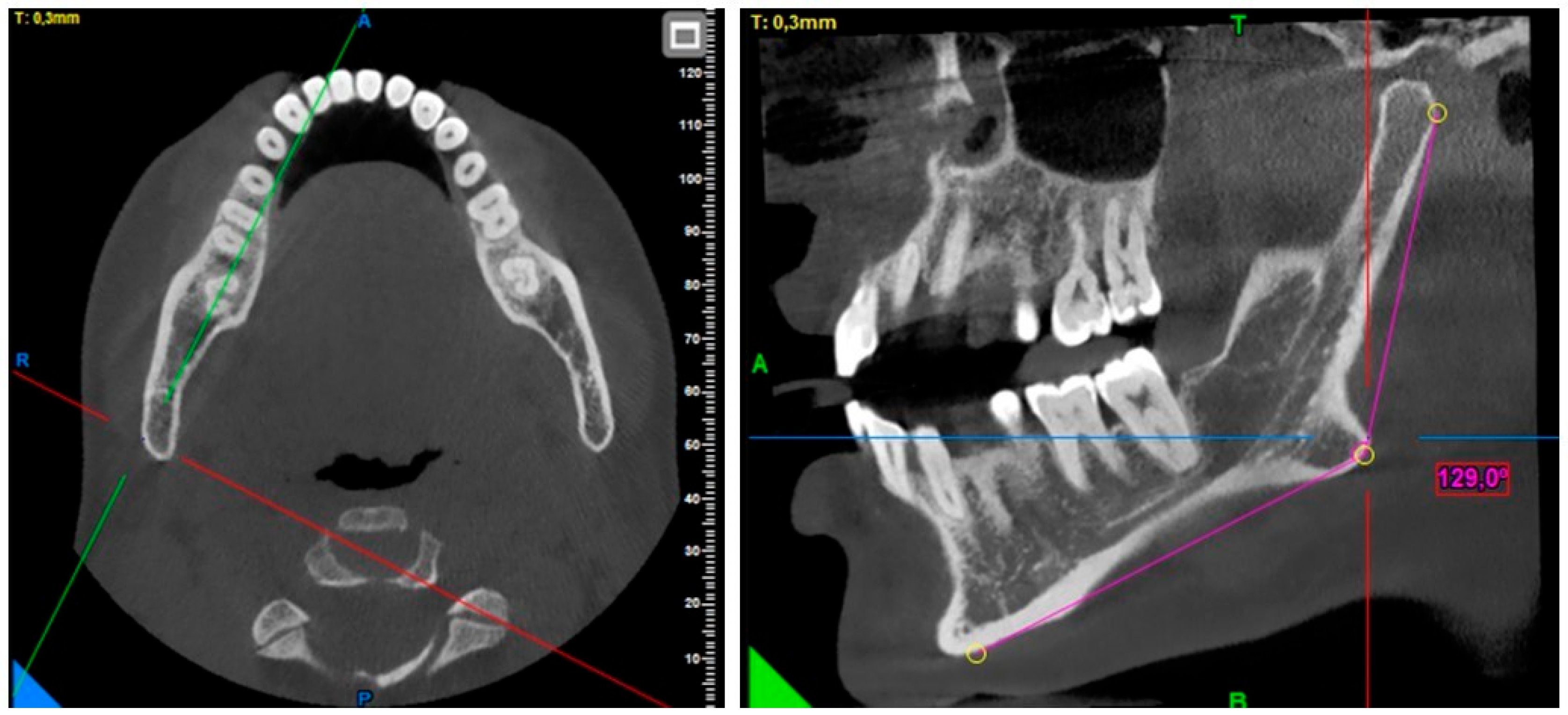
2. Materials and Methods
3. Results
4. Discussion
5. Conclusions
Author Contributions
Funding
Institutional Review Board Statement
Informed Consent Statement
Data Availability Statement
Conflicts of Interest
References
- Soares, A.P.; Fischer, H.; Aydin, S.; Steffen, C.; Schmidt-Bleek, K.; Rendenbach, C. Uncovering the unique characteristics of the mandible to improve clinical approaches to mandibular regeneration. Front. Physiol. 2023, 14, 1152301. [Google Scholar] [CrossRef] [PubMed]
- Jency Evanjelin, P.; Umamaheswari, T.N. Distinctive Anatomical Patterns of the Mandibular Coronoid Process, Condyle, and Sigmoid Notch: Cone Beam Computed Tomography (CBCT) Imaging for Advanced Personal Identification. Cureus 2024, 16, e60978. [Google Scholar]
- Gomes, A.F.; Nejaim, Y.; Brasil, D.M.; Groppo, F.C.; Ferreira-Caria, P.H.; Haiter-Neto, F. Assessment of Volume and Height of the Coronoid Process in Patients With Different Facial Types and Skeletal Classes: A Cone-Beam Computed Tomography Study. J. Oral. Maxillofac. Surg. 2015, 73, e1–e5. [Google Scholar] [CrossRef] [PubMed]
- Bains, S.K.; Bhatia, A.; Kumar, N.; Kataria, A.; Balmuchu, I.; Srivastava, S. Assessment of Morphological Variations of the Coronoid Process, Condyle, and Sigmoid Notch as an Adjunct in Personal Identification Using Orthopantomograms Among the North Indian Population. Cureus 2023, 15, e40275. [Google Scholar] [CrossRef]
- López-Ramírez, J.C.; Mariel-Cárdenas, J.; Gutiérrez-Cantú, F.J.; Salas-Orozco, M.F., Sr.; Medina-Solís, C.E.; Hernández-Molinar, Y.; Trejo-Rivero, E.; Patiño-Marín, N. Association Between Gender, Age, and Skeletal Class With Mandibular Condyle Morphology: A Retrospective Study. Cureus 2023, 15, e49043. [Google Scholar] [CrossRef]
- Mahapatra, S.; Hebbale, M.; Mhapuskar, A.; Halli, R.; Mittal, A. Anatomical Variants of Condylar Process, Coronoid Process, and Sigmoid Notch in a Maharashtrian Population: A Radiographic Study. Cureus 2023, 15, e40434. [Google Scholar] [CrossRef]
- Esfehani, M.; Ghasemi, M.; Katiraee, A.; Tofangchiha, M.; Alizadeh, A.; Taghavi-Damghani, F.; Testarelli, L.; Reda, R. Forensic Gender Determination by Using Mandibular Morphometric Indices an Iranian Population: A Panoramic Radiographic Cross-Sectional Study. J. Imaging 2023, 9, 40. [Google Scholar] [CrossRef]
- Bakan, A.; Kervancıoğlu, P.; Bahşi, İ.; Yalçın, E.D. Comparison of the Gonial Angle With Age and Gender Using Cone-Beam Computed Tomography Images. Cureus 2022, 14, e24997. [Google Scholar] [CrossRef]
- Tavassol, F.; Spalthoff, S.; Essig, H.; Bredt, M.; Gellrich, N.C.; Kokemüller, H. Elongated coronoid process: CT-based quantitative analysis of the coronoid process and review of literature. Int. J. Oral. Maxillofac. Surg. 2012, 41, 331–338. [Google Scholar] [CrossRef]
- Gürleyük, A.C.; Yeler, D.Y.; Eninanç, İ.; Yeler, H. The relationship of medial sigmoid depression and sigmoid notch morphology with vertical and sagittal growth patterns in Turkish population. Eur. J. Anat. 2024, 28, 357–364. [Google Scholar] [CrossRef]
- Fan, Y.; Penington, A.; Kilpatrick, N.; Hardiman, R.; Schneider, P.; Clement, J.; Claes, P.; Matthews, H. Quantification of mandibular sexual dimorphism during adolescence. J. Anat. 2019, 234, 709–717. [Google Scholar] [CrossRef] [PubMed]
- Dutra, V.; Yang, J.; Devlin, H.; Susin, C. Mandibular bone remodelling in adults: Evaluation of panoramic radiographs. Dentomaxillofac. Radiol. 2004, 33, 323–328. [Google Scholar] [CrossRef] [PubMed]
- Chole, R.H.; Patil, R.N.; Balsaraf-Chole, S.; Gondivkar, S.; Gadbail, A.R.; Yuwanati, M.B. Association of mandible anatomy with age, gender, and dental status: A radiographic study. ISRN Radiol. 2013, 2013, 453763. [Google Scholar] [CrossRef] [PubMed]
- Taleb, N.S.A.; Beshlawy, M.E. Mandibular ramus and gonial angle measurements as predictors of sex and age in an Egyptian population sample: A digital panoramic study. J. Forensic Res. 2015, 6, 5. [Google Scholar]
- Ingaleshwar, P.; Bhosale, S.; Nimbulkar, G.; Smitha, T.; Deepak, V.; Britto, F. Assessment of condyle-coronoid angle and gonial angle for gender determination: A digital panoramic study in Bagalkot population. J. Oral. Maxillofac. Pathol. 2022, 26, 414–418. [Google Scholar] [CrossRef]
- Direk, F.; Uysal, I.I.; Kivrak, A.S.; Unver-Dogan, N.; Fazliogullari, Z.; Karabulut, A.K. Reevaluation of Mandibular Morphometry According to Age, Gender, and Side. J. Craniofac. Surg. 2018, 29, 1054–1059. [Google Scholar] [CrossRef]
- Gamba, T.d.O.; Alves, M.C.; Haiter-Neto, F. Mandibular sexual dimorphism analysis in CBCT scans. J. Forensic Leg. Med. 2016, 38, 106–110. [Google Scholar] [CrossRef]
- Bulut, O.; Freudenstein, N.; Hekimoglu, B.; Gurcan, S. Dilemma of Gonial Angle in Sex Determination: Sexually Dimorphic or Not? Am. J. Forensic Med. Pathol. 2019, 40, 361–365. [Google Scholar] [CrossRef] [PubMed]
- Jyothsna, M.; Ranjith, K.; Sarat, G. Determination of gender using condylar height and coronoid height-an orthopantomographic study. Ann. Essences Dent. 2017, 9, 5a. [Google Scholar]
- Mohsen, A.M.; Ye, J.; Al-Nasri, A.; Chu, C.; Zhang, W.B.; Lin-Wang. Three-dimensional evaluation of the mandibular condyle in adults with various skeletal patterns. Korean J. Orthod. 2023, 53, 67–76. [Google Scholar] [CrossRef]
- Saini, V.; Srivastava, R.; Rai, R.K.; Shamal, S.N.; Singh, T.B.; Tripathi, S.K. Mandibular ramus: An indicator for sex in fragmentary mandible. J. Forensic Sci. 2011, 56 (Suppl. 1), S13–S16. [Google Scholar] [CrossRef] [PubMed]
- Ramesh, A.; Velpula, N.; Tandon, R.; Zardi, F.T.; Kanakagiri, M. Determination of age and gender using condylar height and coronoid height- An orthopantomographic study. IP Int. J. Maxillofac. Imaging 2020, 4, 87–90. [Google Scholar]
- Samatha, K.; Byahatti, S.M.; Ammanagi, R.A.; Tantradi, P.; Sarang, C.K.; Shivpuje, P. Sex determination by mandibular ramus: A digital orthopantomographic study. J. Forensic Dent. Sci. 2016, 8, 95–98. [Google Scholar]
- Horowitz, S.L.; Shapiro, H.H. Modifications of mandibular architecture following removal of temporalis muscle in the rat. J. Dent. Res. 1951, 30, 276. [Google Scholar] [CrossRef]
- Manoj, M.; Mathew, L.; Natarajan, S.; Yellapurkar, S.; Shetty, S.; Denny, C.; Dahal, S. Morphometric anlaysis of mandibular coronoid, condyle and sigmoid shape using panoromic view for personal identification in south Indian population. J. Clin. Imaging Sci. 2022, 12, 25. [Google Scholar] [CrossRef]
- Lyu, Y.S.; Li, Z.H. Cone beam CT imaging findings in patients with temporomandibular joint disorder syndrome and unilateral chewing. Shanghai Kou Qiang Yi Xue 2022, 31, 653–656. [Google Scholar] [PubMed]
- Cheong, Y.W.; Lo, L.J. Facial asymmetry: Etiology, evaluation, and management. Chang. Gung Med. J. 2011, 34, 341–351. [Google Scholar] [PubMed]
- Larrazabal-Moron, C.; Sanchis-Gimeno, J.A. Gonial angle growth patterns according to age and gender. Ann. Anat. 2018, 215, 93–96. [Google Scholar] [CrossRef]
- Assari, A.; Alasmari, B.; Aleid, M.; Salem, M. Characteristics of mandibular parameters in different age groups a CBCT assessment. EC Dent. Sci. 2017, 14, 95–103. [Google Scholar]


| Gender | N | Mean | Std. Deviation | p | |
|---|---|---|---|---|---|
| Right gonial angle | female | 78 | 128.6615 | 5.50129 | 0.001 |
| male | 63 | 125.6841 | 5.10352 | ||
| Right coronoid height | female | 78 | 11.1654 | 2.94560 | 0.852 |
| male | 63 | 11.2714 | 3.63120 | ||
| Right sigmoid notch depth | female | 78 | 13.1295 | 1.79943 | 0.044 |
| male | 63 | 13.8841 | 2.46283 | ||
| Right condyle height | female | 78 | 16.2769 | 3.32624 | 0.006 |
| male | 63 | 17.8571 | 3.36527 | ||
| Distance between right condyle and coronoid | female | 78 | 33.9244 | 3.26040 | 0.007 |
| male | 63 | 35.5222 | 3.60392 | ||
| Left gonial angle | female | 78 | 128.8364 | 5.97237 | 0.000 |
| male | 63 | 125.2635 | 4.89326 | ||
| Left coronoid height | female | 78 | 10.8923 | 2.68167 | 0.220 |
| male | 63 | 11.6095 | 3.92879 | ||
| Left sigmoid notch depth | female | 78 | 13.1397 | 1.61054 | 0.020 |
| male | 63 | 14.0111 | 2.55369 | ||
| Left condyle height | female | 78 | 16.4885 | 3.38115 | 0.025 |
| male | 63 | 17.7635 | 3.25697 | ||
| Distance between left condyle and coronoid | female | 78 | 33.8885 | 3.38257 | 0.101 |
| male | 63 | 34.8476 | 3.46921 |
| N | Age Groups | p | |||||
|---|---|---|---|---|---|---|---|
| 20–29 | 30–39 | 40–49 | 50–59 | 60 and over | |||
| 30 | 30 | 24 | 30 | 27 | |||
| Right gonial angle | (Mean) | 126.765 | 126.450 | 128.108 | 127.863 | 128.300 | 0.629 |
| (SD) | 5.8384 | 5.3092 | 6.5360 | 5.5068 | 4.9373 | ||
| Right coronoid height | (Mean) | 11.0921 | 11.6735 | 11.0250 | 11.1400 | 10.3000 | 0.141 |
| (SD) | 3.2874 | 3.0329 | 4.4634 | 2.9019 | 3.0775 | ||
| Right sigmoid notch depth | (Mean) | 13.4026 | 13.7206 | 13.0500 | 13.1867 | 12.6222 | 0.072 |
| (SD) | 1.8919 | 2.4210 | 2.2581 | 2.1071 | 1.9146 | ||
| Right condyle height | (Mean) | 16.9737 | 16.5000 | 17.8750 | 17.5400 | 16.5889 | 0.625 |
| (SD) | 3.8518 | 3.3181 | 3.4431 | 3.3255 | 3.0806 | ||
| Right condyle–coronoid distance | (Mean) | 33.5263 | 34.7353 | 35.7667 | 35.4700 | 34.6556 | 0.145 |
| (SD) | 3.7899 | 3.5671 | 1.4022 | 3.8768 | 2.9127 | ||
| Left gonial angle | (Mean) | 126.252 | 126.717 | 128.316 | 127.833 | 129.276 | 0.270 |
| (SD) | 5.8411 | 6.4570 | 6.0936 | 5.2597 | 4.9626 | ||
| Left coronoid height | (Mean) | 11.1711 | 11.9294 | 10.4500 | 11.7800 | 10.0778 | 0.176 |
| (SD) | 3.4463 | 3.1343 | 4.9458 | 3.0573 | 2.4489 | ||
| Left sigmoid notch depth | (Mean) | 13.4184 | 14.0206 | 13.3333 | 13.9833 | 12.6481 | 0.087 |
| (SD) | 1.9656 | 2.2569 | 2.3784 | 2.2028 | 1.7566 | ||
| Left condyle height | (Mean) | 16.9579 | 16.9706 | 17.7000 | 16.8800 | 16.7778 | 0.536 |
| (SD) | 3.5679 | 3.5178 | 3.1343 | 2.9202 | 3.5290 | ||
| Left condyle–coronoid distance | (Mean) | 33.2526 | 34.4147 | 34.7500 | 34.2100 | 34.0630 | 0.093 |
| (SD) | 3.3842 | 3.5865 | 2.6729 | 3.5747 | 3.2117 | ||
Disclaimer/Publisher’s Note: The statements, opinions and data contained in all publications are solely those of the individual author(s) and contributor(s) and not of MDPI and/or the editor(s). MDPI and/or the editor(s) disclaim responsibility for any injury to people or property resulting from any ideas, methods, instructions or products referred to in the content. |
© 2025 by the authors. Licensee MDPI, Basel, Switzerland. This article is an open access article distributed under the terms and conditions of the Creative Commons Attribution (CC BY) license (https://creativecommons.org/licenses/by/4.0/).
Share and Cite
Dogan, M.E.; Turkoglu, B.N.; Şengul, I. A Morphometric Evaluation of the Mandibular Condyle, Coronoid Process, and Gonial Angle: Age and Gender Differences in CBCT Imaging. Diagnostics 2025, 15, 1459. https://doi.org/10.3390/diagnostics15121459
Dogan ME, Turkoglu BN, Şengul I. A Morphometric Evaluation of the Mandibular Condyle, Coronoid Process, and Gonial Angle: Age and Gender Differences in CBCT Imaging. Diagnostics. 2025; 15(12):1459. https://doi.org/10.3390/diagnostics15121459
Chicago/Turabian StyleDogan, Mehmet Emin, Burcu Nur Turkoglu, and Ilhan Şengul. 2025. "A Morphometric Evaluation of the Mandibular Condyle, Coronoid Process, and Gonial Angle: Age and Gender Differences in CBCT Imaging" Diagnostics 15, no. 12: 1459. https://doi.org/10.3390/diagnostics15121459
APA StyleDogan, M. E., Turkoglu, B. N., & Şengul, I. (2025). A Morphometric Evaluation of the Mandibular Condyle, Coronoid Process, and Gonial Angle: Age and Gender Differences in CBCT Imaging. Diagnostics, 15(12), 1459. https://doi.org/10.3390/diagnostics15121459