
Malignant Transformation of Renal Cyst with Bosniak I Features

 , , , , and
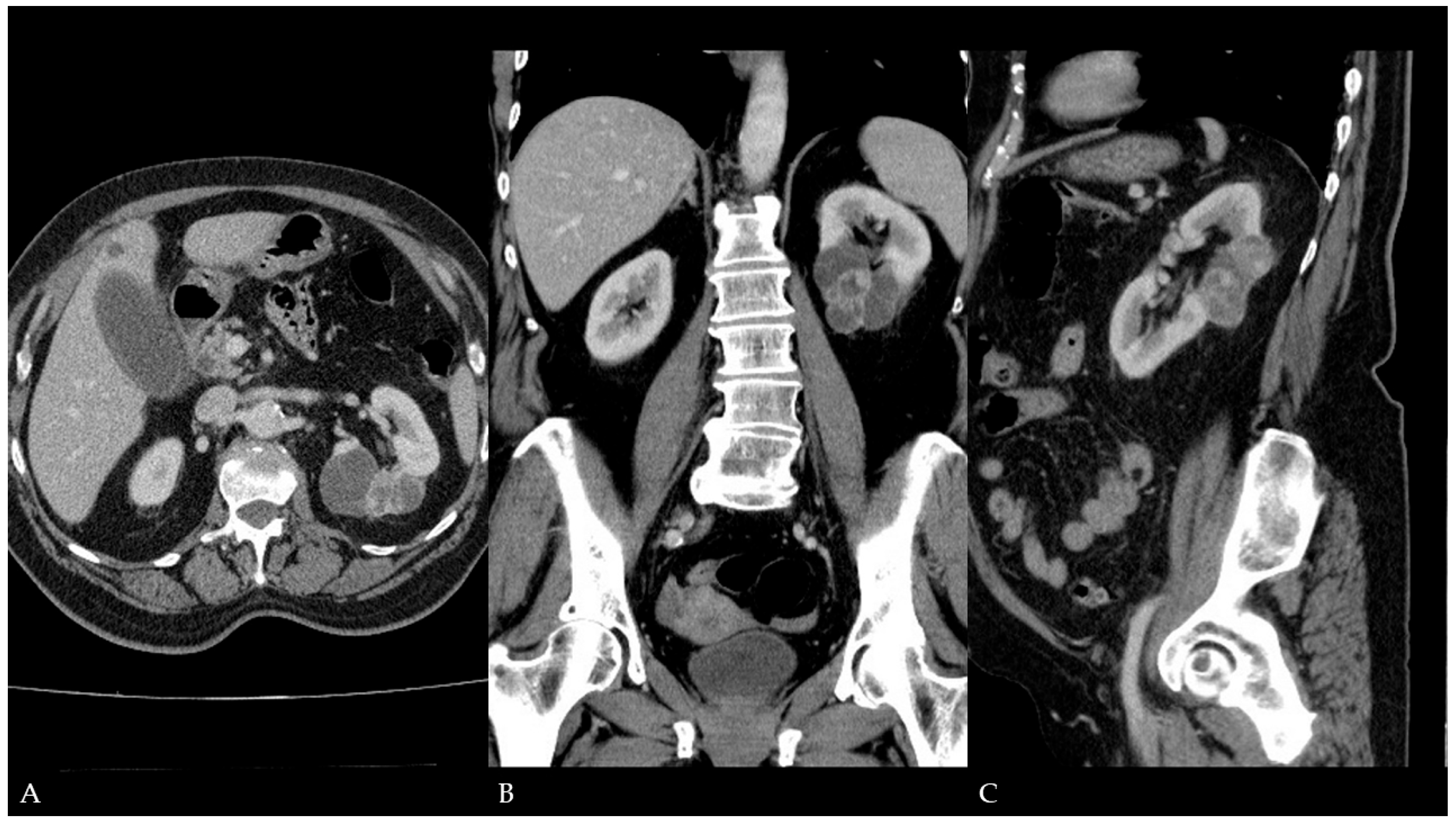
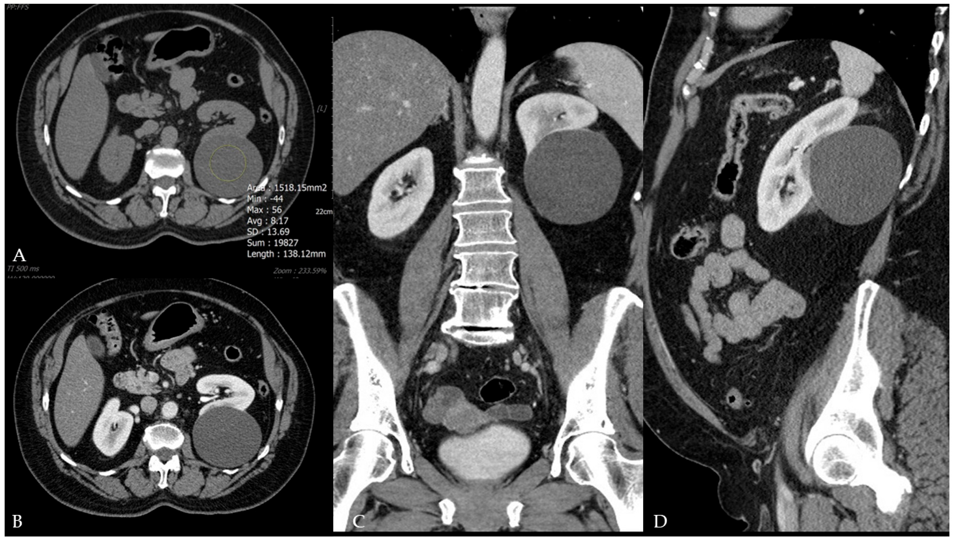
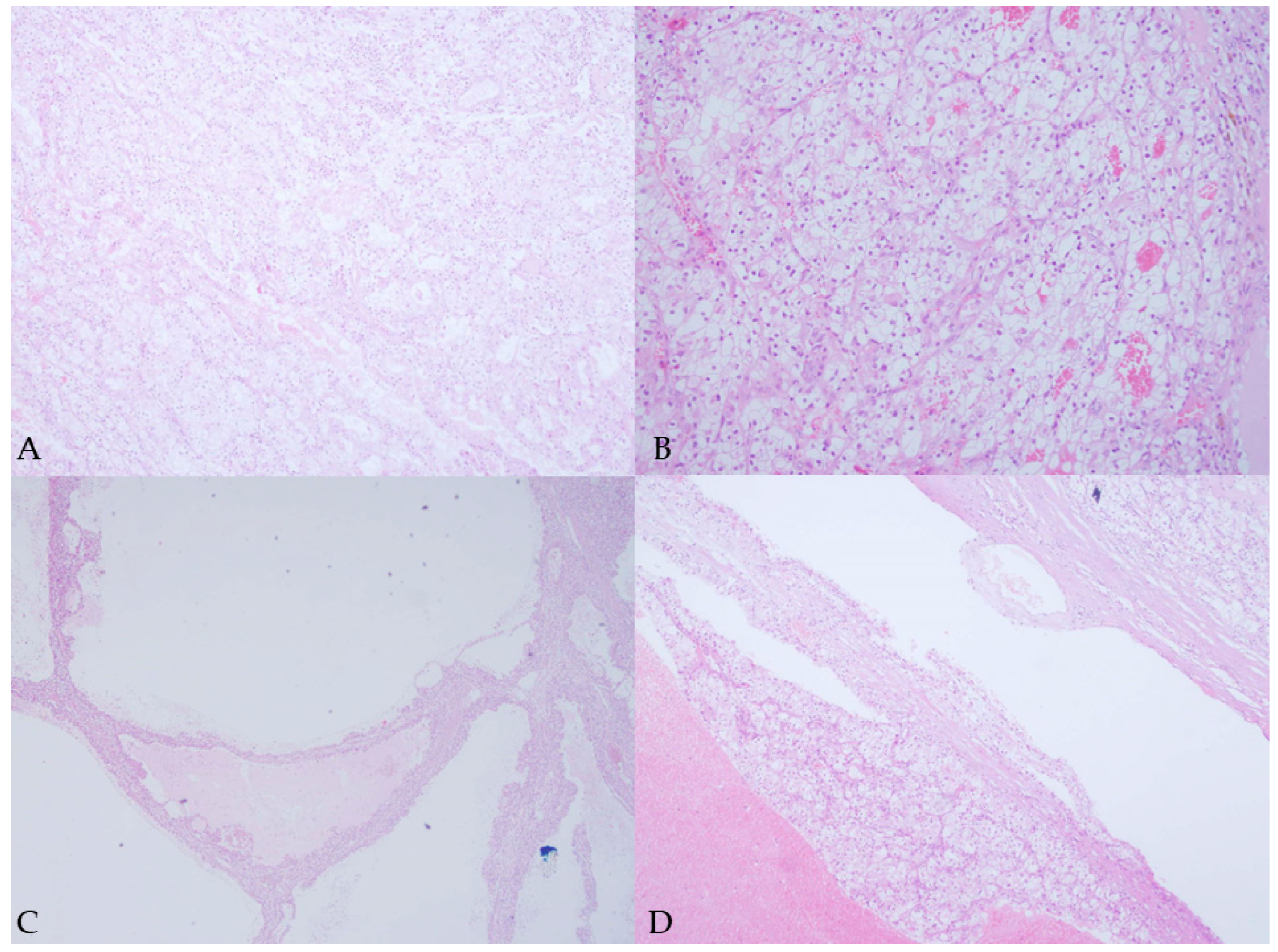
Abstract
Author Contributions
Funding
Institutional Review Board Statement
Informed Consent Statement
Data Availability Statement
Conflicts of Interest
References
- Silverman, S.G.; Pedrosa, I.; Ellis, J.H.; Hindman, N.M.; Schieda, N.; Smith, A.D.; Remer, E.M.; Shinagare, A.B.; Curci, N.E.; Raman, S.S.; et al. Bosniak Classification of Cystic Renal Masses, Version 2019: An Update Proposal and Needs Assessment. Radiology 2019, 292, 475–488. [Google Scholar] [CrossRef] [PubMed]
- Cantisani, V.; Bertolotto, M.; Clevert, D.A.; Correas, J.M.; Drudi, F.M.; Fischer, T.; Gilja, O.H.; Granata, A.; Graumann, O.; Harvey, C.J.; et al. EFSUMB 2020 Proposal for a Contrast-Enhanced Ultrasound-Adapted Bosniak Cyst Categorization—Position Statement. Ultraschall Med. 2021, 42, 154–166. [Google Scholar] [CrossRef] [PubMed]
- Sevcenco, S.; Spick, C.; Helbich, T.H.; Heinz, G.; Shariat, S.F.; Klingler, H.C.; Rauchenwald, M.; Baltzer, P.A. Malignancy rates and diagnostic performance of the Bosniak classification for the diagnosis of cystic renal lesions in computed tomography—A systematic review and meta-analysis. Eur. Radiol. 2017, 27, 2239–2247. [Google Scholar] [CrossRef] [PubMed]
- Chen, S.; Jin, B.; Xu, L.; Fu, G.; Meng, H.; Liu, B.; Li, J.; Xia, D. Cystic renal cell carcinoma: A report of 67 cases including 4 cases with concurrent renal cell carcinoma. BMC Urol. 2014, 14, 87. [Google Scholar] [CrossRef] [PubMed]
- Graumann, O.; Osther, S.S.; Osther, P.J. Characterization of complex renal cysts: A critical evaluation of the Bosniak classification. Scand. J. Urol. Nephrol. 2011, 45, 84–90. [Google Scholar] [CrossRef] [PubMed]
- Ferro, M.; Crocetto, F.; Barone, B.; Del Giudice, F.; Maggi, M.; Lucarelli, G.; Busetto, G.M.; Autorino, R.; Marchioni, M.; Cantiello, F.; et al. Artificial intelligence and radiomics in evaluation of kidney lesions: A comprehensive literature review. Ther. Adv. Urol. 2023, 15, 17562872231164803. [Google Scholar] [CrossRef] [PubMed]
- Sheikhy, A.; Dehghani Firouzabadi, F.; Lay, N.; Jarrah, N.; Yazdian Anari, P.; Malayeri, A. State of the art review of AI in renal imaging. Abdom. Radiol. 2025, epub ahead of print. [Google Scholar] [CrossRef]
Disclaimer/Publisher’s Note: The statements, opinions and data contained in all publications are solely those of the individual author(s) and contributor(s) and not of MDPI and/or the editor(s). MDPI and/or the editor(s) disclaim responsibility for any injury to people or property resulting from any ideas, methods, instructions or products referred to in the content. |
© 2025 by the authors. Licensee MDPI, Basel, Switzerland. This article is an open access article distributed under the terms and conditions of the Creative Commons Attribution (CC BY) license (https://creativecommons.org/licenses/by/4.0/).
Share and Cite
Ćulap, S.; Brkić, F.; Matković, A.; Svetec, J.; Jurjević, N.; Horvat Pavlov, K.; Vidjak, V.; Ferenc, T. Malignant Transformation of Renal Cyst with Bosniak I Features. Diagnostics 2025, 15, 1326. https://doi.org/10.3390/diagnostics15111326
Ćulap S, Brkić F, Matković A, Svetec J, Jurjević N, Horvat Pavlov K, Vidjak V, Ferenc T. Malignant Transformation of Renal Cyst with Bosniak I Features. Diagnostics. 2025; 15(11):1326. https://doi.org/10.3390/diagnostics15111326
Chicago/Turabian StyleĆulap, Sandra, Filip Brkić, Andro Matković, Jelena Svetec, Nikolina Jurjević, Katarina Horvat Pavlov, Vinko Vidjak, and Thomas Ferenc. 2025. "Malignant Transformation of Renal Cyst with Bosniak I Features" Diagnostics 15, no. 11: 1326. https://doi.org/10.3390/diagnostics15111326
APA StyleĆulap, S., Brkić, F., Matković, A., Svetec, J., Jurjević, N., Horvat Pavlov, K., Vidjak, V., & Ferenc, T. (2025). Malignant Transformation of Renal Cyst with Bosniak I Features. Diagnostics, 15(11), 1326. https://doi.org/10.3390/diagnostics15111326