
Screening and Grading of Textural Interface Opacities in DSAEK Grafts with the M-TIO Scale for Predicting Visual Outcomes
 , ,
, ,
Abstract
:1. Introduction
2. Materials and Methods
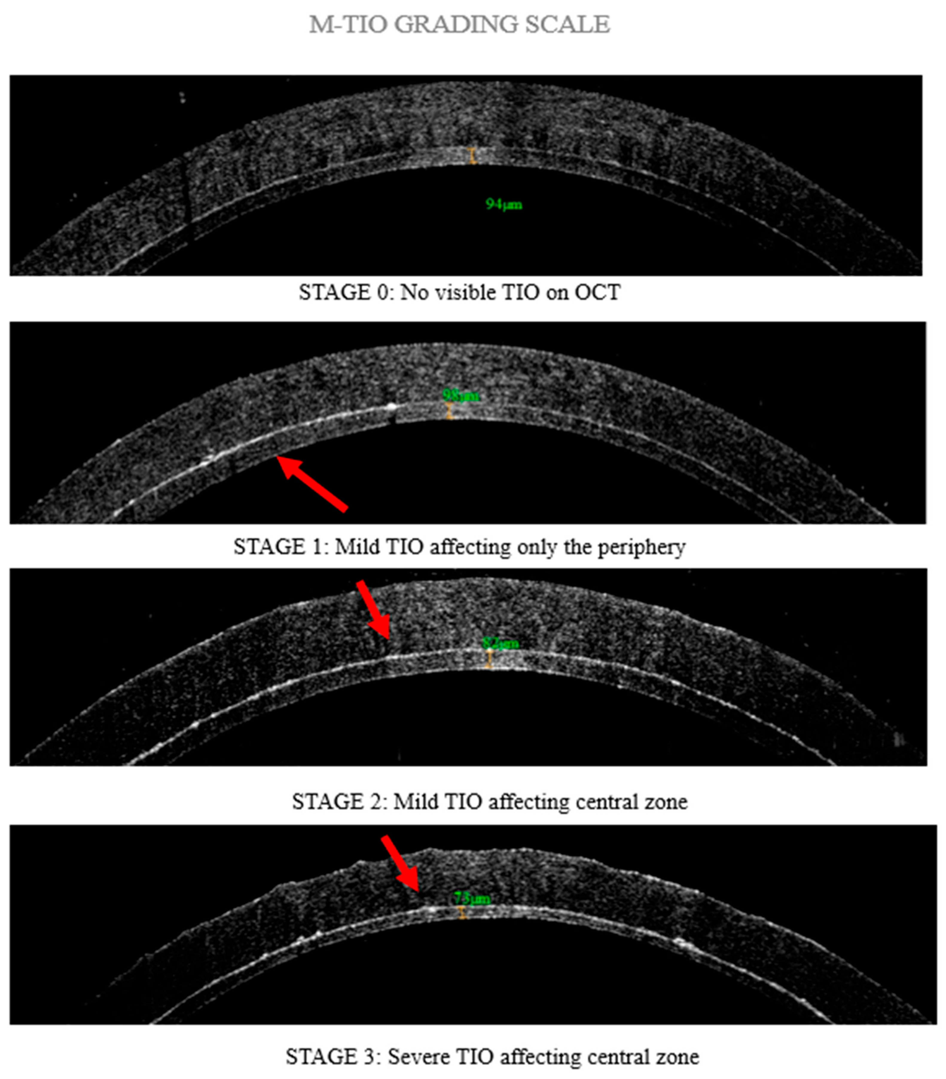
2.1. DSAEK Graft Preparation
2.2. Data Analysis
2.3. Statistical Analysis
3. Results
4. Discussion
5. Conclusions
Author Contributions
Funding
Institutional Review Board Statement
Informed Consent Statement
Data Availability Statement
Acknowledgments
Conflicts of Interest
References
- Van Meter, W.; Mathews, P.; Philippy, B.; DeMatteo, J. 2023 Eye Banking Statistical Report—Executive Summary. Eye Bank. Corneal Transplant. 2024, 3, e0034. [Google Scholar] [CrossRef]
- Stuart, A.J.; Romano, V.; Virgili, G.; Shortt, A.J. Descemet’s Membrane Endothelial Keratoplasty (DMEK) versus Descemet’s Stripping Automated Endothelial Keratoplasty (DSAEK) for Corneal Endothelial Failure. Cochrane Database Syst. Rev. 2018, 2018, 6. [Google Scholar] [CrossRef]
- Busin, M.; Patel, A.K.; Scorcia, V.; Ponzin, D. Microkeratome-Assisted Preparation of Ultrathin Grafts for Descemet Stripping Automated Endothelial Keratoplasty. Investig. Opthalmology Vis. Sci. 2012, 53, 521. [Google Scholar] [CrossRef] [PubMed]
- Dickman, M.M.; Kruit, P.J.; Remeijer, L.; van Rooij, J.; Van der Lelij, A.; Wijdh, R.H.J.; van den Biggelaar, F.J.H.M.; Berendschot, T.T.J.M.; Nuijts, R.M.M.A. A Randomized Multicenter Clinical Trial of Ultrathin Descemet Stripping Automated Endothelial Keratoplasty (DSAEK) versus DSAEK. Ophthalmology 2016, 123, 2276–2284. [Google Scholar] [CrossRef]
- Busin, M.; Madi, S.; Santorum, P.; Scorcia, V.; Beltz, J. Ultrathin Descemet’s Stripping Automated Endothelial Keratoplasty with the Microkeratome Double-Pass Technique. Ophthalmology 2013, 120, 1186–1194. [Google Scholar] [CrossRef]
- Droutsas, K.; Petrelli, M.; Miltsakakis, D.; Andreanos, K.; Karagianni, A.; Lazaridis, A.; Koutsandrea, C.; Kymionis, G. Visual Outcomes of Ultrathin-Descemet Stripping Endothelial Keratoplasty versus Descemet Stripping Endothelial Keratoplasty. J. Ophthalmol. 2018, 2018, 5924058. [Google Scholar] [CrossRef]
- Weisenthal, R.W.; Yin, H.Y.; Jarstad, A.R.; Wang, D.; Verdier, D.D. Long-Term Outcomes in Fellow Eyes Comparing DSAEK and DMEK for Treatment of Fuchs Corneal Dystrophy. Am. J. Ophthalmol. 2022, 233, 216–226. [Google Scholar] [CrossRef] [PubMed]
- Lee, W.B.; Jacobs, D.S.; Musch, D.C.; Kaufman, S.C.; Reinhart, W.J.; Shtein, R.M. Descemet’s Stripping Endothelial Keratoplasty: Safety and Outcomes. Ophthalmology 2009, 116, 1818–1830. [Google Scholar] [CrossRef]
- Basak, S.; Basak, S. Complications and Management in Descemet′s Stripping Endothelial Keratoplasty: Analysis of Consecutive 430 Cases. Indian J. Ophthalmol. 2014, 62, 209–218. [Google Scholar] [CrossRef]
- Shih, C.Y.; Ritterband, D.C.; Rubino, S.; Palmiero, P.-M.; Jangi, A.; Liebmann, J.; Ritch, R.; Seedor, J.A. Visually Significant and Nonsignificant Complications Arising From Descemet Stripping Automated Endothelial Keratoplasty. Am. J. Ophthalmol. 2009, 148, 837–843. [Google Scholar] [CrossRef]
- Vira, S.; Shih, C.Y.; Ragusa, N.; Sheyman, A.; Feder, R.; Weisenthal, R.W.; Rosenwasser, G.O.D.; Hannush, S.B.; Udell, I.J.; Bouchard, C.S. Textural Interface Opacity After Descemet Stripping Automated Endothelial Keratoplasty. Cornea 2013, 32, e54–e59. [Google Scholar] [CrossRef] [PubMed]
- Kymionis, G.D. Interface Wavelike Deposits After Descemet Stripping Automated Endothelial Keratoplasty. Arch. Ophthalmol. 2009, 127, 1389. [Google Scholar] [CrossRef] [PubMed]
- Kim, K.; Alder, B.; Vora, G.K.; Carlson, A.N.; Afshari, N.A.; Kuo, A.N.; Kim, T. Textural Interface Opacity after Descemet-Stripping Automated Endothelial Keratoplasty. J. Cataract. Refract. Surg. 2014, 40, 1514–1520. [Google Scholar] [CrossRef]
- Juthani, V.V.; Goshe, J.M.; Srivastava, S.K.; Ehlers, J.P. Association Between Transient Interface Fluid on Intraoperative OCT and Textural Interface Opacity After DSAEK Surgery in the PIONEER Study. Cornea 2014, 33, 887–892. [Google Scholar] [CrossRef]
- Newman, L.R.; Rosenwasser, G.O.D.; Dubovy, S.R.; Matthews, J.L. Clinicopathologic Correlation of Textural Interface Opacities in Descemet Stripping Automated Endothelial Keratoplasty. Cornea 2014, 33, 306–309. [Google Scholar] [CrossRef]
- Kymionis, G.; Voulgari, N.; Kontadakis, G.; Mikropoulos, D.; Petrovic, A.; Droutsas, K. Surgical Management of Post-Descemet Stripping Automated Endothelial Keratoplasty Interface Haze Associated with Interface Deposits. Indian J. Ophthalmol. 2020, 68, 174. [Google Scholar] [CrossRef] [PubMed]
- Kontadakis, G.A.; Palioura, S.; Yoo, S.H. Wavelike Interface Opacities After Descemet-Stripping Automated Endothelial Keratoplasty: 7-Year Follow-Up. Eye Contact Lens Sci. Clin. Pract. 2017, 43, e13–e15. [Google Scholar] [CrossRef]
- Graffi, S.; Leon, P.; Mimouni, M.; Nahum, Y.; Spena, R.; Mattioli, L.; Busin, M. Anterior Segment Optical Coherence Tomography of Post-Descemet Stripping Automated Endothelial Keratoplasty Eyes to Evaluate Graft Morphology and Its Association With Visual Outcome. Cornea 2018, 37, 1087–1092. [Google Scholar] [CrossRef]
- Shih, C.Y.; Ritterband, D.C.; Palmiero, P.-M.; Seedor, J.A.; Papachristou, G.; Harizman, N.; Liebmann, J.M.; Ritch, R. The Use of Postoperative Slit-Lamp Optical Coherence Tomography to Predict Primary Failure in Descemet Stripping Automated Endothelial Keratoplasty. Am. J. Ophthalmol. 2009, 147, 796–800.e1. [Google Scholar] [CrossRef]
- Neokleous, A.; Michail, N.; Herodotou, F.; Athanasiadou, A.; Christodoulou, S.; Kola, D.; Panayidou, K.; Hadjilouka, G.; Palioura, S. Long-Term Monitoring of Corneal Grafts Via Anterior Segment OCT Pachymetry Maps. Ophthalmol. Sci. 2025, 5, 100724. [Google Scholar] [CrossRef]
- Vajpayee, R.B.; Maharana, P.K.; Jain, S.; Sharma, N.; Jhanji, V. Thin Lenticule <scp>D</Scp> Escemet’s Stripping Automated Endothelial Keratoplasty: Single, Slow Pass Technique. Clin. Exp. Ophthalmol. 2014, 42, 411–416. [Google Scholar] [CrossRef]
- Clerici, R.; Ceccuzzi, R.; Fausto, R.; Tinelli, C.; Di Palma, M.R.; Mantegna, G.; Riva, I.; Busin, M.; De Angelis, G.; Quaranta, L. Single-Pass Mikrokeratome and Anterior Chamber Pressurizer for the Ultrathin Descemet-Stripping Automated Endothelial Keratoplasty Graft Preparation. Cornea 2021, 40, 755–763. [Google Scholar] [CrossRef]
- Eye Bank Association of America Medical Standards 2023. Eye Bank. Corneal Transplant. 2024, 3, e0027. [CrossRef]
- Wang, Q.; Stoakes, I.M.; Moshirfar, M.; Harvey, D.H.; Hoopes, P.C. Assessment of Pupil Size and Angle Kappa in Refractive Surgery: A Population-Based Epidemiological Study in Predominantly American Caucasians. Cureus 2023, 15, 43998. [Google Scholar] [CrossRef] [PubMed]
- Duggan, M.J.; Rose-Nussbaumer, J.; Lin, C.C.; Austin, A.; Labadzinzki, P.C.; Chamberlain, W.D. Corneal Higher-Order Aberrations in Descemet Membrane Endothelial Keratoplasty versus Ultrathin DSAEK in the Descemet Endothelial Thickness Comparison Trial. Ophthalmology 2019, 126, 946–957. [Google Scholar] [CrossRef] [PubMed]
- Sami Khan, F.; Khalid, M. Examining Higher Order Aberration in Eyes after DSEK. Punjab Univ. J. Math. 2022, 54, 89–101. [Google Scholar] [CrossRef]
- Chaudhury, P.; Singh, P. Role of Pin Hole in Rapid Recognition of Refractive Error as Cause of Diminished Vision. J. Evol. Med. Dent. Sci. 2022, 11, 452–454. [Google Scholar] [CrossRef]
- Melki, S.A.; Safar, A.; Martin, J.; Ivanova, A.; Adi, M. Potential Acuity Pinhole. Ophthalmology 1999, 106, 1262–1267. [Google Scholar] [CrossRef]
- Narang, P.; Agarwal, A.; Kumar, D.A.; Agarwal, A. Pinhole Pupilloplasty: Small-Aperture Optics for Higher-Order Corneal Aberrations. J. Cataract. Refract. Surg. 2019, 45, 539–543. [Google Scholar] [CrossRef]
- Nakagawa, S.; Schielzeth, H. A General and Simple Method for Obtaining R 2 from Generalized Linear Mixed-effects Models. Methods Ecol. Evol. 2013, 4, 133–142. [Google Scholar] [CrossRef]
- Tang, M.; Stoeger, C.; Galloway, J.; Holiman, J.; Bald, M.R.; Huang, D. Evaluating DSAEK Graft Deturgescence in Preservation Medium After Microkeratome Cut With Optical Coherence Tomography. Cornea 2013, 32, 847–850. [Google Scholar] [CrossRef] [PubMed]
- Eguchi, H.; Hotta, F.; Kusaka, S.; Shimomura, Y. Intraoperative Optical Coherence Tomography Imaging in Corneal Surgery: A Literature Review and Proposal of Novel Applications. J. Ophthalmol. 2020, 2020, 1497089. [Google Scholar] [CrossRef] [PubMed]
- Hindman, H.B.; Huxlin, K.R.; Pantanelli, S.M.; Callan, C.L.; Sabesan, R.; Ching, S.S.T.; Miller, B.E.; Martin, T.; Yoon, G. Post-DSAEK Optical Changes. Cornea 2013, 32, 1567–1577. [Google Scholar] [CrossRef] [PubMed]


| Linear Mixed Model 1 | |||
|---|---|---|---|
| N = 221 | Mean logMAR (99% CI) | p-Value | |
| TIO | <0.001 | ||
| 0 | 44 | 0.151 (0.077, 0.225) | |
| 1 | 110 | 0.311 (0.264, 0.359) | |
| 2 | 56 | 0.424 (0.358, 0.490) | |
| 3 | 11 | 0.680 (0.532, 0.828) | |
| Linear Mixed Model 1 | ||
|---|---|---|
| Mean Diff logMAR (99% CI 2) | p-Value 2 | |
| TIO | ||
| Grade 1–Grade 0 | 0.160 (0.054, 0.266) | <0.001 |
| Grade 2–Grade 0 | 0.273 (0.154, 0.392) | <0.001 |
| Grade 2–Grade 1 | 0.113 (0.016, 0.210) | 0.002 |
| Grade 3–Grade 0 | 0.529 (0.331, 0.727) | <0.001 |
| Grade 3–Grade 1 | 0.369 (0.183, 0.556) | <0.001 |
| Grade 3–Grade 2 | 0.256 (0.062, 0.451) | <0.001 |
Disclaimer/Publisher’s Note: The statements, opinions and data contained in all publications are solely those of the individual author(s) and contributor(s) and not of MDPI and/or the editor(s). MDPI and/or the editor(s) disclaim responsibility for any injury to people or property resulting from any ideas, methods, instructions or products referred to in the content. |
© 2025 by the authors. Licensee MDPI, Basel, Switzerland. This article is an open access article distributed under the terms and conditions of the Creative Commons Attribution (CC BY) license (https://creativecommons.org/licenses/by/4.0/).
Share and Cite
Chatzea, M.S.; Kymionis, G.D.; Vakalopoulos, D.G.; O’Brien, R.C.; Mora, D.; Llanes, K.; Fout, E.; Buras, W.; Triglia, C.; Tonk, R.S.; et al. Screening and Grading of Textural Interface Opacities in DSAEK Grafts with the M-TIO Scale for Predicting Visual Outcomes. Diagnostics 2025, 15, 1241. https://doi.org/10.3390/diagnostics15101241
Chatzea MS, Kymionis GD, Vakalopoulos DG, O’Brien RC, Mora D, Llanes K, Fout E, Buras W, Triglia C, Tonk RS, et al. Screening and Grading of Textural Interface Opacities in DSAEK Grafts with the M-TIO Scale for Predicting Visual Outcomes. Diagnostics. 2025; 15(10):1241. https://doi.org/10.3390/diagnostics15101241
Chicago/Turabian StyleChatzea, Marina S., George D. Kymionis, Dionysios G. Vakalopoulos, Robert C. O’Brien, Daniella Mora, Katrina Llanes, Elizabeth Fout, William Buras, Concetta Triglia, Rahul S. Tonk, and et al. 2025. "Screening and Grading of Textural Interface Opacities in DSAEK Grafts with the M-TIO Scale for Predicting Visual Outcomes" Diagnostics 15, no. 10: 1241. https://doi.org/10.3390/diagnostics15101241
APA StyleChatzea, M. S., Kymionis, G. D., Vakalopoulos, D. G., O’Brien, R. C., Mora, D., Llanes, K., Fout, E., Buras, W., Triglia, C., Tonk, R. S., & Yoo, S. H. (2025). Screening and Grading of Textural Interface Opacities in DSAEK Grafts with the M-TIO Scale for Predicting Visual Outcomes. Diagnostics, 15(10), 1241. https://doi.org/10.3390/diagnostics15101241