
The Contribution of Vessel Wall Magnetic Resonance Imaging to the Diagnosis of Primary and Secondary Central Nervous System Vasculitis
 , , ,
, , ,
Abstract
1. Introduction
2. Materials and Methods
2.1. Study Design
2.2. Imaging Assessment
2.3. Image Evaluation
2.4. Statistical Analyses
3. Results
3.1. Patient Population
3.2. Primary Central Nervous System Vasculitis
3.3. MRI Findings
3.4. Secondary Central Nervous System Vasculitis
3.5. Secondary Central Nervous System Vasculitis—Systemic Immunomediated
3.6. Infectious Central Nervous System Vasculitis
4. Discussion
5. Limitations
6. Conclusions
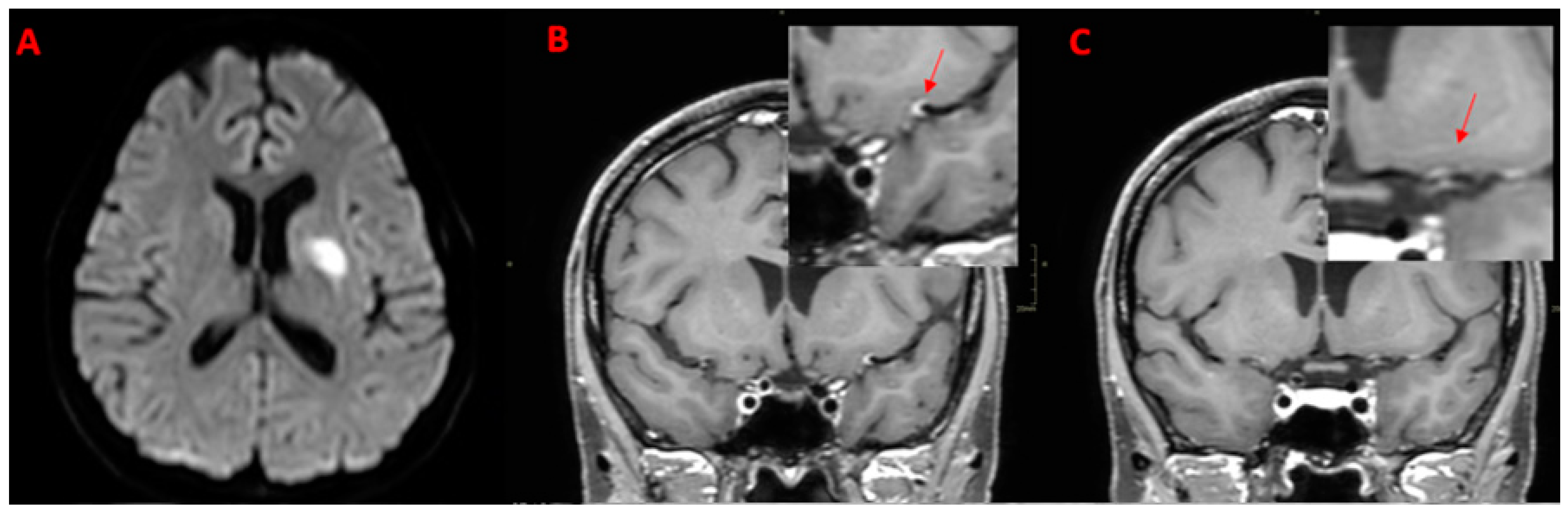
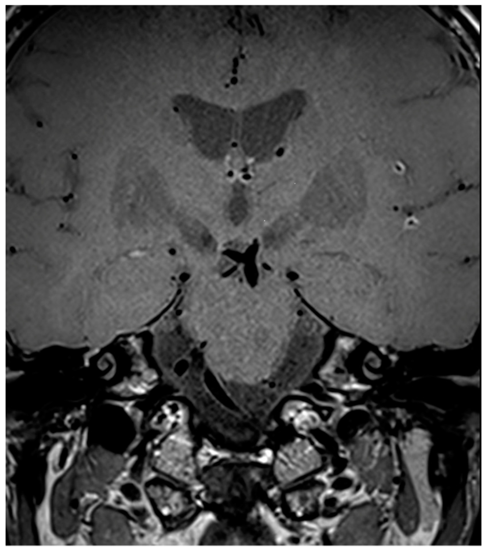
7. Legend
Author Contributions
Funding
Institutional Review Board Statement
Informed Consent Statement
Data Availability Statement
Conflicts of Interest
References
- Salvarani, C.; Brown, R.D., Jr.; Hunder, G.G. Adult primary central nervous system vasculitis. Lancet 2012, 380, 767–777. [Google Scholar] [CrossRef] [PubMed]
- Calabrese, L.H.; Mallek, J.A. Primary angiitis of the central nervous system. Report of 8 new cases, review of the literature, and proposal for diagnostic criteria. Medicine 1988, 67, 20–39. [Google Scholar] [CrossRef] [PubMed]
- Hajj-Ali, R.A.; Calabrese, L.H. Central nervous system vasculitis: Advances in diagnosis. Curr. Opin. Rheumatol. 2020, 32, 41–46. [Google Scholar] [CrossRef] [PubMed]
- Godasi, R.; Pang, G.; Chauhan, S.; Bollu, P.C. Primary Central Nervous System Vasculitis. In StatPearls; StatPearls Publishing Copyright © 2024, StatPearls Publishing LLC.: Treasure Island, FL, USA, 2024. [Google Scholar]
- Ferro, F.; Quartuccio, L.; Monti, S.; Delvino, P.; Di Cianni, F.; Fonzetti, S.; La Rocca, G.; Posarelli, C.; Treppo, E.; Talarico, R.; et al. One year in review 2021: Systemic vasculitis. Clin. Exp. Rheumatol. 2021, 39 (Suppl. 129), 3–12. [Google Scholar] [CrossRef] [PubMed]
- Abdel Razek, A.A.; Alvarez, H.; Bagg, S.; Refaat, S.; Castillo, M. Imaging spectrum of CNS vasculitis. Radiographics 2014, 34, 873–894. [Google Scholar] [CrossRef] [PubMed]
- Néel, A.; Pagnoux, C. Primary angiitis of the central nervous system. Clin. Exp. Rheumatol. 2009, 27, S95–S107. [Google Scholar] [PubMed]
- Merli, E.; Rustici, A.; Gramegna, L.L.; Di Donato, M.; Agati, R.; Tonon, C.; Lodi, R.; Favoni, V.; Pierangeli, G.; Cortelli, P.; et al. Vessel-wall MRI in primary headaches: The role of neurogenic inflammation. Headache 2023, 63, 1372–1379. [Google Scholar] [CrossRef] [PubMed]
- Rustici, A.; Merli, E.; Cevoli, S.; Donato, M.D.; Pierangeli, G.; Favoni, V.; Bortolotti, C.; Sturiale, C.; Cortelli, P.; Cirillo, L. Vessel-wall MRI in thunderclap headache: A useful tool to answer the riddle? Interv. Neuroradiol. 2021, 27, 219–224. [Google Scholar] [CrossRef] [PubMed]
- Mossa-Basha, M.; Hwang, W.D.; De Havenon, A.; Hippe, D.; Balu, N.; Becker, K.J.; Tirschwell, D.T.; Hatsukami, T.; Anzai, Y.; Yuan, C. Multicontrast high-resolution vessel wall magnetic resonance imaging and its value in differentiating intracranial vasculopathic processes. Stroke 2015, 46, 1567–1573. [Google Scholar] [CrossRef] [PubMed]
- Arnett, N.; Pavlou, A.; Burke, M.P.; Cucchiara, B.L.; Rhee, R.L.; Song, J.W. Vessel wall MR imaging of central nervous system vasculitis: A systematic review. Neuroradiology 2022, 64, 43–58. [Google Scholar] [CrossRef]
- Wu, F.; Ma, Q.; Song, H.; Guo, X.; Diniz, M.A.; Song, S.S.; Gonzalez, N.R.; Bi, X.; Ji, X.; Li, D.; et al. Differential Features of Culprit Intracranial Atherosclerotic Lesions: A Whole-Brain Vessel Wall Imaging Study in Patients With Acute Ischemic Stroke. J. Am. Heart Assoc. 2018, 7, e009705. [Google Scholar] [CrossRef] [PubMed]
- Berlit, P.; Kraemer, M. Cerebral vasculitis in adults: What are the steps in order to establish the diagnosis? Red flags and pitfalls. Clin. Exp. Immunol. 2014, 175, 419–424. [Google Scholar] [CrossRef]
- Cirillo, L.; Rustici, A.; Toni, F.; Zoli, M.; Bartiromo, F.; Gramegna, L.L.; Cicala, D.; Tonon, C.; Caranci, F.; Lodi, R. Vessel Wall MRI: Clinical implementation in cerebrovascular disorders-technical aspects. Radiol. Med. 2022, 127, 645–651. [Google Scholar] [CrossRef] [PubMed]
- Godi, C.; Destro, F.; Garofalo, P.; Tombetti, E.; Ambrosi, A.; Iadanza, A.; Michelozzi, C.; Falini, A.; Anzalone, N. Hemodynamic nature of black-blood enhancement in long-term coiled cerebral aneurysms. Neuroradiology 2023, 65, 1685–1694. [Google Scholar] [CrossRef] [PubMed]
- Fazekas, F.; Chawluk, J.B.; Alavi, A.; Hurtig, H.I.; Zimmerman, R.A. MR signal abnormalities at 1.5 T in Alzheimer’s dementia and normal aging. AJR Am. J. Roentgenol. 1987, 149, 351–356. [Google Scholar] [CrossRef] [PubMed]
- Venkataraman, P.; Tadi, P.; Lui, F. Lacunar Syndromes. Available online: https://www.ncbi.nlm.nih.gov/books/NBK534206/ (accessed on 20 December 2023).
- Mandell, D.M.; Mossa-Basha, M.; Qiao, Y.; Hess, C.P.; Hui, F.; Matouk, C.; Johnson, M.H.; Daemen, M.J.; Vossough, A.; Edjlali, M.; et al. Intracranial Vessel Wall MRI: Principles and Expert Consensus Recommendations of the American Society of Neuroradiology. AJNR Am. J. Neuroradiol. 2017, 38, 218–229. [Google Scholar] [CrossRef] [PubMed]
- Pascarella, R.; Antonenko, K.; Boulouis, G.; De Boysson, H.; Giannini, C.; Heldner, M.R.; Kargiotis, O.; Nguyen, T.N.; Rice, C.M.; Salvarani, C.; et al. European Stroke Organisation (ESO) guidelines on Primary Angiitis of the Central Nervous System (PACNS). Eur. Stroke J. 2023, 8, 842–879. [Google Scholar] [CrossRef] [PubMed]
- Miller, D.V.; Salvarani, C.; Hunder, G.G.; Brown, R.D.; Parisi, J.E.; Christianson, T.J.; Giannini, C. Biopsy findings in primary angiitis of the central nervous system. Am. J. Surg. Pathol. 2009, 33, 35–43. [Google Scholar] [CrossRef] [PubMed]
- Salvarani, C.; Brown, R.D., Jr.; Hunder, G.G. Adult primary central nervous system vasculitis: An update. Curr. Opin. Rheumatol. 2012, 24, 46–52. [Google Scholar] [CrossRef]
- Harland, T.A.; Seinfeld, J.; Cava, L.F.; Neumann, R.T.; Roark, C.; Kumpe, D.; Case, D. Anti-neutrophil cytoplasmic antibody associated central nervous system vasculitis with brain and spinal cord subarachnoid hemorrhage: A rare case report and review of the literature. J. Clin. Neurosci. 2019, 62, 253–255. [Google Scholar] [CrossRef]
- Pipitone, N.; Salvarani, C. The role of infectious agents in the pathogenesis of vasculitis. Best. Pract. Res. Clin. Rheumatol. 2008, 22, 897–911. [Google Scholar] [CrossRef] [PubMed]
- Graf, J. Central Nervous System Disease in Antineutrophil Cytoplasmic Antibodies-Associated Vasculitis. Rheum. Dis. Clin. N. Am. 2017, 43, 573–578. [Google Scholar] [CrossRef] [PubMed]
- Rodrigues, M.; Galego, O.; Costa, C.; Jesus, D.; Carvalho, P.; Santiago, M.; Malcata, A.; Ines, L. Central nervous system vasculitis in systemic lupus erythematosus: A case series report in a tertiary referral centre. Lupus 2017, 26, 1440–1447. [Google Scholar] [CrossRef] [PubMed]
- Rowshani, A.T.; Remans, P.; Rozemuller, A.; Tak, P.P. Cerebral vasculitis as a primary manifestation of systemic lupus erythematosus. Ann. Rheum. Dis. 2005, 64, 784–786. [Google Scholar] [CrossRef] [PubMed]
- Mazzacane, F.; Mazzoleni, V.; Scola, E.; Mancini, S.; Lombardo, I.; Busto, G.; Rognone, E.; Pichiecchio, A.; Padovani, A.; Morotti, A.; et al. Vessel Wall Magnetic Resonance Imaging in Cerebrovascular Diseases. Diagnostics 2022, 12, 258. [Google Scholar] [CrossRef] [PubMed]
- Küker, W. Imaging of cerebral vasculitis. Int. J. Stroke 2007, 2, 184–190. [Google Scholar] [CrossRef] [PubMed]
- Jennette, J.C.; Falk, R.J.; Bacon, P.A.; Basu, N.; Cid, M.C.; Ferrario, F.; Flores-Suarez, L.F.; Gross, W.L.; Guillevin, L.; Hagen, E.C.; et al. 2012 revised International Chapel Hill Consensus Conference Nomenclature of Vasculitides. Arthritis Rheum 2013, 65, 1–11. [Google Scholar] [CrossRef] [PubMed]
- Guggenberger, K.V.; Torre, G.D.; Ludwig, U.; Vogel, P.; Weng, A.M.; Vogt, M.L.; Fröhlich, M.; Schmalzing, M.; Raithel, E.; Forman, C.; et al. Vasa vasorum of proximal cerebral arteries after dural crossing–potential imaging confounder in diagnosing intracranial vasculitis in elderly subjects on black-blood MRI. Eur. Radiol. 2022, 32, 1276–1284. [Google Scholar] [CrossRef] [PubMed]
- Birnbaum, J.; Hellmann, D.B. Primary angiitis of the central nervous system. Arch. Neurol. 2009, 66, 704–709. [Google Scholar] [CrossRef]
- Mossa-Basha, M.; Alexander, M.; Gaddikeri, S.; Yuan, C.; Gandhi, D. Vessel wall imaging for intracranial vascular disease evaluation. J. Neurointerv. Surg. 2016, 8, 1154–1159. [Google Scholar] [CrossRef]
- Edjlali, M.; Qiao, Y.; Boulouis, G.; Menjot, N.; Saba, L.; Wasserman, B.A.; Romero, J.M. Vessel wall MR imaging for the detection of intracranial inflammatory vasculopathies. Cardiovasc. Diagn. Ther. 2020, 10, 1108–1119. [Google Scholar] [CrossRef] [PubMed]
- Blitstein, M.K.; Tung, G.A. MRI of cerebral microhemorrhages. AJR Am. J. Roentgenol. 2007, 189, 720–725. [Google Scholar] [CrossRef] [PubMed]
- Haller, S.; Vernooij, M.W.; Kuijer, J.P.A.; Larsson, E.M.; Jäger, H.R.; Barkhof, F. Cerebral Microbleeds: Imaging and Clinical Significance. Radiology 2018, 287, 11–28. [Google Scholar] [CrossRef] [PubMed]


| Characteristic | N (%) [PCNSV, 6] | N (%) [Systemic SCNSV, 4] | N (%) [Infectious SCNSV, 4] |
|---|---|---|---|
| Gender, male | 3 (50.0%) | 2 (50.0%) | 3 (75.0%) |
| Age, <50 years | 2 (33.3%) | 2 (50.0%) | 2 (50.0%) |
| Risk factors | PCNSV (6) | Systemic SCNSV (4) | Infectious SCNSV (4) |
| Smoking | 1 (16.7%) | 2 (50.0%) | 2 (50.0%) |
| Hypertension | 1 (16.7%) | 2 (50.0%) | 1 (25.0%) |
| Diabetes | 0 (0.0%) | 3 (75.0%) | 0 (0.0%) |
| Dyslipidemia | 1 (16.7%) | 3 (75.0%) | 1 (25.0%) |
| Atrial fibrillation | 0 (0.0%) | 0 (0.0%) | 0 (0.0%) |
| Onset Event | CSF Test | Autoimmune Panel Tests | Other Serological Tests | PET Imaging | Clinical Diagnosis | |
|---|---|---|---|---|---|---|
| 50Y F | Episode suggestive of stroke | Positive (inflammatory pattern) proteins 364 (nv < 50), WBC 96 (nv < 5). Presence of oligoclonal bands | Negative | Negative | (n/a) | PCNSV |
| 52Y, F | Episode suggestive of stroke | Negative | Positive (ANA 1:160, anti-gastric parietal cells 44 (nv <10) | CRP (−) ESR (+) 72 (nv < 20) | Negative | PCNSV |
| 53Y M | Episode suggestive of stroke | Non-significative positive | Positive ENA+, anti-SSA RO52 | Negative | Negative | PCNSV |
| 63Y, M | Episode suggestive of stroke | Positive (inflammatory pattern) proteins 549 (nv < 50), WBC 171 (nv < 5). Presence of oligoclonal bands (some only intrathecal, some both in serum and in CSF in a mirror pattern) | Negative | Negative | Negative | PCNSV |
| 29Y, F | Episode of TIA | Not performed (lumbar puncture contraindicated) | Negative | Negative | Negative | PCNSV |
| 32Y, M | Episode suggestive of stroke | Negative | Negative | Negative | Negative | PCNSV |
| WM Hyperintensities (Fazekas Scale) | Cortical/Subcortical Infarcts | Present/Absent DWI Hyperintensities (Subacute Events) | Site of Ischemic Lesions | Site of VWI CE | Secondary Sites of VWI CE | Present/Absent SWI Alterations (Microbleeds) | Site of SWI Alteration | |
|---|---|---|---|---|---|---|---|---|
| 50Y, F | 1 | Present | Present | Parietal lobe | MCA (M1) | None | Present | Supratentorial, subtentorial |
| 52Y, F | 1 | Present | Present | Multiple subcortical in MCA territories | MCA M2 | None | Absent | // |
| 53Y, M | 1 | Present | Present | Fight corona radiata | ICA | ACOP | Present | Supratentorial |
| 63Y, M | 1 | Present | Present | Multiple Subcortical in ACA, MCA Territories and Pons | ACA (A2) | None | Present | Supratentorial, subtentorial |
| 29Y, F | // | Absent | Absent | No | ICA | ACA (A1), MCA (M1) | Absent | // |
| 32, M | // | Absent | Present | No | ICA | ACA (A1), MCA (M1) | Absent | // |
| FLAIR CWM Hyperintensity (Fazekas Scale) | FLAIR Chronic Infarct (Cortical) | Leptomeningeal Enhancement | Parenchymal Enhancement | DWI Subacute Infarct | SWI Microbleed | MRA TOF Stenosis | Concentric VWI Wall Thickening | Eccentric VWI Wall Thickening | Concentric VWI Wall Enhancement | Eccentric VWI Wall Enhancement | Sites of CE | |
|---|---|---|---|---|---|---|---|---|---|---|---|---|
| PCNSV (n = 6) | 4 (66.7%) | 3 (50.0%) | 0 (0.0%) | 2 (33.33%) | 5 (83.3%) | 3 (50.0%) | 5 (83.3%) multiple: 2 (40.0%) | 4 (66.7%) | 0 (%) | 5 (83.3%) | 1 (16.7%) | MCA (M1, M2), ACA (A1, A2), ICA, ACOP |
| Onset Event | CSF Test | Autoimmune Panel Tests | Other Serological Tests | PET Imaging | Clinical Diagnosis | |
|---|---|---|---|---|---|---|
| 66Y, M | Episode suggestive of stroke | Negative | Positive ANA + (low title) | PCR (+) | Large-vessel uptake (aortic-supra-aortic vessels) | Systemic CNSV (Takayasu-like syndrome) |
| 32Y, F | Seizures | Positive (inflammatory pattern) proteins 141 (nv < 50), WBC 9 (nv < 5), | Positive ANA 1:320, ENA + (SSA, anti-RNP) | CRP (+) ESR (+) 106 (nv < 20) | (n/a) | Systemic CNSV (mixed connective tissue disease) |
| 49Y, M | Episode suggestive of stroke | Negative | Positive Anti-ENA (+) | (n/a) | Negative | Systemic CNSV (ulcerative colitis + vasculitis) |
| 83Y, F | Episode suggestive of stroke | (n/a) | Negative | CRP (+) ESR (+) 46 (nv < 20) | Negative | Systemic CNSV (GCA flare-up with CNS involvement) |
| WM Hyperintensities (Fazekas Scale) | Cortical/Subcortical Infarcts | Present/Absent DWI Hyperintensities (Subacute Events) | Site of Ischemic Lesions | Site of VWI CE | Secondary Sites of VWI CE | Present/Absent SWI Alterations (Microbleeds) | Site of SWI Alteration | |
|---|---|---|---|---|---|---|---|---|
| 66Y, M | 3 | Present | Present | Bilateral Basal Ganglia, Left SCA | SCA | ACM (M1) ICA BA | Present | Subtentorial |
| 32Y, F | 1 | Absent | Absent | No | None | None | Absent | // |
| 49Y, M | // | Absent | Absent | No | ACM (M2) | ACM (M2) | Present | Supratentorial |
| 83Y, F | 2 | Present | Present | Cortical-Subcortical in PCA Territory | PCA (P2) | None | Absent | // |
| Onset Event | CSF Test | Autoimmune Panel Tests | Other Serological Tests | PET Imaging | Clinical Diagnosis | |
|---|---|---|---|---|---|---|
| 49y, M | Headache, hearing disorders | Positive (inflammatory pattern + T. Pallidum positivity) proteins 82 (nv < 50), WBC 73 (nv < 5), TPHA liquor + 1:640 | Negative | T. Pallidum antibody-positive (TPHA > 1:640, RPR +) | (n/a) | Luetic CNSV |
| 58Y, M | Episode suggestive of stroke | Positive (inflammatory pattern + T. Pallidum positivity) proteins 103 (nv < 50), WBC 17 (nv < 5), TPHA liquor + 1:640 | Negative | CRP (+), T. Pallidum antibody-positive CRP 1,7 (nv < 0.5), ESR 52 (nv < 20), T. Pallidum antibody-positive (TPHA 1:640, RPR +) | Large-vessel uptake | Luetic CNSV |
| 5Y, M | Episode suggestive of stroke | (n/a) | Negative | VZV-IgM antibody-positive CRP (+) | (n/a) | VZV-CNSV |
| 39Y, F | Episode suggestive of stroke | Positive (inflammatory pattern + HIV antibodies + Epstein–Barr virus positivity 490 copies/mL) | Negative | CRP (+) | (n/a) | HIV/EBV-related CNSV |
| WM Hyperintensities (Fazekas Scale) | Cortical/Subcortical Infarcts | Present/Absent DWI Hyperintensities (Subacute Events) | Site of Ischemic Lesions | Site of VWI CE | Secondary Sites of VWI CE | Present/Absent SWI Alterations (Microbleeds) | Site of SWI Alteration | |
|---|---|---|---|---|---|---|---|---|
| 49Y, M | 1 | Present | Present | Multiple subcortical in MCA territories | ICA | MCA (M1) ACA (A1) | Present | Subtentorial |
| 58Y, M | 2 | Present | Present | Multiple subcortical in ACA and MCA territories and left SCA | ICA | None | Present | Supratentorial |
| 5Y, M | // | Absent | Absent | No | VA | BA | Present | Subtentorial |
| 39Y, F | 1 | Absent | Present | No | M1 | PCA (P1) | Absent | // |
| FLAIR CWM Hyperintensity (Fazekas Scale) | FLAIR Chronic Infarct (Cortical) | Leptomeningeal Enhancement | Parenchymal Enhancement | DWI Subacute Infarct | SWI Microbleed | MRA TOF Stenosis | Concentric VWI Wall Thickening | Eccentric VWI Wall Thickening | Concentric VWI Wall Enhancement | Eccentric VWI Wall Enhancement | Sites of VWI CE | |
|---|---|---|---|---|---|---|---|---|---|---|---|---|
| SCNSV Systemic disease (n = 4) | 3 (75.0%) | 2 (50.0%) | 0 (0.0%) | 2 (50.0%) | 2 (50.0%) | 2 (50.0%) | 3 (75.0%) | 2 (50.0%) | 0 (0%) | 3 (75.0%) | 0 (0%) | BA, VA, SCA, ICA, ACM (M1), PCA (P2) |
| multiple: 3 (100.0%) | ||||||||||||
| FLAIR CWM Hyperintensity (Fazekas Scale) | FLAIR Chronic Infarct (Cortical) | Leptomeningeal Enhancement | Parenchymal Enhancement | DWI subacute Infarct | SWI Microbleed | MRA TOF Stenosis | Concentric VWI Wall Thickening | Eccentric VWI Wall Thickening | Concentric VWI Wall Enhancement | Eccentric VWI Wall Enhancement | Sites of VWI CE | |
| SCNSV Luetic (n = 2) | 2 (100.0%) | 2 (100.0%) | 0 (0.0%) | 0 (0.0%) | 2 (100.0%) | 2 (100.0%) | 1 (50.0%) multiple: 1 (100.0%) | 1 (50.0%) | 0 (0%) | 2 (100.0%) | 0 (0%) | ACM(M1), ACA(A1), ICA |
| SCNSV VZV-related (n = 1) | 0 (0.0%) | 0 (0.0%) | 0 (0.0%) | 0 (0.0%) | 0 (0.0%) | 1 (100.0%) | 1 (100.0%) | 1 (100.0%) | 0 (0%) | 1 (100.0%) | 0 (0%) | BA, VA |
| multiple: 1 (100.0%) | ||||||||||||
| SCNSV HIV/EBV-related (n = 1) | 1 (100.0%) | 0 (0.0%) | 0 (0.0%) | 0 (0.0%) | 1 (100.0%) | 0 (0.0%) | 1 (100.0%) multiple: 1 (100.0%) | 1 (100.0%) | 0 (0.0%) | 1 (100.0%) | 0 (0.0%) | ICA, ACA (A1), MCA (M1) |
Disclaimer/Publisher’s Note: The statements, opinions and data contained in all publications are solely those of the individual author(s) and contributor(s) and not of MDPI and/or the editor(s). MDPI and/or the editor(s) disclaim responsibility for any injury to people or property resulting from any ideas, methods, instructions or products referred to in the content. |
© 2024 by the authors. Licensee MDPI, Basel, Switzerland. This article is an open access article distributed under the terms and conditions of the Creative Commons Attribution (CC BY) license (https://creativecommons.org/licenses/by/4.0/).
Share and Cite
D’Aniello, S.; Rustici, A.; Gramegna, L.L.; Godi, C.; Piccolo, L.; Gentile, M.; Zini, A.; Carrozzi, A.; Lodi, R.; Tonon, C.; et al. The Contribution of Vessel Wall Magnetic Resonance Imaging to the Diagnosis of Primary and Secondary Central Nervous System Vasculitis. Diagnostics 2024, 14, 927. https://doi.org/10.3390/diagnostics14090927
D’Aniello S, Rustici A, Gramegna LL, Godi C, Piccolo L, Gentile M, Zini A, Carrozzi A, Lodi R, Tonon C, et al. The Contribution of Vessel Wall Magnetic Resonance Imaging to the Diagnosis of Primary and Secondary Central Nervous System Vasculitis. Diagnostics. 2024; 14(9):927. https://doi.org/10.3390/diagnostics14090927
Chicago/Turabian StyleD’Aniello, Serena, Arianna Rustici, Laura Ludovica Gramegna, Claudia Godi, Laura Piccolo, Mauro Gentile, Andrea Zini, Alessandro Carrozzi, Raffaele Lodi, Caterina Tonon, and et al. 2024. "The Contribution of Vessel Wall Magnetic Resonance Imaging to the Diagnosis of Primary and Secondary Central Nervous System Vasculitis" Diagnostics 14, no. 9: 927. https://doi.org/10.3390/diagnostics14090927
APA StyleD’Aniello, S., Rustici, A., Gramegna, L. L., Godi, C., Piccolo, L., Gentile, M., Zini, A., Carrozzi, A., Lodi, R., Tonon, C., Dall’Olio, M., Simonetti, L., Chieffo, R., Anzalone, N., & Cirillo, L. (2024). The Contribution of Vessel Wall Magnetic Resonance Imaging to the Diagnosis of Primary and Secondary Central Nervous System Vasculitis. Diagnostics, 14(9), 927. https://doi.org/10.3390/diagnostics14090927