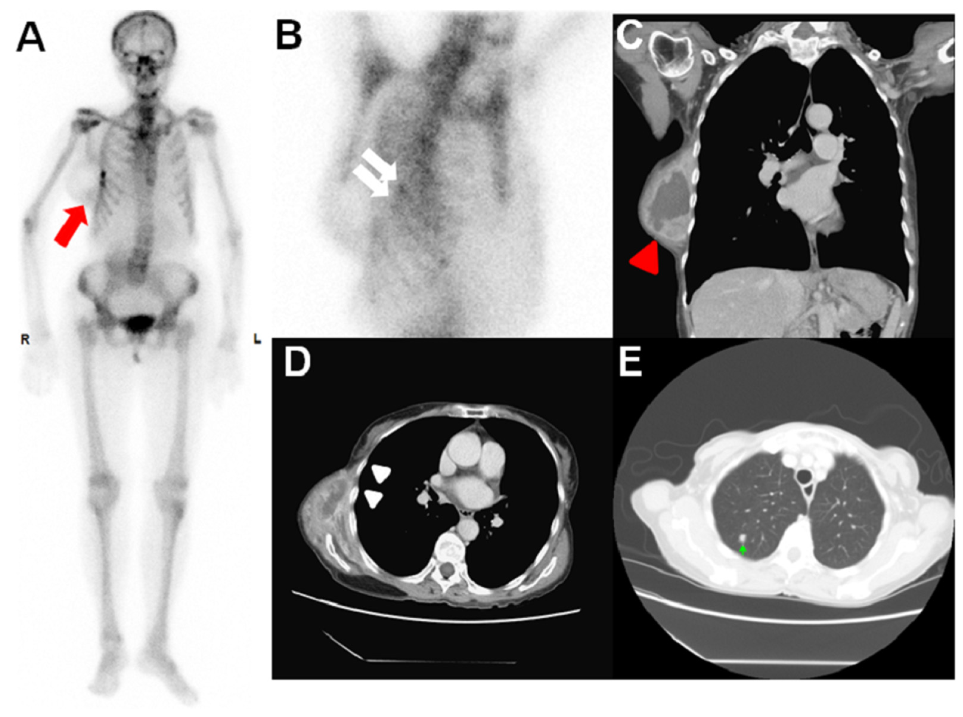
Myxofibrosarcoma of the Chest Wall Detected on 99mTc-MDP Whole-Body Bone Scan

 ,
, Abstract
Share and Cite
Lee, C.-H.; Shen, H.-Y.; Shiue, Y.-L.; Chan, H.-Y.; Chan, H.-P. Myxofibrosarcoma of the Chest Wall Detected on 99mTc-MDP Whole-Body Bone Scan. Diagnostics 2024, 14, 857. https://doi.org/10.3390/diagnostics14080857
Lee C-H, Shen H-Y, Shiue Y-L, Chan H-Y, Chan H-P. Myxofibrosarcoma of the Chest Wall Detected on 99mTc-MDP Whole-Body Bone Scan. Diagnostics. 2024; 14(8):857. https://doi.org/10.3390/diagnostics14080857
Chicago/Turabian StyleLee, Chia-Hsuan, Hueng-Yuan (Daniel) Shen, Yow-Ling (Shirley) Shiue, Hung-Yen Chan, and Hung-Pin Chan. 2024. "Myxofibrosarcoma of the Chest Wall Detected on 99mTc-MDP Whole-Body Bone Scan" Diagnostics 14, no. 8: 857. https://doi.org/10.3390/diagnostics14080857
APA StyleLee, C.-H., Shen, H.-Y., Shiue, Y.-L., Chan, H.-Y., & Chan, H.-P. (2024). Myxofibrosarcoma of the Chest Wall Detected on 99mTc-MDP Whole-Body Bone Scan. Diagnostics, 14(8), 857. https://doi.org/10.3390/diagnostics14080857