
A Rare Case of Dirofilariasis in the Genian Region

 , ,
, , 
Abstract
1. Introduction
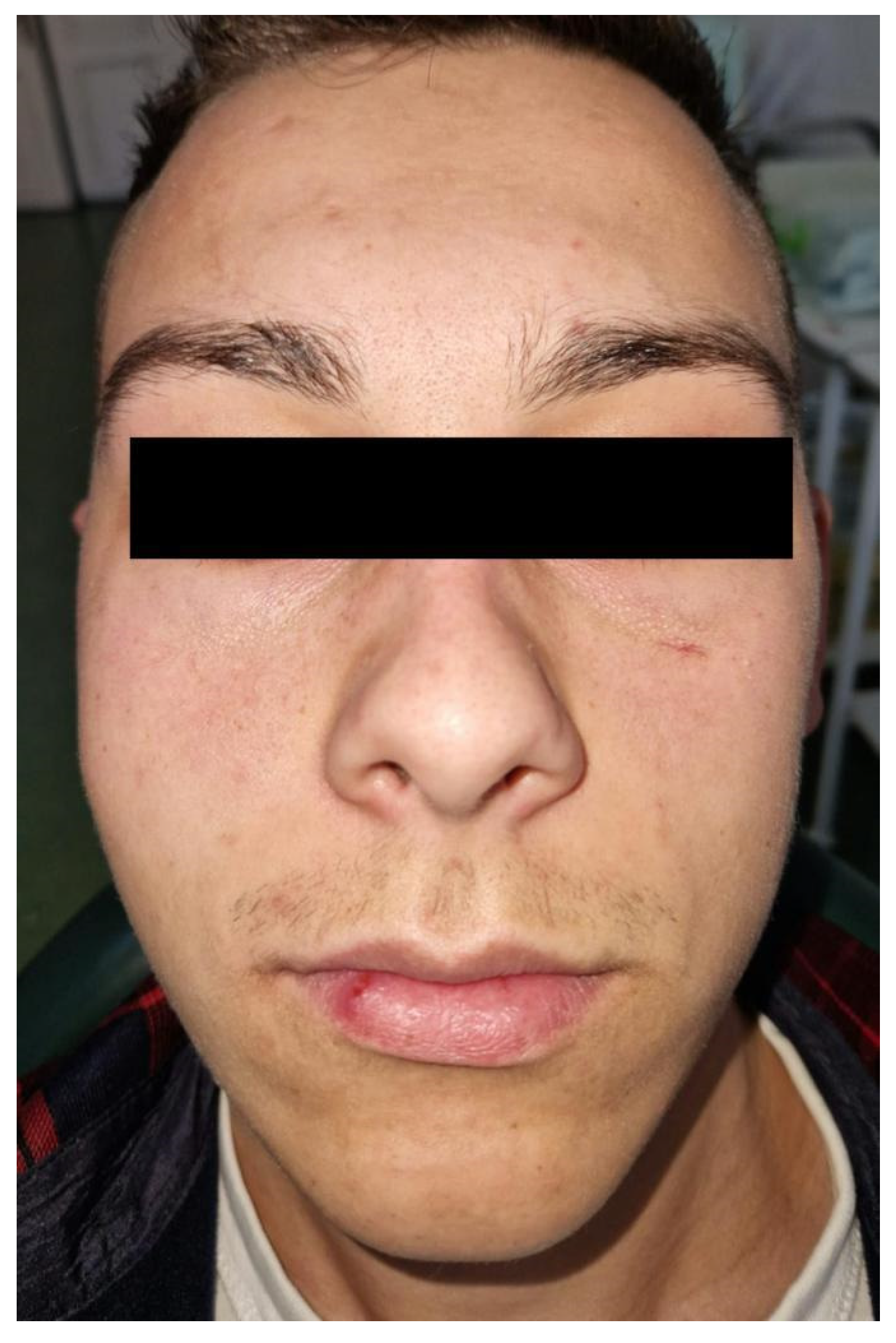
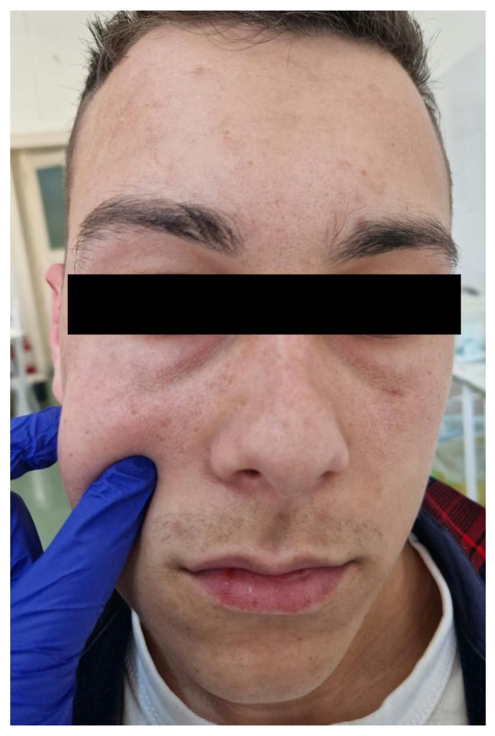
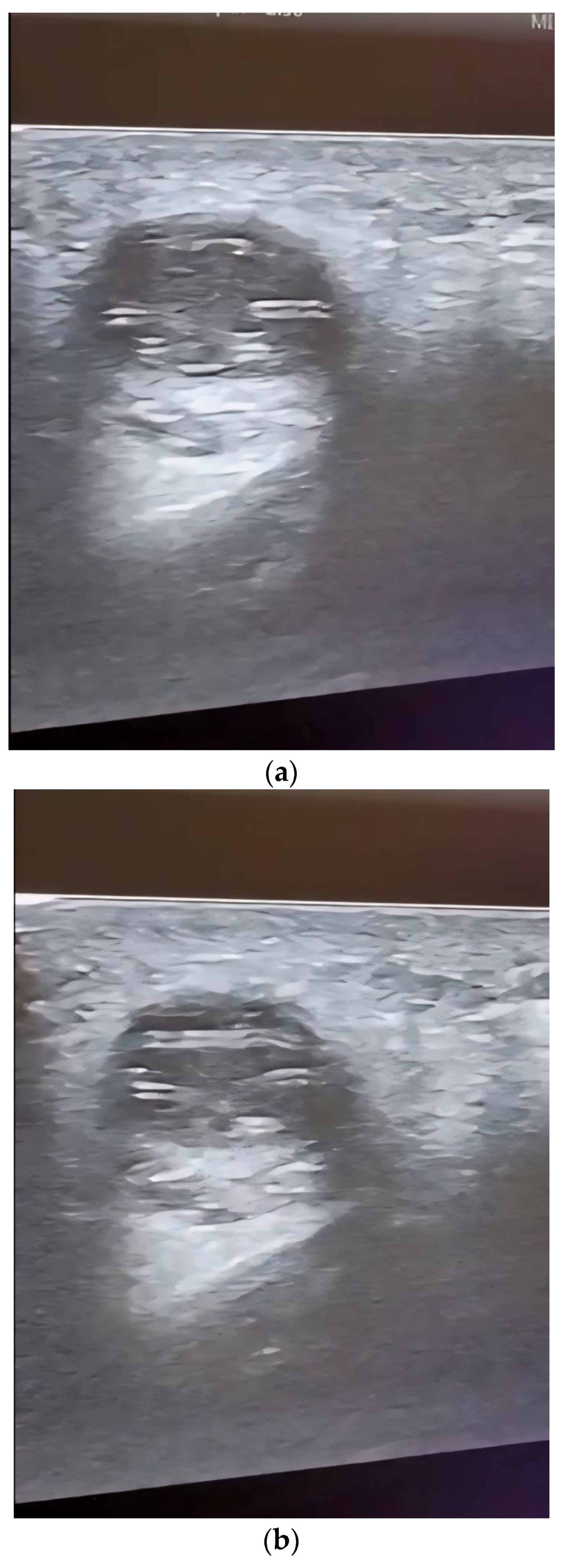
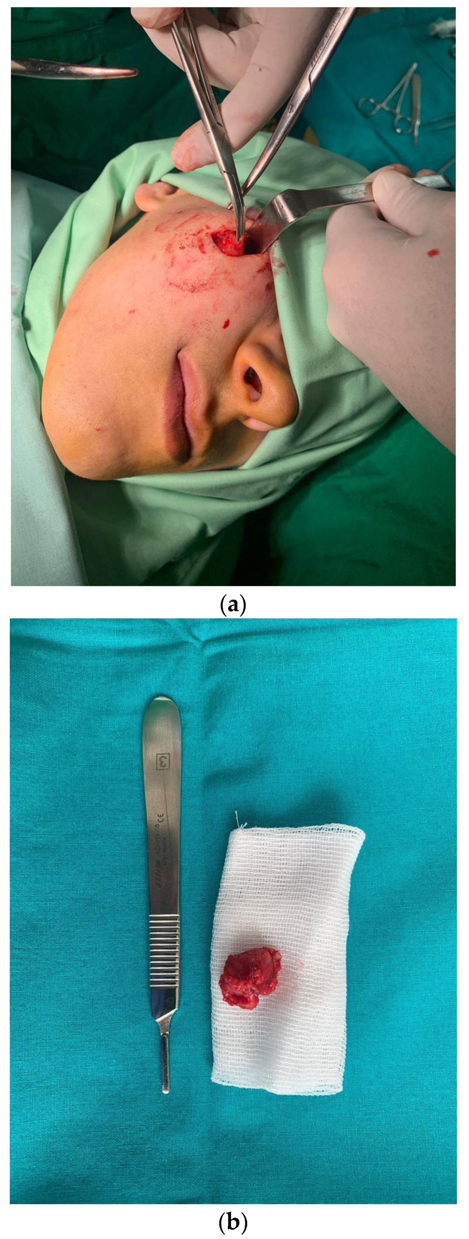
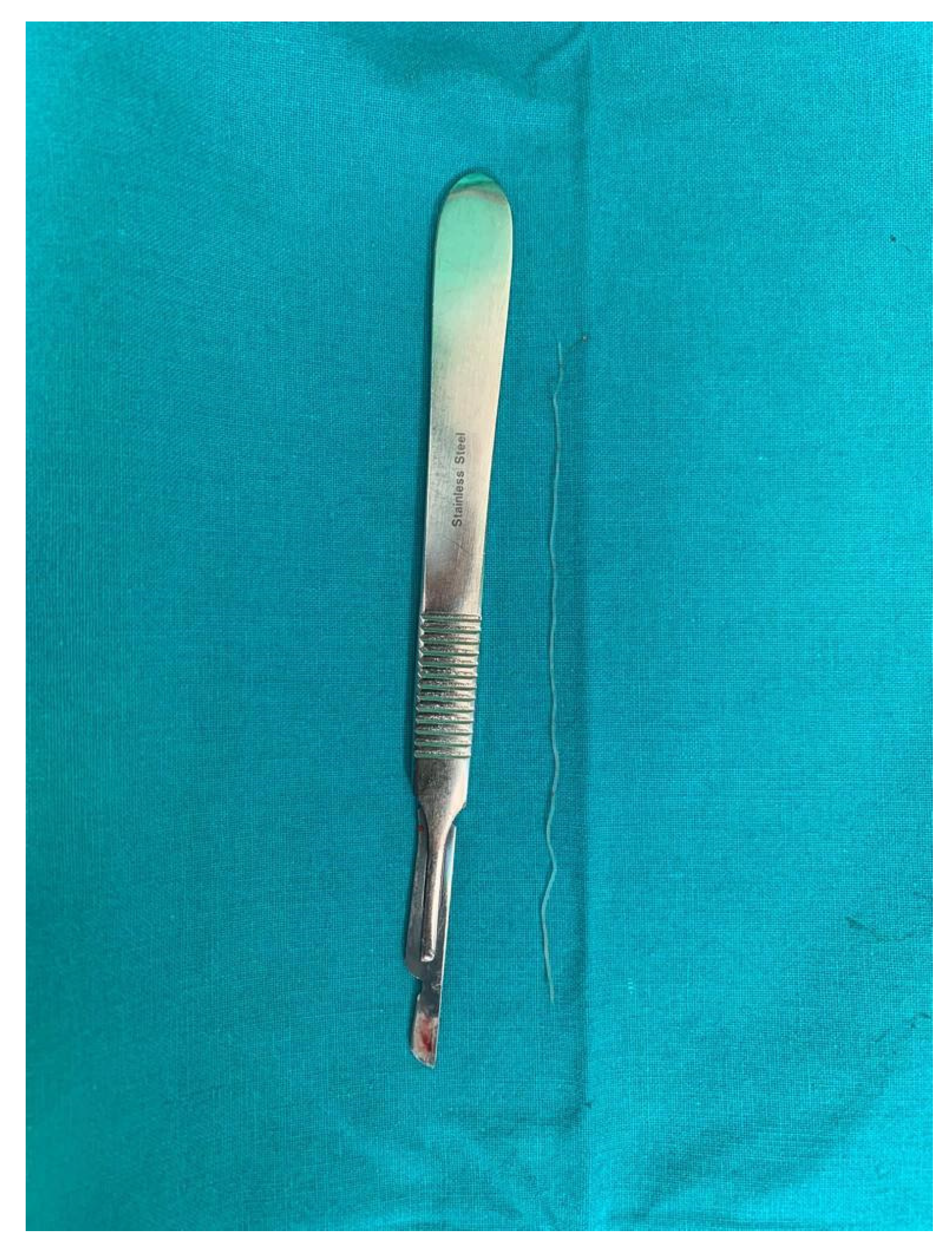
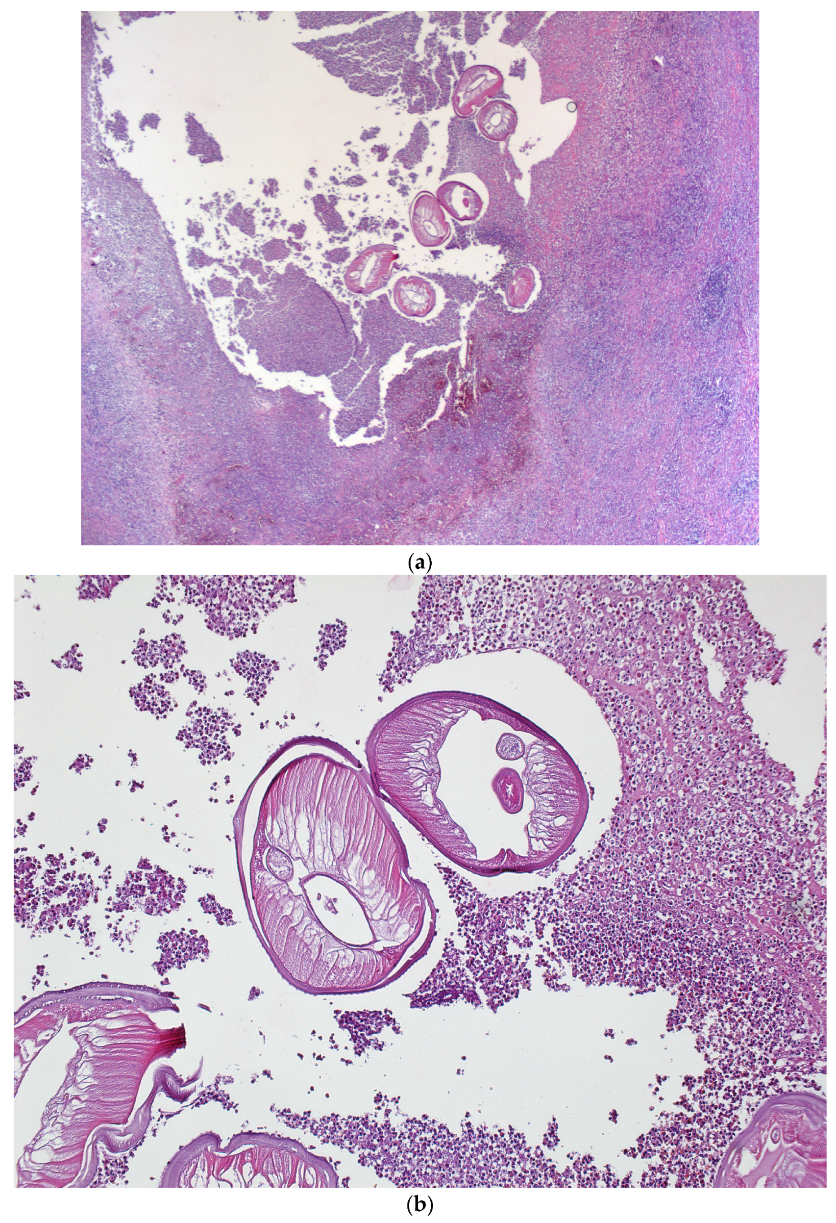
2. Case Report
3. Discussion
4. Conclusions
Author Contributions
Funding
Institutional Review Board Statement
Informed Consent Statement
Data Availability Statement
Conflicts of Interest
References
- Chan, C.; Kermanshahi, M.; Mathew, B.; England, R. A rare case of Dirofilaria repens infection. J. Laryngol. Otol. 2013, 127, 607–609. [Google Scholar] [CrossRef]
- Latifoglu, O.; Zmen, S.; Sezer, C. Dirofilaria repens presenting as a premasseteric nodule. Oral Surg. Oral Med. Oral Pathol. Oral Radiol. Endod. 2002, 94, 217–220. [Google Scholar] [CrossRef] [PubMed]
- Ganushkina, L.A.; Rakova, V.M.; Ivanova, I.B.; Supriaga, V.G.; Sergiev, V.P. Entomological monitoring of an area to assess Dirofilaria transmission risk. Med. Parazitol. 2014, 3, 9–12. [Google Scholar]
- Pupić-Bakrač, A.; Pupić-Bakrač, J.; Beck, A.; Jurković, D.; Polkinghorne, A.; Beck, R. Dirofilaria repens microfilaremia in humans: Case description and literature review. One Health 2021, 13, 100306. [Google Scholar] [CrossRef] [PubMed]
- Simon, F.; Siles-Lucas, M.; Morchon, R.; Gonzalez-Miguel, J.; Mellado, L.; Carreton, E.; Montoya-Alonso, J.A. Human and animal dirofilariasis: The emergence of a zoonotic mosaic. Clin. Microbiol. Rev. 2012, 25, 507–544. [Google Scholar] [CrossRef] [PubMed]
- Sabunas, V.; Radzijevskaja, J.; Sakalauskas, P.; Petkevičius, S.; Karveliene, B.; Ziliukiene, J.; Lipatova, I.; Paulauskas, A. Dirofilaria repens in dogs and humans in Lithuania, Parasit. Vectors 2019, 12, 177. [Google Scholar] [CrossRef]
- Fuehrer, H.P.; Auer, H.; Leschnik, M.; Silbermayr, K.; Duscher, G.; Joachim, A. Dirofilaria in Humans, Dogs, and Vectors in Austria (1978–2014)-From Imported Pathogens to the Endemicity of Dirofilaria repens. PLoS Negl. Trop. Dis. 2016, 10, e0004547. [Google Scholar] [CrossRef]
- Simon, F.; Morchon, R.; Gonzalez-Miguel, J.; Maecos-Atxutegi, M.; Siles-Lucas, F.M. What is new about animal and human dirofilariasis? Trends Parasitol. 2009, 25, 404–509. [Google Scholar] [CrossRef]
- Kondrashin, A.V.; Morozova, L.F.; Stepanova, E.V.; Turbabina, N.A.; Maksimova, M.S.; Morozov, E.N. Anthology of Dirofilariasis in Russia (1915–2017). Pathogens 2020, 9, 275. [Google Scholar] [CrossRef]
- Mateju, J.; Chanova, M.; Modry, D.; Mitkova, B.; Hrazdilova, K.; Zampachova, V.; Kolarova, L. Dirofilaria repens: Emergence of autochthonous human infections in the Czech Republic (case reports). BMC Infect. Dis. 2016, 16, 171. [Google Scholar] [CrossRef] [PubMed]
- Capelli, G.; Genchi, C.; Baneth, G.; Bourdeau, P.; Brianti, E.; Cardoso, L.; Danesi, P.; Fuehrer, H.P.; Giannelli, A.; Ionică, A.M. Recent advances on Dirofilaria repens in dogs and humans in Europe. Parasit. Vectors 2018, 11, 663. [Google Scholar] [CrossRef]
- Potters, I.; Vanfraechem, G.; Bottieau, E. Dirofilaria repens Nematode Infection with Microfilaremia in Traveler Returning to Belgium from Senegal, Emerg. Infect. Dis. 2018, 24, 1761–1763. [Google Scholar] [CrossRef]
- Kludkowska, M.; Pielok, L.; Frackowiak, K.; Masny, A.; Golab, E.; Paul, M. Dirofilaria repens infection as a cause of intensive peripheral microfilaraemia in a Polish patient: Process description and cases review. Acta Parasitol. 2018, 63, 657–663. [Google Scholar] [CrossRef] [PubMed]
- Antolová, D.; Miterpakova, M.; Paralicova, Z. Case of human Dirofilaria repens infection manifested by cutaneous larva migrans syndrome. Parasitol. Res. 2015, 114, 2969–2973. [Google Scholar] [CrossRef]
- Mendoza-Roldan, J.A.; Gabrielli, S.; Cascio, A.; Manoj, R.R.; Bezerra-Santos, M.A.; Benelli, G.; Brianti, E.; Latrofa, M.S.; Otranto, D. Zoonotic Dirofilaria immitis and Dirofilaria repens infection in humans and an integrative approach to the diagnosis. Acta Trop. 2021, 223, 106083. [Google Scholar] [CrossRef]
- Mistry, M.A.; Hoejvig, J.; Helleberg, M.; Stensvold, C.R.; Jokelainen, P.; Noehr, A.; Bonde, C. Human subcutaneous dirofilariasis: The ‘migrating’ skin tumor. Case Rep. Plast. Surg. Hand Surg. 2021, 8, 181–185. [Google Scholar] [CrossRef]
- Rose, R.; Kleitsch, K.U.; Born, D.; Heye, P. Dirofilaria repens in a Pediatric Patient—First Case Report from Switzerland. Eur. J. Pediatr. Surg. 2023, 11, 29–31. [Google Scholar] [CrossRef] [PubMed]
- Melasom, H.A.; Kurtzhals, J.A.L.; Qvortrup, K.; Bargum, R.; Barfod, T.S.; la Cour, M.; Heegaard, S. Subconjunctival Dirofilaria repens Infestation: A Light and Scanning Electron Microscopy Study. Open Ophtalmol. J. 2011, 5, 21–24. [Google Scholar] [CrossRef] [PubMed]
- Lindner, A.K.; Tappe, D.; Gertler, M.; Equihua Martinez, G.; Richter, J. A live worm emerging from the eyelid. J. Travel Med. 2018, 25, 66. [Google Scholar] [CrossRef] [PubMed]
- Pampiglione, S.; Rivasi, F. Human dirofilariasis due to Dirofilaria (Nochtiella) repens: An update of world literature from 1995 to 2000. Parassitologia 2000, 42, 231–254. [Google Scholar] [PubMed]
- Taylor, M.J.; Bandi, C.; Hoerauf, A. Wolbachia bacterial endosymbionts of filarial nematodes. Adv. Parasitol. 2005, 60, 245–284. [Google Scholar] [PubMed]
- Landmann, F. The Wolbachia Endosymbionts. Microbiol Spectr. 2019, 7, 1028–1128. [Google Scholar] [CrossRef]
- Taylor, M.J.; Voronin, D.; Johnston, K.L.; Ford, L. Wolbachia filarial interactions. Cell. Microbiol. 2013, 15, 520–526. [Google Scholar] [CrossRef]
- Werren, J.H.; Baldo, L.; Clark, M.E. Wolbachia: Master manipulators of invertebrate biology. Nat. Rev. Microbiol. 2008, 6, 741–751. [Google Scholar] [CrossRef] [PubMed]
- Cancrini, G.; Romi, R.; Gabrielli, S.; Toma, L.; Di Paolo, M.; Scaramozzino, P. First finding of Dirofilaria repens in a natural population of Aedes albopictus. Med. Vet. Entomol. 2003, 17, 448–451. [Google Scholar] [CrossRef] [PubMed]
- Cancrini, G.; Magi, M.; Gabrielli, S.; Arispici, M.; Tolari, F.; Dell’Omodarme, M.; Prati, M.C. Natural vectors of dirofilariasis in rural and urban areas of the Tuscan region, central Italy. J. Med. Entomol. 2006, 43, 574–579. [Google Scholar] [CrossRef] [PubMed]
- Cancrini, G.; Gabrielli, S. Vectors of Dirofilaria nematodes: Biology, behavior and host/parasite relationships. In Dirofilaria immitis and D. repens in Dog and Cat and Human Infections; Genchi, C., Rinaldi, L., Cringoli, G., Eds.; Veterinary Parasitology and Parasitic Diseases, Department of Pathology and Animal Health, Faculty of Veterinary Medicine, University of Naples Federico II: Napoli, Italy, 2007; pp. 48–58. ISBN 9788889132142. [Google Scholar]
- Giangaspero, A.; Marangi, M.; Latrofa, M.S.; Martinelli, D.; Traversa, D.; Otranto, D.; Genchi, C. Evidences of increasing risk of dirofilariasis in southern Italy. Parasitol. Res. 2013, 112, 1357–1361. [Google Scholar] [CrossRef]
- Sulesco, T.; von Thien, H.; Toderas, L.; Toderas, I.; Lühken, R.; Tannich, E. Detection of Dirofilaria repens and Dirofilaria immitis DNA in mosquitoes from Belarus. Parasitol. Res. 2016, 115, 3535–3541. [Google Scholar] [CrossRef]
- Slatko, B.E.; Taylor, M.J.; Foster, J.M. The Wolbachia endosymbiont as an anti-filarial nematode target. Symbiosis 2010, 51, 55–65. [Google Scholar] [CrossRef]
- Foster, J.; Ganatra, M.; Kamal, I.; Ware, J.; Makarova, K.; Ivanova, N.; Bhattacharyya, A.; Kapatral, V.; Kumar, S.; Posfai, J.; et al. The Wolbachia genome of Brugia malayi: Endosymbiont evolution within a human pathogenic nematode. PLoS Biol. 2005, 3, e121. [Google Scholar] [CrossRef]
- Foster, J.M.; Hoerauf, A.; Slatko, B.E.; Taylor, M.J. The Wolbachia bacterial endosymbionts of filarial nematodes. In Parasitic Nematodes: Molecular Biology, Biochemistry and Immunology, 2nd ed.; Kennedy, M.W., Harnett, W., Eds.; CABI: Wallingford, UK, 2013; pp. 308–336. [Google Scholar]
- Genchi, C.; Kramer, L.H.; Sassera, D.; Bandi, C. Wolbachia and its implications for the immunopathology of filariasis. Endocr. Metab. Immune Disord.-Drug Targets 2012, 12, 53–56. [Google Scholar] [CrossRef]
- Petry, G.; Genchi, M.; Schmidt, H.; Schaper, R.; Lawrenz, B.; Genchi, C. Evaluation of the adulticidal efficacy of imidacloprid 10%/moxidectin 2.5% (w/v) spot-on (advocate, advantage multi) against Dirofilaria repens in experimentally infected dogs. Parasitol. Res. 2015, 114, 131–144. [Google Scholar] [CrossRef]
- Genchi, M.; Pengo, G.; Genchi, C. Efficacy of moxidectin microsphere sustained release formulation for the prevention of subcutaneous filarial (Dirofilaria repens) infection in dogs. Vet. Parasitol. 2010, 170, 167–169. [Google Scholar] [CrossRef] [PubMed]
- Albanese, F.; Abramo, F.; Braglia, C.; Caporali, C.; Venco, L.; Vercelli, A.; Ghibaudo, G.; Leone, F.; Carrani, F.; Giannelli, A.; et al. Nodular lesions due to infestation by Dirofilaria repens in dogs from Italy. Vet. Dermatol. 2013, 24, 255-e56. [Google Scholar] [CrossRef] [PubMed]
- Rocconi, F.; Di Tommaso, M.; Traversa, D.; Palmieri, C.; Pampurini, F.; Boari, A. Allergic dermatitis by Dirofilaria repens in a dog: Clinical picture and treatment. Parasitol. Res. 2012, 111, 493–496. [Google Scholar] [CrossRef] [PubMed]
- Permi, H.S.; Veena, S.; Prasad, H.K.; Kumar, Y.S.; Mohan, R.; Shetty, K.J. Subcutaneous human dirofilariasis due to Dirofilaria repens: Report of two cases. J. Glob. Infect. Dis. 2011, 3, 199–201. [Google Scholar] [PubMed]
- Jelinek, T.; Schulte-Hillen, J.; Loscher, T. Human dirofilariasis. Int. J. Dermatol. 1996, 35, 872–875. [Google Scholar] [CrossRef]
- Tahir, D.; Bittar, F.; Barré-Cardi, H.; Sow, D.; Dahmani, M.; Mediannikov, O.; Raoult, D.; Davoust, B.; Parola, P. Molecular survey of Dirofilaria immitis and Dirofilaria repens by new realtime TaqMan PCR assay in dogs and mosquitoes (Diptera: Culicidae) in Corsica (France). Vet. Parasitol. 2017, 235, 1–7. [Google Scholar] [CrossRef] [PubMed]
- Skrinjar, I.; Brailo, V.; Loncar Brzak, B.; Lozic Erent, J.; Bukovski, S.; Juras, D.V. Live Intraoral Dirofilaria repens of Lower Lip Mimicking Mucocele—First Reported Case from Croatia. Int. J. Environ. Res. Public Health 2022, 19, 4330. [Google Scholar] [CrossRef]
- Arbune, M.; Dobre, M. Dirofilariasis—An emergent human parasitosis in Romania. Acta Parasitol. 2015, 60, 485–487. [Google Scholar] [CrossRef]
- Ciuca, L.; Simòn, F.; Rinaldi, L.; Kramer, L.; Genchi, M.; Cringoli, G.; Acatrinei, D.; Miron, L.; Morchon, R. Seroepidemiological survey of human exposure to Dirofilaria spp. in Romania and Moldova. Acta Trop. 2018, 187, 169–174. [Google Scholar] [CrossRef] [PubMed]
- Mănescu, R.; Bărăscu, D.; Mocanu, C.; Pîrvănescu, H.; Mîndriă, I.; Bălăşoiu, M.; Turculeanu, A. Subconjunctival nodule with Dirofilaria repens. Chirurgia 2009, 104, 95–97. [Google Scholar]
- Popescu, I.; Tudose, I.; Racz, P.; Muntau, B.; Giurcaneanu, C.; Poppert, S. Human Dirofilaria repens Infection in Romania: A Case Report. Case Rep. Infect. Dis. 2012, 2012, 472976. [Google Scholar] [PubMed]
- Nissen, T.; Wynn, R. The clinical case report: A review of its merits and limitations. BMC Res. Notes 2014, 7, 264. [Google Scholar] [CrossRef] [PubMed]
- Sałamatin, R.V.; Pavlikovska, T.M.; Sagach, O.; Nikolayenko, S.; Kornyushin, V.; Kharchenko, V.; Masny, A.; Cielecka, D.; Konieczna-Sałamatin, J.; Conn, D.; et al. Human dirofilariasis due to Dirofilaria repens in Ukraine, an emergent zoonosis: Epidemiological report of 1465 cases. Acta Parasitol. 2013, 58, 592–598. [Google Scholar] [CrossRef]
- Hennocq, Q.; Helary, A.; Debelmas, A.; Monsel, G.; Labat, A.; Bertolus, C.; Martin, C.; Caumes, E. Oral migration of Dirofilaria repens after creeping dermatitis. Parasite 2020, 27, 16. [Google Scholar] [CrossRef]

| WBCs | 5.05 × 10³/µL |
| RBCs | 4.5 × 1003/µL |
| Hb | 14.4 g/dL |
| Platelets | 145 × 103/µL |
| Neutrophils | 3.11 × 10³/µL |
| Lymphocytes | 1.26 × 10³/µL |
| Monocytes | 0.61 × 10³/µL |
| Eosinophils | 0.06 × 10³/µL |
| Basophils | 0.02 × 10³/µL |
Disclaimer/Publisher’s Note: The statements, opinions and data contained in all publications are solely those of the individual author(s) and contributor(s) and not of MDPI and/or the editor(s). MDPI and/or the editor(s) disclaim responsibility for any injury to people or property resulting from any ideas, methods, instructions or products referred to in the content. |
© 2024 by the authors. Licensee MDPI, Basel, Switzerland. This article is an open access article distributed under the terms and conditions of the Creative Commons Attribution (CC BY) license (https://creativecommons.org/licenses/by/4.0/).
Share and Cite
Nicolau, A.; Sava, F.P.; Severin, F.; Ciofu, M.L.; Ferariu, D.; Dodu, D.; Costan, V.V. A Rare Case of Dirofilariasis in the Genian Region. Diagnostics 2024, 14, 628. https://doi.org/10.3390/diagnostics14060628
Nicolau A, Sava FP, Severin F, Ciofu ML, Ferariu D, Dodu D, Costan VV. A Rare Case of Dirofilariasis in the Genian Region. Diagnostics. 2024; 14(6):628. https://doi.org/10.3390/diagnostics14060628
Chicago/Turabian StyleNicolau, Andrei, Florin Petrică Sava, Florentina Severin, Mihai Liviu Ciofu, Dan Ferariu, Daniela Dodu, and Victor Vlad Costan. 2024. "A Rare Case of Dirofilariasis in the Genian Region" Diagnostics 14, no. 6: 628. https://doi.org/10.3390/diagnostics14060628
APA StyleNicolau, A., Sava, F. P., Severin, F., Ciofu, M. L., Ferariu, D., Dodu, D., & Costan, V. V. (2024). A Rare Case of Dirofilariasis in the Genian Region. Diagnostics, 14(6), 628. https://doi.org/10.3390/diagnostics14060628