
A Unique Case of Appendiceal Intussusception (Inversion): A Case in Bloom

 , , , , ,
  , , ,
, , , 
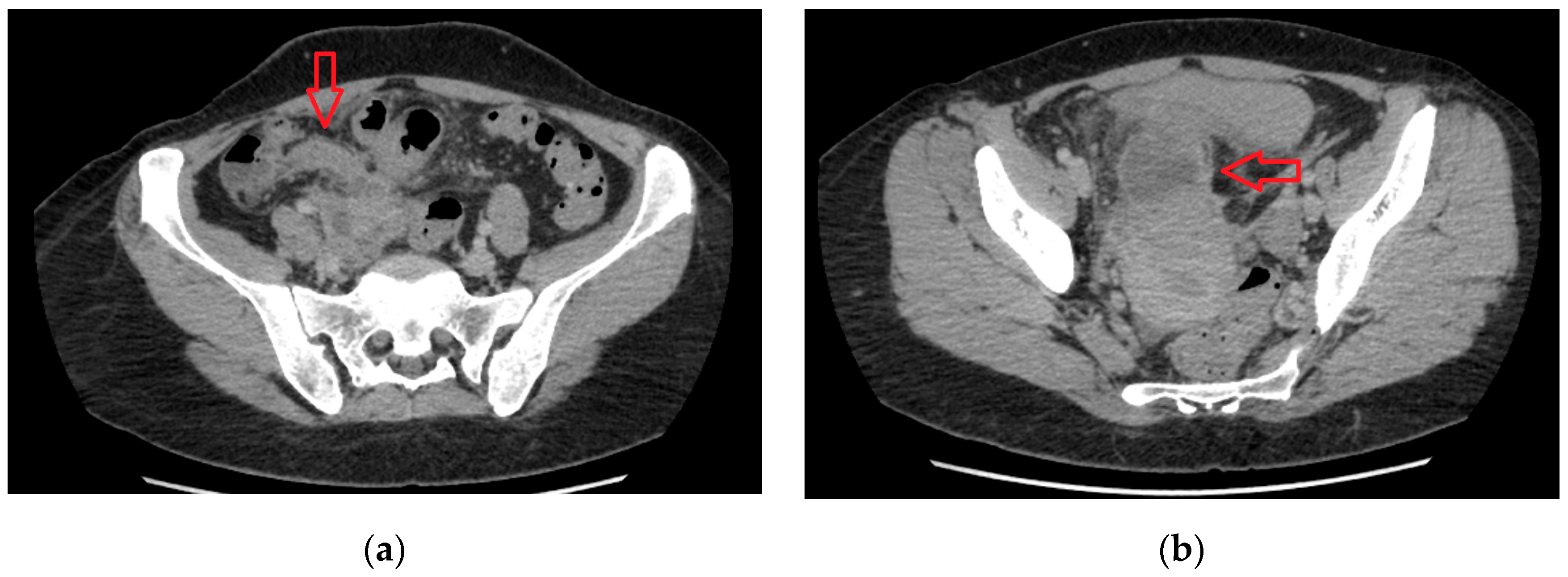
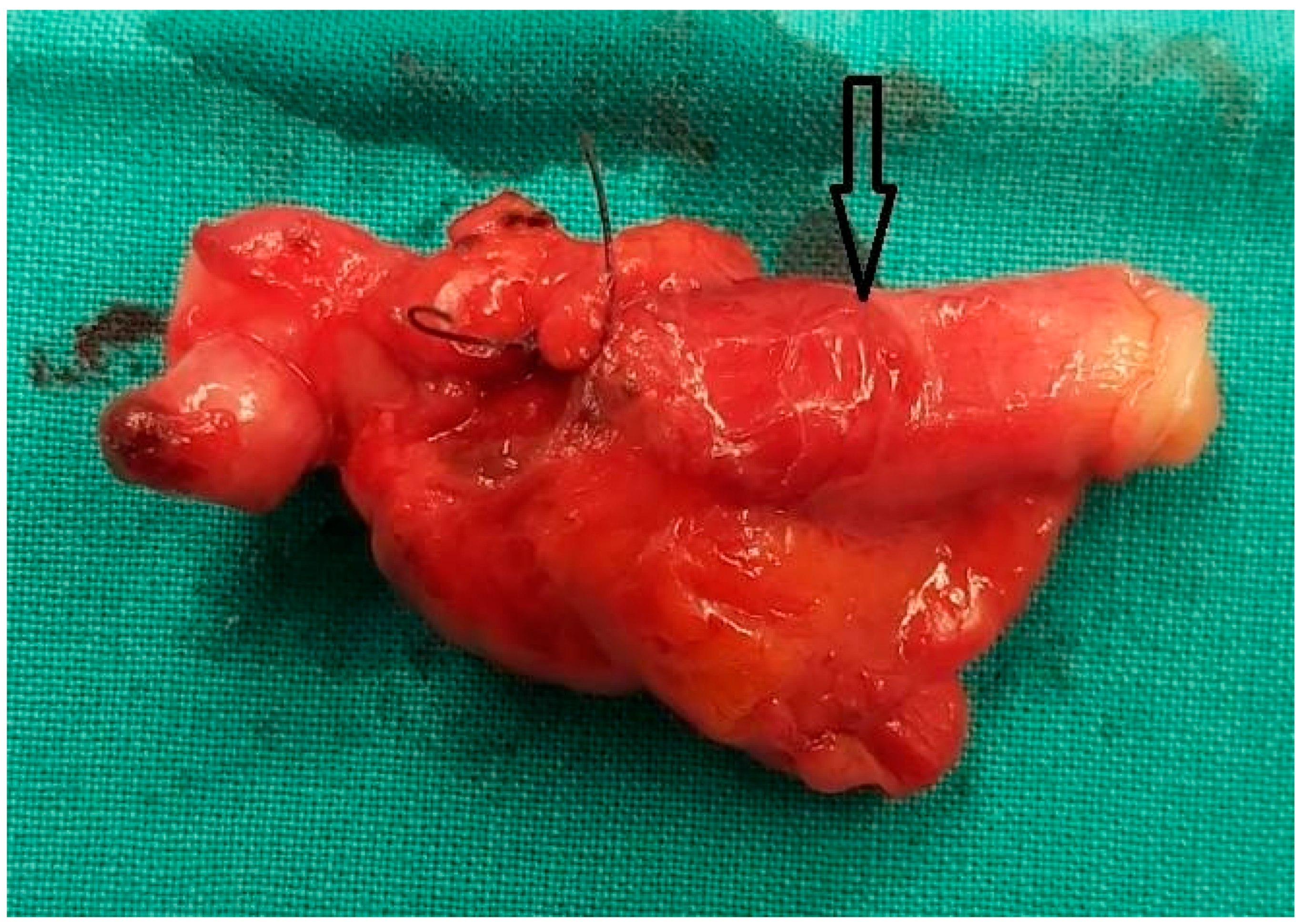
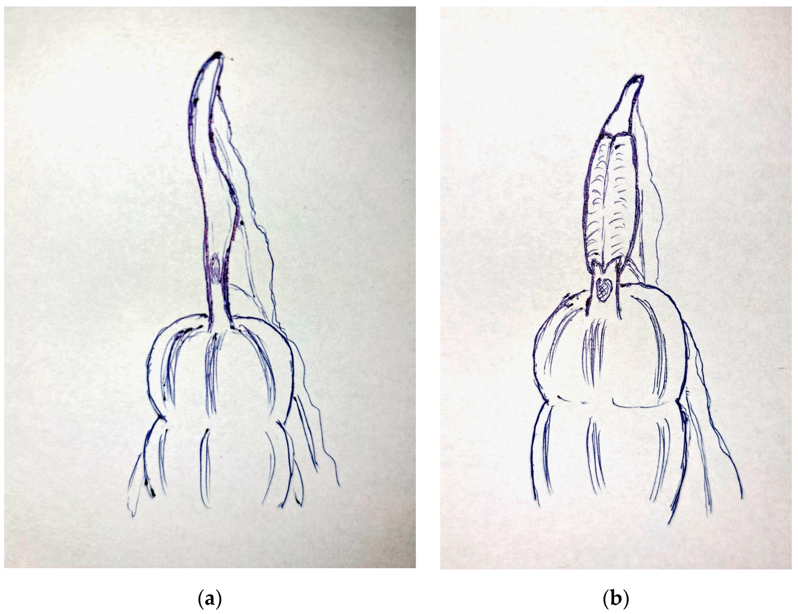
Abstract
Author Contributions
Funding
Institutional Review Board Statement
Informed Consent Statement
Data Availability Statement
Conflicts of Interest
References
- Schildberg, C.W.; Reissig, K. Diagnostic, Therapy and Complications in Acute Appendicitis of 19,749 Cases Based on Routine Data: A Retrospective Multicenter Observational Study. J. Clin. Med. 2022, 11, 4495. [Google Scholar] [CrossRef]
- Collins, D.C. 71,000 Human Appendix Specimens. A Final Report Summarizing Forty Year’s Study. Am. J. Proctol. 1963, 14, 265–281. [Google Scholar] [PubMed]
- Park, B.S.; Shin, D.H. Appendiceal intussusception requiring an ileocecectomy: A case report and comment on the optimal surgery. BMC Surg. 2018, 18, 48. [Google Scholar] [CrossRef] [PubMed]
- Chaar, C.I.; Wexelman, B. Intussusception of the appendix: Comprehensive review of the literature. Am. J. Surg. 2009, 198, 122–128. [Google Scholar] [CrossRef] [PubMed]
- Forshal, I. Intussusception of the vermiform appendix with a report of seven cases in children. Br. J. Surg. 1953, 40, 305–312. [Google Scholar] [CrossRef] [PubMed]
- Danielson, K.S. Cecocolic intussusception--a postoperative complication of appendectomy. N. Engl. J. Med. 1969, 280, 35–36. [Google Scholar] [CrossRef] [PubMed]
- Lipskar, A.; Telem, D. Failure of appendectomy to resolve appendiceal intussusception. J. Ped. Surg. 2008, 43, 1554–1556. [Google Scholar] [CrossRef] [PubMed]
- Sivanandham, S.; Jakate, S. Impacted golf tee shaped appendiceal fecalith causing appendiceal non-visualization and mimicking cecal mass: A case report. Int. J. Case Rep. Images 2022, 13, 147–151. [Google Scholar] [CrossRef]
- Jaschinski, T.; Mosch, C.G. Laparoscopic versus open surgery for suspected appendicitis. Cochrane Database Syst. Rev. 2018, 11, CD001546. [Google Scholar] [CrossRef] [PubMed]


Disclaimer/Publisher’s Note: The statements, opinions and data contained in all publications are solely those of the individual author(s) and contributor(s) and not of MDPI and/or the editor(s). MDPI and/or the editor(s) disclaim responsibility for any injury to people or property resulting from any ideas, methods, instructions or products referred to in the content. |
© 2024 by the authors. Licensee MDPI, Basel, Switzerland. This article is an open access article distributed under the terms and conditions of the Creative Commons Attribution (CC BY) license (https://creativecommons.org/licenses/by/4.0/).
Share and Cite
Mantalovas, S.; Paschou, E.; Kountouri, I.; Sevva, C.; Papadopoulos, K.; Roulia, P.; Dagher, M.; Laskou, S.; Lagopoulos, V.; Koulouris, C.; et al. A Unique Case of Appendiceal Intussusception (Inversion): A Case in Bloom. Diagnostics 2024, 14, 555. https://doi.org/10.3390/diagnostics14050555
Mantalovas S, Paschou E, Kountouri I, Sevva C, Papadopoulos K, Roulia P, Dagher M, Laskou S, Lagopoulos V, Koulouris C, et al. A Unique Case of Appendiceal Intussusception (Inversion): A Case in Bloom. Diagnostics. 2024; 14(5):555. https://doi.org/10.3390/diagnostics14050555
Chicago/Turabian StyleMantalovas, Stylianos, Eleni Paschou, Ismini Kountouri, Christina Sevva, Konstantinos Papadopoulos, Panagiota Roulia, Marios Dagher, Styliani Laskou, Vasileios Lagopoulos, Charilaos Koulouris, and et al. 2024. "A Unique Case of Appendiceal Intussusception (Inversion): A Case in Bloom" Diagnostics 14, no. 5: 555. https://doi.org/10.3390/diagnostics14050555
APA StyleMantalovas, S., Paschou, E., Kountouri, I., Sevva, C., Papadopoulos, K., Roulia, P., Dagher, M., Laskou, S., Lagopoulos, V., Koulouris, C., Louloudopoulou, F., Kopsidas, P., Sapalidis, K., Kesisoglou, I., & Kosmidis, C. (2024). A Unique Case of Appendiceal Intussusception (Inversion): A Case in Bloom. Diagnostics, 14(5), 555. https://doi.org/10.3390/diagnostics14050555