Article

The Evaluation of Radiolabeled Prostate-Specific Membrane Antigen Positron Emission Tomography/Computed Tomography for Initial Staging in Intermediate-Risk Prostate Cancer Patients: A Retrospective Multicenter Analysis

1
Nuclear Medicine Unit, IRCCS Humanitas Research Hospital, 20089 Rozzano, Italy
2
Department of Biomedical Sciences, Humanitas University, 20072 Pieve Emanuele, Italy
3
Nuclear Medicine Unit, Humanitas Gavazzeni, 24125 Bergamo, Italy
4
Nuclear Medicine Unit, Humanitas Istituto Clinico Catanese, 95045 Misterbianco, Italy
5
Urology Unit, IRCCS Humanitas Research Hospital, 20089 Rozzano, Italy
6
Urology Unit, Humanitas Mater Domini, 21100 Castellanza, Italy
7
Urology Unit, Humanitas Istituto Clinico Catanese, 95045 Catania, Italy
*
Author to whom correspondence should be addressed.
Diagnostics 2024, 14(23), 2751; https://doi.org/10.3390/diagnostics14232751
Submission received: 6 November 2024 / Revised: 3 December 2024 / Accepted: 4 December 2024 / Published: 6 December 2024

Abstract

:
Objectives. The aim of the present study was to assess the performance of radiolabeled-PSMA PET/CT in a cohort of intermediate-risk prostate cancer (PCa) patients for initial staging. Methods. This is a retrospective, multicenter analysis of patients diagnosed with intermediate-risk PCa who were staged using radiolabeled PSMA PET/CT to evaluate the extent of the disease before initiating appropriate treatment. The study included patients from the Nuclear Medicine Units of the Humanitas group between 2021 and 2024. The change in management due to the PSMA PET/CT examination was assessed. Results. A total of 181 patients were enrolled across all three centers. Histopathological assessment from biopsy revealed that 51.4% of patients had favorable PCa, while 48.6% had unfavorable disease. PET/CT was positive for the primary lesions in all patients, but it revealed a positivity rate in 23 (12.7%) patients for nodes and distant organs, with a positivity rate of 0.21 in the unfavorable group and 0.05 in the favorable group (p < 0.005). Based on follow-up data, diagnostic accuracy was higher than 90% in both the favorable and unfavorable groups for lymph node and distant metastases. The inclusion of PSMA PET/CT in the diagnostic algorithm for patients with intermediate-risk PCa impacted patient management in 24 (13.3%) cases, based on the multidisciplinary team decision. Conclusions. PSMA PET/CT can affect the management of patients with intermediate-risk PCa in up to 13% of cases, mainly for unfavorable diseases. New imaging techniques as a first-line imaging procedure can help to plan the correct therapeutic approach in the intermediate-risk PCa group.

1. Introduction

Prostate cancer (PCa) represents the most diagnosed cancer in men and the second most common cause of cancer death in Western countries [1]. While in most cases localized PCa can be potentially eradicated with radical prostatectomy or definitive radiotherapy, when the disease has already spread to the lymph nodes or to other sites, local treatments are not enough [1,2,3]. Therefore, identifying clinical lymph nodes and metastatic-positive patients is mandatory in order to choose the best therapeutic strategy. Several imaging technologies have been used for PCa staging purposes [4,5,6,7,8,9,10,11,12,13]. Conventional staging, based on abdominopelvic computed tomography (CT) and bone scans, has often been shown to have insufficient sensitivity, especially when staging men with high-risk PCa [5,6,7,8,9,10]. The limited performance of conventional CT and bone scanning in detecting nonlocalized PCa during primary staging can lead to inadequate treatment decisions. Due to the limitations of conventional imaging, several preoperative tools, including the Briganti, Partin, and Memorial Sloan Kettering Cancer Center (MSKCC) nomograms, as well as the Roach formula, have been developed to better predict the individual risk of lymph node metastasis [1]. However, since these tools have also demonstrated suboptimal accuracy, pelvic lymph node dissection (LND) is currently considered the most reliable approach to assessing nodal involvement [1].
The field of non-invasive nodal and metastatic staging of PCa is evolving very rapidly. New imaging technologies, such as radiolabeled prostate-specific membrane antigen positron emission tomography with computed tomography (PSMA PET/CT) and multiparametric magnetic resonance imaging (mpMRI), provide a more sensitive detection of LN and bone metastases than the classical work-up with bone scanning and abdominopelvic CT [11,12,13]. PET/CT with [68Ga]-[18F]-PSMA agents have emerged as a highly sensitive and specific modality for the initial staging of PCa, mainly in patients at high and very high risk for the disease [11,13]. Therefore, there is growing evidence supporting the use of PSMA PET/CT for primary staging in the replacement of conventional imaging. However, there are still some open questions about the use of this technology in the specific risk category of patients, such as patients with intermediate-risk disease [14,15]; indeed, most published studies are relative to mixed populations (i.e., intermediate and high risks). Furthermore, the emerging use of specific scores from PSMA PET/CT images, such as primary score, PSMA-RADS, and others [16,17], for subclassifying recurrence risk and histopathological patterns (favorable vs. unfavorable) [18], complicates the initial evaluation of these patients.
The aim of the present study was to assess the performance of radiolabeled PSMA PET/CT in a cohort of intermediate-risk PCa patients for initial staging.

2. Materials and Methods

2.1. Study Design and Patient Selection

This is a retrospective, multicenter analysis of patients diagnosed with intermediate-risk PCa (both favorable and unfavorable, following the guidelines by Mohler et al. [19] and Sanda et al. [20]). These patients were staged using radiolabeled PSMA PET/CT to evaluate the extent of the disease before initiating appropriate treatment. The study included patients from the Nuclear Medicine Units of the Humanitas group (Cancer Center in Rozzano, Milan, Italy; Gavazzeni in Bergamo, Italy; and Istituto Clinico Catanese in Catania, Italy) between 2021 and 2024. Inclusion criteria were as follows: (1) age > 18 years; (2) patients with a well-established diagnosis of intermediate-risk PCa (ISUP grade: 2 or 3); and (3) patients who underwent 68Ga-PSMA-11 or 18F-PSMA-1007 PET/CT. Exclusion criteria were as follows: (1) previous history of other oncological diseases; (2) previous radiation treatments, androgen-based therapies, or chemotherapies; and (3) missing or unavailable clinical and follow-up data. This study was conducted in accordance with the principles of the Declaration of Helsinki (1964) and received approval from the Ethical Committee (n.169/24, date: 19 March 2024).

2.2. PSMA PET/CT Protocol

All patients underwent PSMA-ligand PET/CT scan with either [68Ga]Ga-PSMA-11 or [18F]F-PSMA1007. The 18F-PSMA-1007 precursor, cassettes, and reagents for the synthesis were acquired from ABX Advanced Biochemical Compounds (Radeberg, Germany). 18F-PSMA-1007 was synthesized in a TRASIS AllInOne Module following the prescribed step-by-step procedure. The entire synthesis process, from start to transfer, takes approximately 45 min, yielding a final product volume of 20 ± 1.0 mL. The average synthesis yield of 18F-PSMA-1007 is approximately ≥40% ± 10%, without correction for decay. The radiochemical purity of the final product exceeds 95%. The MiniAllInOne module was used to synthesize 68Ga-PSMA-11. The precursor, cassettes, and reagents for this synthesis were acquired from Trasis (Ans, Belgium). The entire synthesis process, from start to transfer, takes approximately 20 min, yielding a final product volume of 10 ± 0.5 mL. The average synthesis yield of 68Ga-PSMA-11 is approximately ≥65%, without correction for decay. The radiochemical purity of the final product exceeds 98%, as estimated by high-performance liquid chromatography. Supplementary Table S1 reports the information about tracer production at each center.
PSMA-ligand PET/CT was performed according to versions 1.0 and 2.0 of the European Association of Nuclear Medicine (EANM) guidelines for prostate cancer imaging [21]. PET/CT images were acquired between 60 and 90 and between 90 and 120 min after [68Ga]Ga-PSMA-11 or [18F]F-PSMA-1007, respectively. All patients underwent oral hydration and bladder voiding prior to PET/CT imaging. In selected cases, furosemide was administered 30 min after PSMA-ligand injection (20 mg intravenously). PET/CT images were acquired using different PET/CT scanners (Supplementary Table S2). Co-registered CT data were used for the attenuation correction of PET images.

2.3. Image Analysis

All images were reviewed by two nuclear medicine physicians with at least 5 years of experience with PET using dedicated software PET/CT scans. PET/CT images were defined as negative if no area of increased radiopharmaceutical uptake was observed compared to the background. The criterion for positivity was at least one abnormal area of radiopharmaceutical uptake outside the physiological distribution or higher than the surrounding physiological activity, respectively, for 68GaPSMA-11 and 18F-PSMA-1007. Both visual and qualitative analyses were used. Semiquantitative analysis in terms of maximum standardized uptake value (SUVmax) was calculated by using an isocontour voxel of interest (VOI). Moreover, in the primary prostate lesions, the primary score was documented as previously defined, with 5 categories: score 1, no significant pattern within the prostate; score 2, a diffuse transition or central zone pattern; score 3, focal transition zone activity above twice the background transition zone counts; score 4, focal peripheral zone activity of any intensity; and score 5, an SUV of more than 12 [16].
All imaging studies were classified in terms of diagnostic accuracy for lymph node (N) and distant metastasis (M) using per-patient-based analysis, by using the following criteria: true positive (TP), patients with a positive PSMA PET, and the evidence of disease on histopathology in the case of radical prostatectomy ± LAD, or follow-up PSA trend or any imaging after treatments; true negative (TN), patients with a negative PSMA PET/CT and no evidence of disease at histopathology or follow-up; false positive (FP), patients without any evidence of disease on histopathology/follow-up, but a positive PSMA PET/CT; false negative (FN), patients with evidence of disease on histopathology/follow-up but a negative PSMA PET/CT.

2.4. Multiparametric MRI Imaging

Reports from mpMRI examinations for each patient were evaluated, and if available, the images were reassessed by a dedicated radiologist. Prostate Imaging Reporting and Data System (PI-RADS) Version 2.1 [22] was used as the reference to score each lesion.

2.5. Change in Management

The change in management was defined as a change of therapeutic strategies (i.e., from the local to systemic treatment, defined as major) or a change of therapeutic approach (i.e., radiation planning, tailoring of LND template based on PSMA PET/CT results, or a combination of local and/or systemic treatments, defined as minor).

2.6. Statistical Analysis

The primary purpose of the present study was to assess the effect of radiolabeled PSMA PET/CT on patient management in a cohort of intermediate-risk PCa patients referring to the Humanitas group’s hospitals for initial staging. The secondary endpoints were (1) to determine the additional diagnostic value provided by the primary score from PSMA PET/CT in this cohort of patients and (2) to compare the detection of disease provided by radiolabeled PSMA PET/CT with that by mpMRI, in a subgroup of patients.
A sample size of approximately 200 patients was determined to achieve a power of over 80% to detect a 10–15% change in therapeutic approach due to the inclusion of PSMA PET/CT in the diagnostic algorithm. The distribution of data was assessed by using the Shapiro–Wilk test. Descriptive statistics regarding patient, imaging, and tumor characteristics as well as preoperative and postoperative data were provided for the whole population. Medians and ranges were reported for normally and non-normally distributed continuous variables. Categorical variables were presented as frequencies and proportions. Statistically significant differences between groups were assessed using Mann–Whitney and Wilcoxon rank-sum tests for non-normally distributed noncontinuous variables and Pearson chi-square tests for categorical variables. Sensitivity, specificity, positive predictive value, and negative predictive value of PSMA PET/CT for N and M staging purposes were determined and their 95% confidence intervals were calculated. Concordance between different imaging tools (PSMA PET/CT and MRI) was measured using Cohen’s kappa coefficient. The significance was set at 0.05. All analyses were performed with MedCalc®.

3. Results

A total of 181 patients were enrolled across all three centers. The characteristics of the study population are reported in Table 1. Histopathological assessment from biopsy revealed that 51.4% of patients had favorable PCa, while 48.6% had unfavorable disease. The PSA levels were significantly higher in the unfavorable group compared to the favorable group (p < 0.05), and treatment types also differed, with local treatment being more common in the favorable group than the unfavorable group (94.7% vs. 69.3%, p < 0.05). All patients underwent PET/CT before any treatment approach, with 78 (43.1%) receiving a [68Ga]Ga-PSMA-11 scan and 103 (56.9%) undergoing an [18F]F-PSMA-1007 examination. PET/CT was positive for the primary lesions in all patients, but it revealed positivity in 23 (12.7%) patients for nodes and distant organs. In particular, PSMA PET/CT identified lymph node and distant metastases with a higher positivity rate in the unfavorable group than in the favorable group (14.8% vs. 2.2%, p < 0.005, and 13.6% vs. 5.4%, p = 0.0576, respectively). The primary tumor was monofocal in 124 (68.5%) patients, occurring more often in favorable than in unfavorable patients (34.4% vs. 28.4%), although this difference was not statistically significant (p = 0.386). The primary scores were distributed as follows: score 1 in 17 (9.4%), score 2A and 2B in 19 (10.5%), score 3 in 16 (8.8%), score 4 in 75 (41.4%), and score 5 in 54 (29.8%). The rate of a primary score ≥4 was higher in the unfavorable group compared to the favorable group (n = 74; 84.1% vs. n = 75; 59.1%, p < 0.005). PSMA positivity at the lymph node level was found in patients with a primary score ≥ 4 both in favorable and unfavorable risk groups. Conversely, distant metastasis positivity at PSMA PET/CT was found in 80% and 92% of patients with primary scores ≥ 4, respectively, for favorable and unfavorable risk groups. Based on follow-up data (histopathology was available in 105 patients), diagnostic performance was high in both the favorable and unfavorable groups, for both lymph node and distant metastases (Table 2). The positivity rates in lymph node and distant metastasis for [18F]F-PSMA-1007 vs. [68Ga]Ga-PSMA-11 PET/CT were 8/103 (7.8%) vs. 7/78 (8.9%) and 6/103 (5.8%) vs. 11/78 (14.1%), respectively. However, in patients with the availability of the standard of reference, false positive findings were equally distributed between [18F]F-PSMA-1007 and [68Ga]Ga-PSMA-11 PET/CT, either for lymph node or distant metastases.

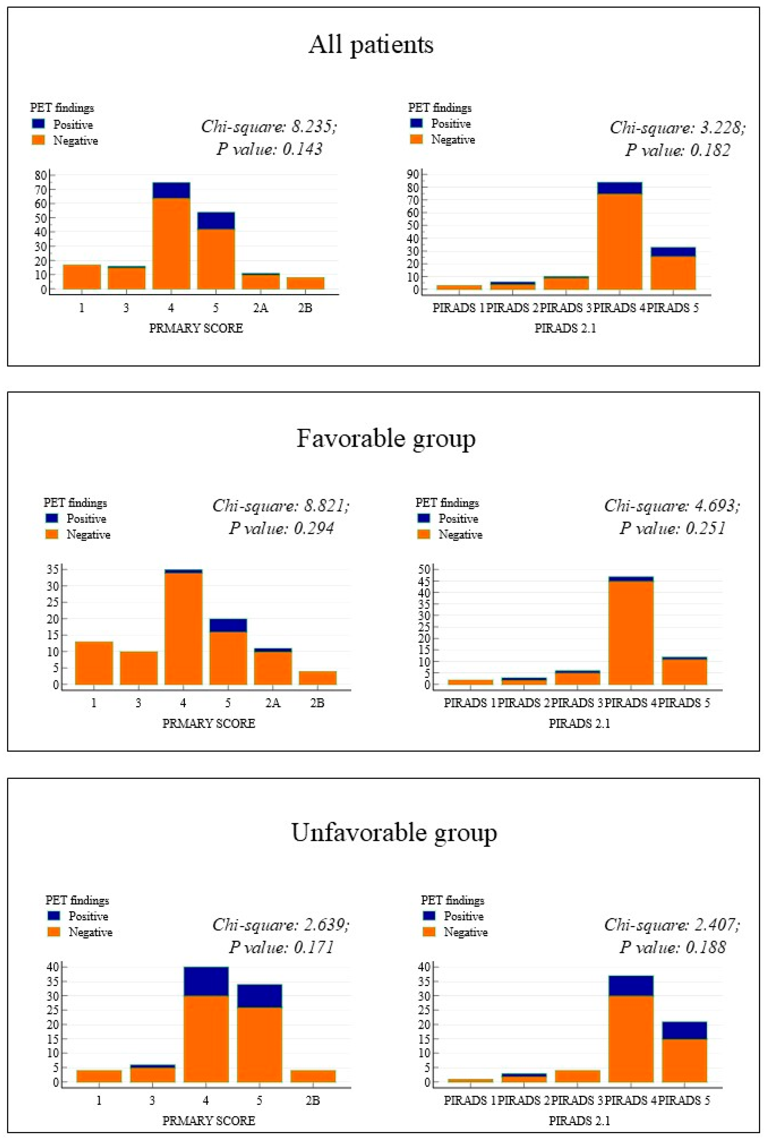
3.1. MpMRI and PSMA PET/CT

Both MRI and PSMA PET/CT images were available for 136 (75%) patients. At MRI, the PIRADS 2.1 scores were distributed as follows: score 1 in 3 (2.2%) patients, score 2 in 6 (4.4%), score 3 in 10 (7.4%), score 4 in 84 (61.8%), and score 5 in 33 (24.3%) patients. The primary tumor was unifocal in 100 (79.4%) patients according to MRI, showing poor agreement with PSMA PET/CT (K-Cohen = 0.14; CI95%: 0.03–0.31). Specifically, 29/100 (29%) patients identified as having monofocal tumors upon MRI were considered to have plurifocal tumors on PSMA PET/CT. MRI detected capsular invasion in 10 (7.4%) cases and seminal vesicle invasion in 6 (4.4%) cases. The rates of capsular and seminal vesicle invasion were higher in the unfavorable group compared to the favorable group (10.6% vs. 4.3% for capsular invasion and 7.6% vs. 1.4% for seminal vesicle invasion), although these differences were not statistically significant. Notably, a PIRADS 2.1 score of ≥4 was slightly more frequent in the unfavorable group than in the favorable group (87.9% vs. 84.2%), but this was also not statistically significant. Figure 1 illustrates the distribution of PIRADS 2.1 and primary scores in the favorable and unfavorable patient groups and their correlation with PSMA PET/CT findings.
No findings suggestive of lymph node or distant metastases were reported upon MRI for any of the 136 patients. Neither demographic nor MRI findings were significantly associated with a positive PSMA PET/CT in either the favorable or unfavorable risk categories. There was a slightly increased number of patients at unfavorable risk showing vesicle and capsular invasion (Table 3).

3.2. PSMA PET/CT and Change Management

The inclusion of PSMA PET/CT in the diagnostic algorithm for patients with intermediate-risk PCa impacted patient management in 24 (13.3%) cases, based on the multidisciplinary team decision. The impact was minor in 10 (41.7%) cases and major in 14 (58.3%) cases. Notably, PSMA PET/CT affected the management of patients with both favorable and unfavorable diseases in similar proportions (12% vs. 15%, respectively). However, major changes were more frequently observed in the unfavorable risk group compared to the favorable risk group (n = 8, 61.5% vs. n = 2, 18.2%; p < 0.05; Figure 2). Additional information about the change in management before and after PSMA PET/CT is reported in Table 4.

4. Discussion

The present study found that PSMA PET/CT had a positivity rate for nodal and distant organs for 12% of patients with intermediate-risk PCa, with a higher rate in the unfavorable risk group compared to the favorable risk group (21% vs. 5%). The inclusion of PSMA PET/CT in the diagnostic workflow affected therapeutic management in 13% of cases, with a major impact on 62% of these patients.
This underscores the utility of PSMA PET/CT in intermediate-risk unfavorable PCa, suggesting an additional indication for this advanced imaging modality. Conversely, the effect of PSMA PET/CT in favorable intermediate-risk PCa patients was limited in terms of positivity rate and management changes, with major changes in about 18% of cases. Dekalo et al. [23] reported a PSMA PET/CT positivity rate for lymph node involvement of 5% in patients with favorable intermediate-risk PCa, with a sensitivity of 50% and a PPV of 25% based on histopathological analysis. The authors concluded against using PSMA PET/CT in this cohort due to its low clinical relevance compared to current practices such as nomograms. Conversely, Hagens et al. [15], in a study of 396 patients with newly diagnosed unfavorable intermediate-risk PCa, found a PSMA PET positivity rate of 9.3% for both lymph node and distant metastases but did not report diagnostic performance data. Chikatamarla et al. [24] found higher incidence rates for metastatic disease using [18F]F-PSMA-1007 PET/CT for primary PCa staging. Within their intermediate-risk PCa population, 8.5% (6/71) of men had bone metastases on PSMA PET/CT, which could be attributed to [18F]F-PSMA-1007’s higher rates of nonspecific bone lesions [25,26,27]. Similarly, we found that 8/181 (4.4%) patients had false positive results on PSMA PET/CT, mainly for bone metastases, with similar frequencies for both 68Ga and 18F PSMA agents. The interpretation of the imaging should consider both clinical and experience data; in the present study, the images were carefully reinterpreted by nuclear medicine physicians with more than 10 years of experience in PCa imaging. The PIRADS score is widely used for defining PCa risk at initial diagnosis, but the primary score has also been described [16]. In this study, the primary score was used to assess the prediction of distant metastases in patients with favorable and unfavorable diseases. Patients with unfavorable diseases had both a higher positivity rate and a higher primary score. In contrast, no difference was observed in PIRADS scores, suggesting the potential utility of the primary score as a “biomarker” for predicting widespread disease in intermediate-risk PCa.
Current EAU guidelines recommend that initial staging for intermediate-risk patients include at least cross-sectional abdominopelvic CT imaging and a bone scan for metastatic screening, with PSMA PET/CT being used if available to increase accuracy [28]. However, these guidelines do not differentiate between favorable and unfavorable risk, and the evidence is considered weak. NCCN guidelines suggest using conventional imaging or new imaging technologies such as PSMA PET/CT only in patients with unfavorable PCa risk, while advocating for active surveillance in favorable PCa risk patients [29]. Our study found a change in management in up to 13% of patients with intermediate-risk PCa, highlighting the importance of accurate staging, as intermediate-risk is the most prevalent, accounting for over 40% of PCa cases.
Our study has some limitations. First, given the retrospective nature of this study, the presence of selection bias cannot be ruled out, meaning results should be interpreted with caution. Second, there was a lack of histopathologic evaluation in all patients, because the majority of patients with a PSMA PET/CT positivity were treated with radiotherapy (with/without a hormonal agent), and no re-evaluation by a central anatomical pathologist for the biopsy results was made. Furthermore, although PSMA PET/CT scans were performed in three hospitals, the scan protocols were standardized to limit their influence on the final findings. Additionally, two different PSMA agents were used; however, the PSMA images were reassessed by a reader with solid experience in PCa imaging. Moreover, only a per-patient-based analysis was performed. Finally, the number of patients needed to have statistical power was not reached (181 vs. 200), which makes the article, at best, hypothesis-generating.

5. Conclusions

In conclusion, PSMA PET/CT can affect the patient management of patients with intermediate-risk PCa in up to 13% of cases, mainly in cases of unfavorable diseases. The present study underlined the need to opt for new imaging techniques as a first-line imaging procedure to plan the correct therapeutic approach on a large scale. Future prospective studies are needed to further investigate the diagnostic value of PSMA PET/CT in this highly incident PCa patient population. Moreover, the inclusion of PSMA PET/CT, as a diagnostic imaging modality, is strongly recommended in future clinical trials.

Supplementary Materials

The following supporting information can be downloaded at https://www.mdpi.com/article/10.3390/diagnostics14232751/s1. Table S1. Synthesis and quality control for the PSMA tracers’ production in each center. Table S2. PET scanners in each center.

Author Contributions

Conceptualization, L.E., P.G., V.F. and G.L.; methodology, L.E.; formal analysis, L.E.; investigation, L.E. and V.F.; resources, L.E., P.G., L.S., D.A., G.G., M.R., L.M., J.J., A.S., M.B. and K.M.; data curation, A.S., M.B., P.G., L.S., D.A., G.G., M.R., L.M., J.J. and K.M.; writing—original draft preparation, L.E.; writing—review and editing, L.E., P.G., M.L., G.G., V.F. and N.B. All authors have read and agreed to the published version of the manuscript.

Funding

This research received no external funding.

Institutional Review Board Statement

This study was conducted in accordance with the Declaration of Helsinki and approved by the Ethics Committee of REGIONE LOMBARDIA, CET5 (n.169/24, date: 19 March 2024).

Informed Consent Statement

Informed consent was obtained from all subjects involved in the study. Written informed consent has been obtained from the patient(s) to publish this paper.

Data Availability Statement

The original contributions presented in this study are included in the article/Supplementary Material. Further inquiries can be directed to the corresponding author.

Acknowledgments

We would like to thank all the staff at the Humanitas Research Hospital, Rozzano, Humanitas Gavazzeni, Bergamo, Italy, and Humanitas Istituto Clinico Catanese Catania, Italy.

Conflicts of Interest

The authors declare no conflicts of interest.

References

  1. Mottet, N.; Bellmunt, J.; Briers, E.; Bolla, M.; Bourke, L.; Cornford, P.; De Santis, M.; Henry, A.; Joniau, S.; Lam, T.; et al. Members of the EAU—ESTRO—ESUR—SIOG Prostate Cancer Guidelines Panel. In EAU—ESTRO—ESUR—SIOG Guidelines on Prostate Cancer. Edn. Presented at the EAU Annual Congress Milan; EAU Guidelines Office: Arnhem, The Netherlands, 2021; ISBN 978-94-92671-13-4. [Google Scholar]
  2. Zelic, R.; Garmo, H.; Zugna, D.; Stattin, P.; Richiardi, L.; Akre, O.; Pettersson, A. Predicting Prostate Cancer Death with Different Pretreatment Risk Stratification Tools: A Head-to-head Comparison in a Nationwide Cohort Study. Eur. Urol. 2020, 77, 180. [Google Scholar] [CrossRef] [PubMed]
  3. Dess, R.T.; Suresh, K.; Zelefsky, M.J.; Freedland, S.J.; Mahal, B.A.; Cooperberg, M.R.; Davis, B.J.; Horwitz, E.M.; Terris, M.K.; Amling, C.L.; et al. Development and Validation of a Clinical Prognostic Stage Group System for Nonmetastatic Prostate Cancer Using Disease-Specific Mortality Results From the International Staging Collaboration for Cancer of the Prostate. JAMA Oncol. 2020, 6, 1912. [Google Scholar] [CrossRef] [PubMed]
  4. Smith, J.A., Jr.; Scardino, P.T.; Resnick, M.I.; Hernandez, A.D.; Rose, S.C.; Marlene, J. Transrectal ultrasound versus digital rectal examination for the staging of carcinoma of the prostate: Results of a prospective, multi-institutional trial. J. Urol. 1997, 157, 902. [Google Scholar] [CrossRef] [PubMed]
  5. de Rooij, M.; Hamoen, E.H.J.; Witjes, J.A.; Barentsz, J.O.; Rovers, M.M. 2Accuracy of Magnetic Resonance Imaging for Local Staging of Prostate Cancer: A Diagnostic Meta-analysis. Eur. Urol. 2016, 70, 233. [Google Scholar] [CrossRef] [PubMed]
  6. Kiss, B.; Thoeny, H.C.; Urs, E.S. Current Status of Lymph Node Imaging in Bladder and Prostate Cancer. Urology 2016, 96, 1–7. [Google Scholar] [CrossRef]
  7. Hovels, A.M.; Heesakkers, R.A.M.; Adang, E.M.; Jager, G.J.; Strum, S.; Hoogeveen, Y.L.; Severens, J.L.; Barentsz, J.O. The diagnostic accuracy of CT and MRI in the staging of pelvic lymph nodes in patients with prostate cancer: A meta-analysis. Clin. Radiol. 2008, 63, 387. [Google Scholar] [CrossRef]
  8. von Eyben, F.E.; Kairemo, K. Meta-analysis of (11)C-choline and (18)F-choline PET/CT for management of patients with prostate cancer. Nucl. Med. Commun. 2014, 35, 221. [Google Scholar] [CrossRef]
  9. Van den Bergh, L.; Evelyne, L.; Karin, H.; Christophe, M.D.; Raymond, O.; Sofie, S.; Tom, B.; Filip, A.; Felix, M.M.; Kris, B.; et al. Final analysis of a prospective trial on functional imaging for nodal staging in patients with prostate cancer at high risk for lymph node involvement. Urol. Oncol. 2015, 33, e23-31. [Google Scholar] [CrossRef]
  10. Heck, M.M.; Souvatzoglou, M.; Retz, M.; Nawroth, R.; Kübler, H.; Maurer, T.; Thalgott, M.; Gramer, B.M.; Weirich, G.; Rondak, I.-C.; et al. Prospective comparison of computed tomography, diffusion-weighted magnetic resonance imaging and [11C]choline positron emission tomography/computed tomography for preoperative lymph node staging in prostate cancer patients. Eur. J. Nucl. Med. Mol. Imaging 2014, 41, 694. [Google Scholar] [CrossRef]
  11. Abrams-Pompe, R.S.; Fanti, S.; Schoots, I.G.; Moore, C.M.; Turkbey, B.; Vickers, A.J.; Walz, J.; Steuber, T.; James, A. The Role of Magnetic Resonance Imaging and Positron Emission Tomography/Computed Tomography in the Primary Staging of Newly Diagnosed Prostate Cancer: A Systematic Review of the Literature. Eur. Urol. Oncol. 2021, 4, 370–395. [Google Scholar] [CrossRef]
  12. Briganti, A.; Passoni, N.; Ferrari, M.; Capitanio, U.; Suardi, N.; Gallina, A.; Da Pozzo, L.F.; Picchio, M.; Di Girolamo, V.; Salonia, A.; et al. When to perform bone scan in patients with newly diagnosed prostate cancer: External validation of the currently available guidelines and proposal of a novel risk stratification tool. Eur. Urol. 2010, 57, 551. [Google Scholar] [CrossRef] [PubMed]
  13. Hofman, M.S.; Lawrentschuk, N.; Francis, R.J.; Tang, C.; Vela, I.; Thomas, P.; Rutherford, N.; Martin, J.M.; Frydenberg, M.; Shakher, R.; et al. Prostate-specific membrane antigen PET-CT in patients with high-risk prostate cancer before curative-intent surgery or radiotherapy (proPSMA): A prospective, randomised, multicentre study. Lancet 2020, 395, 1208. [Google Scholar] [CrossRef] [PubMed]
  14. Scheltema, M.J.; Chang, J.I.; Stricker, P.D.; van Leeuwen, P.J.; Nguyen, Q.A.; Ho, B.; Delprado, W.; Lee, J.; Thompson, J.E.; Cusick, T.; et al. Diagnostic accuracy of 68 Ga-prostate-specific membrane antigen (PSMA) positron-emission tomography (PET) and multiparametric (mp)MRI to detect intermediategrade intra-prostatic prostate cancer using whole-mount pathology: Impact of the addition of 68Ga-PSMA PET to mpMRI. BJU Int. 2019, 124, 42–49. [Google Scholar] [PubMed]
  15. Hagens, M.J.; Luining, W.I.; Jager, A.; Donswijk, M.L.; Cheung, Z.; Wondergem, M.; Oprea-Lager, D.E.; Vis, A.N.; van Leeuwen, P.J.; van der Poel, H.G. The Diagnostic Value of PSMA PET/CT in Men with Newly Diagnosed Unfavorable Intermediate-Risk Prostate Cancer. J. Nucl. Med. 2023, 64, 1238–1243. [Google Scholar] [CrossRef] [PubMed]
  16. Emmett, L.; Luining, W.I.; Jager, A.; Donswijk, M.L.; Cheung, Z.; Wondergem, M.; Oprea-Lager, D.E.; Vis, A.N.; van Leeuwen, P.J.; van der Poel, A.G. The PRIMARY Score: Using Intraprostatic 68Ga-PSMA PET/CT Patterns to Optimize Prostate Cancer Diagnosis. J. Nucl. Med. 2022, 63, 1644–1650. [Google Scholar]
  17. Rowe, S.P.; Pienta, K.J.; Pomper, M.G.; Gorin, M.A. Proposal for a Structured Reporting System for Prostate-Specific Membrane Antigen-Targeted PET Imaging: PSMA-RADS Version 1.0. J. Nucl. Med. 2018, 59, 479–485. [Google Scholar] [CrossRef]
  18. Sanda, M.G.; Cadeddu, J.A.; Kirkby, E.; Chen, R.C.; Crispino, T.; Fontanarosa, J.; Freedland, S.J.; Greene, K.; Klotz, L.H.; Makarov, D.V.; et al. Clinically Localized Prostate Cancer: AUA/ASTRO/SUO Guideline. Part II: Recommended Approaches and Details of Specific Care Options. J. Urol. 2018, 199, 990–997. [Google Scholar] [CrossRef]
  19. Mohler, J.L.; Antonarakis, E.S.; Armstrong, A.J.; D’Amico, V.A.; Davis, B.J.; Dorff, T.; Eastham, J.A.; Enke, C.A.; Farrington, T.A.; Higano, C.S.; et al. Prostate Cancer, Version 2.2019, NCCN Clinical Practice Guidelines in Oncology. J. Natl. Compr. Canc. Netw. 2019, 17, 479–505. [Google Scholar] [CrossRef]
  20. Sanda, M.G.; Cadeddu, J.A.; Kirkby, E.; Chen, R.C.; Crispino, T.; Fontanarosa, J.; Freedland, S.J.; Greene, K.; Klotz, L.H.; Makarov, D.V.; et al. Clinically Localized Prostate Cancer: AUA/ASTRO/SUO Guideline. Part I: Risk Stratification, Shared Decision Making, and Care Options. J. Urol. 2018, 199, 683–690. [Google Scholar] [CrossRef]
  21. Fendler, W.P.; Eiber, M.; Beheshti, M.; Bomanji, J.; Calais, J.; Ceci, F.; Cho, S.Y.; Fanti, S.; Giesel, F.L.; Goffin, K.; et al. PSMA PET/CT: Joint EANM procedure guideline/SNMMI procedure standard for prostate cancer imaging 2.0. Eur. J. Nucl. Med. Mol. Imaging 2023, 50, 1466–1486. [Google Scholar] [CrossRef] [PubMed] [PubMed Central]
  22. Turkbey, B.; Rosenkrantz, A.B.; Haider, M.A.; Padhani, A.R.; Villeirs, G.; Macura, K.J.; Tempany, C.M.; Choyke, P.L.; Cornud, F.; Margolis, D.J.; et al. Prostate Imaging Reporting and Data System Version 2.1: 2019 Update of Prostate Imaging Reporting and Data System Version 2. Eur. Urol. 2019, 76, 340–351. [Google Scholar] [CrossRef] [PubMed]
  23. Dekalo, S.; Kuten, J.; Campbell, J.; Mintz, I.; Bar-Yosef, Y.; Keizman, D.; Sarid, D.; Even-Sapir, E.; Yossepowitch, O.; Mano, R. 68Ga-prostate-specific membrane antigen positron emission tomography/computed tomography for patients with favorable intermediate-risk prostate cancer. Can. Urol. Assoc. J. 2022, 16, E381–E385. [Google Scholar] [CrossRef] [PubMed] [PubMed Central]
  24. Chikatamarla, V.A.; Okano, S.; Jenvey, P.; Ansaldo, A.; Roberts, M.J.; Ramsay, S.C.; Thomas, P.A.; David, A.P. Risk of metastatic disease using [18F]PSMA-1007 PET/CT for primary prostate cancer staging. EJNMMI Res. 2021, 11, 128. [Google Scholar] [CrossRef] [PubMed]
  25. Arnfield, E.G.; Thomas, P.A.; Roberts, M.J.; Pelecanos, A.M.; Ramsay, S.C.; Lin, C.Y.; Latter, M.J.; Garcia, P.L.; Pattison, D.A. Clinical insignificance of [18F]PSMA-1007 avid non-specific bone Lesions: A Retrosp. Evaluation. Eur. J. Nucl. Med. Mol. Imaging 2021, 48, 4495–4507. [Google Scholar] [CrossRef]
  26. Grunig, H.; Maurer, A.; Thali, Y.; Kovacs, Z.; Strobel, K.; Burger, I.A.; Joachim, M. Focal unspecific bone uptake on [18F]-PSMA-1007 PET: A multicenter retrospective evaluation of the distribution, frequency, and quantitative parameters of a potential pitfall in prostate cancer imaging. Eur. J. Nucl. Med. Mol. Imaging. 2021, 48, 4483–4494. [Google Scholar] [CrossRef]
  27. Hagens, M.J.; Oprea-Lager, D.E.; Vis, A.N.; Wondergem, M.; Donswijk, M.L.; Meijer, D.; Emmett, L.; van Leeuwen, P.J.; van der Poel, H.G. Reproducibility of PSMA PET/CT imaging for primary staging of treatment-naïve prostate cancer patients depends on the applied radiotracer: A retrospective study. J. Nucl. Med. 2022, 63, 1531–1536. [Google Scholar] [CrossRef]
  28. Presented at the EAU Annual Congress Paris; Edn; EAU Guidelines: Arnhem, The Netherlands, 2024; ISBN 978-94-92671-23-3.
  29. NCCN Clinical Practice Guidelines in Oncology (NCCN Guidelines®). Prostate Cancer; Version 4.2024—17 May 2024; NCCN Guidelines for Patients®: Philadelphia, PA, USA, 2024; Available online: https://www.nccn.org/patients (accessed on 15 July 2024).
Figure 1. Distribution of PIRADS 2.1 and primary score in accordance with PSMA PET/CT results (positive vs. negative for lymph node and distant metastases).
Diagnostics 14 02751 g001
Figure 2. Impact of PSMA PET/CT on patient management based on the intermediate-risk group (favorable vs. unfavorable).
Diagnostics 14 02751 g002
Table 1. Characteristics of the study population.
Variables Risk Category
FavorableUnfavorable
N of patients1819388
Median (range) age in years70 (48–84)69 (52–84) *72 (48–83) *
Initial PSA
Median (range), ng/mL6.6 (1.17–20)6.1 (1.9–12.9) *7.2 (1.2–20) *
ISUP (biopsy), n (%)
    Favorable (2)93 (51.4%) -
    Unfavorable (3)88 (48.6%)-
Type of treatment
    ADT + new-generation HT1 (0.5%)0 *1 (1.1%) *
    Surgery108 (59.7%)66 (71%) *42 (47.7%) *
    HIFU6 (3.3%)5 (5.4%) *1 (1.1%) *
    ADT6 (3.3%)0 *6 (6.8%) *
    RT35 (19.3%)17 (18.3%) *18 (20.6%) *
    RT + ADT24 (13.4%)4 (4.3%) *20 (22.7%) *
  TURP + ADT1 (0.5%)1 (1.1%) *0 *
ADT = androgen deprivation therapy; HIFU: high-intensity focused ultrasound; RT = radiotherapy; TURP: transurethral resection of the prostate; * p < 0.05.
Table 2. Diagnostic performance of PSMA PET/CT for N and M in favorable and unfavorable groups.
TPTNFPFNSens
(95%CI)
Spec
(95%CI)
PPV
(95%CI)
NPV
(95%CI)
Acc
(95%CI)
N *–All131662010099 (97–100)87 (68–100)10099 (97–100)
N–Fav1911010099 (97–100)50 (1–100)10099 (97–100)
N–Unf11761010099 (96–100)91 (75–100)10099 (97–100)
M *–All91648010095 (92–98)53 (20–86)10096 (92–98)
M–Fav2883010097 (93–100)40 (27–100)10097 (93–100)
M–Unf7765010093 (88–99)58 (22–94)10094 (89–99)
* For the lymph node assessment, histopathological analysis was available in 105 patients, while follow-up was used in the residual 76 patients. All = all patients (n = 181); Fav = favorable (n = 93); Unf = unfavorable (n = 88); TP = true positive; TN = true negative; FP = false positive; FN = false negative; Sens = sensitivity; Spec = specificity; PPV = positive predictive value; NPV = negative predictive value; Acc = accuracy.
Table 3. Correlations among demographics, MRI findings, and PSMA PET/CT results.
VariablesRisk Category
FavorableUnfavorable
Negative
PET/CT
Positive
PET/CT
p
Value
Negative
PET
Positive
PET
p
Value
N of patients876 6919
Median (range) age in years69 (52–84)74 (58–78)0.21971 (48–83)73 (58–83)0.239
Initial PSA
Median (range), ng/mL
6.2 (1.9–12.9)5.1 (4.2–10)0.5787 (1.2–20)8 (2.9–20)0.156
N patients available for MRI655 5214
MRI
    Monofocal49 (75%)4 (80%)0.15337 (71%)10 (71%)0.976
    Plurifocal11 (17%)0 12 (23%)3 (21%)
    Missing5 (8%)1 (20%) 3 (6%)1 (7%)
MRI–PIRADS 2.1
    12 (3%)0 1 (2%)0
    22 (3%)1 (20%)0.2512 (4%)1 (7%)0.661
    35 (8%)1 (20%) 4 (8%)0
    445 (69%)2 (40%) 30 (58%)7 (50%)
    511 (17%)1 (20%) 15 (28%)6 (43%)
MRI EPE
    No61 (94%)3 (60%)<0.00546 (88%)10 (72%)
    Yes1 (2%)2 (40%) 4 (8%)3 (21%)0.308
    Doubtful3 (4%)0 2 (4%)1 (7%)
MRI SVI
    No64 (98%)5 (100%)0.03349 (94%)12 (86%)0.288
    Yes1 (2%)0 3 (6%)2 (14%)
EPE = extraprostatic extension; SVI = seminal vesicle invasion.
Table 4. Change in treatment strategies before and after PSMA PET/CT.
Patient n#RiskInitial TreatmentPost-PET
Treatment
Change in
Management
1FavorableSurgerySystemic therapyMajor
2FavorableRadiotherapyRT planningMinor
3UnfavorableRadiotherapyRT planningMinor
4UnfavorableRadiotherapySystemic therapyMajor
5UnfavorableRadiotherapySystemic therapyMajor
6FavorableRadiotherapyRT planningMinor
7UnfavorableRadiotherapySystemic therapyMajor
8FavorableSurgeryTailoring of LND templateMinor
9FavorableSurgeryTailoring of LND templateMinor
10FavorableSurgeryTailoring of LND templateMinor
11UnfavorableRadiotherapySystemic therapyMajor
12UnfavorableRadiotherapyRT planningMinor
13FavorableSurgeryTailoring of LND templateMinor
14FavorableRadiotherapyRT planningMinor
15FavorableSurgeryTailoring of LND templateMinor
16UnfavorableRadiotherapyRT planning and systemic treatmentMajor
17UnfavorableSurgeryTailoring of LND templateMinor
18FavorableRadiotherapyRT planningMinor
19UnfavorableRadiotherapySystemic therapyMajor
20UnfavorableRadiotherapySystemic therapyMajor
21UnfavorableRadiotherapyRT planningMinor
22FavorableSurgerySystemic therapyMajor
23UnfavorableRadiotherapyRT planningMinor
24UnfavorableSurgerySystemic therapyMajor
Disclaimer/Publisher’s Note: The statements, opinions and data contained in all publications are solely those of the individual author(s) and contributor(s) and not of MDPI and/or the editor(s). MDPI and/or the editor(s) disclaim responsibility for any injury to people or property resulting from any ideas, methods, instructions or products referred to in the content.

