
Motion Contrast, Phase Gradient, and Simultaneous OCT Images Assist in the Interpretation of Dark-Field Images in Eyes with Retinal Pathology
 , , and
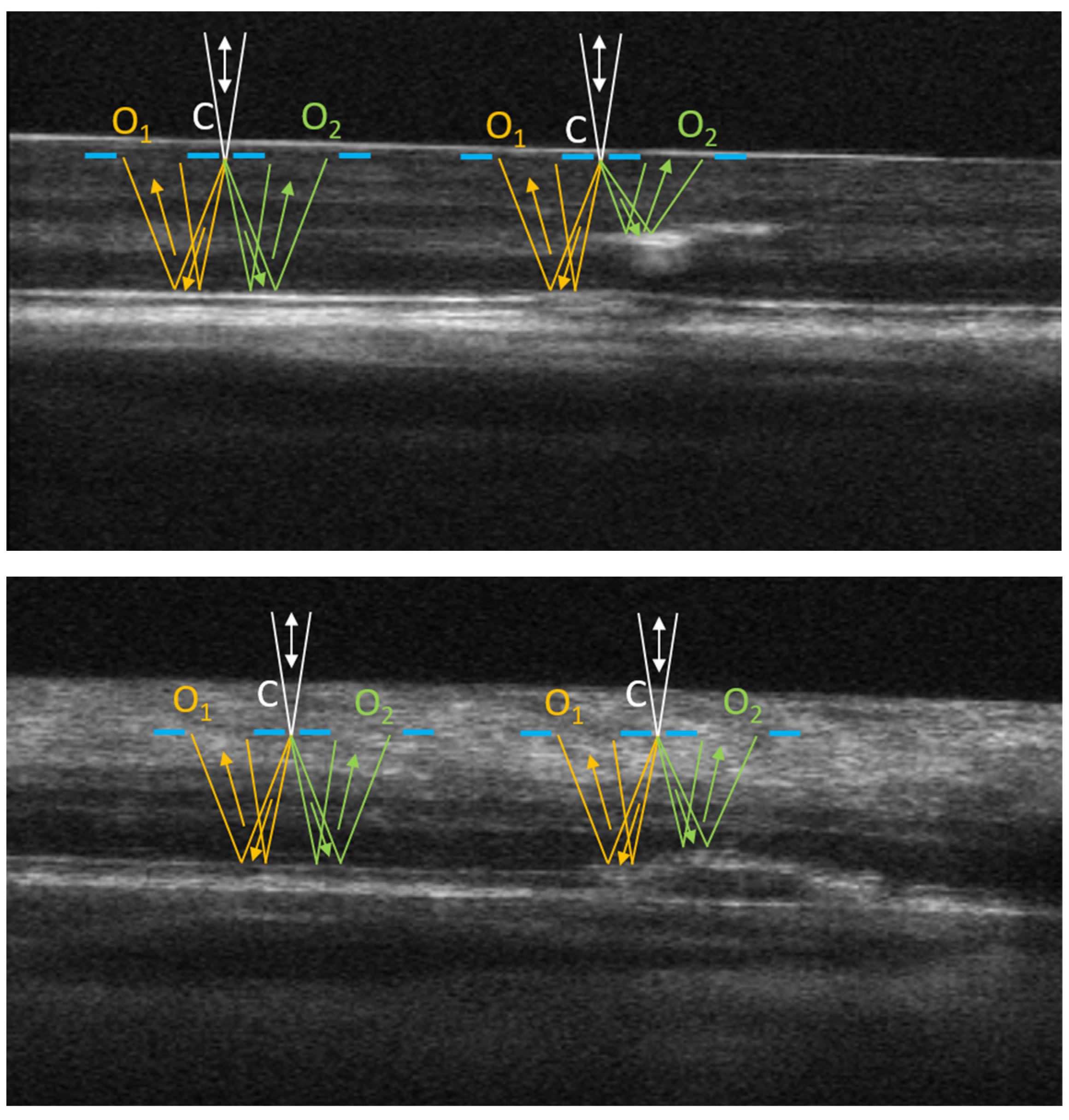
Abstract
1. Introduction
2. Materials and Methods
2.1. Imaging Systems
2.2. Human Subjects and the Imaging Procedure
2.3. Non-Confocal Imaging
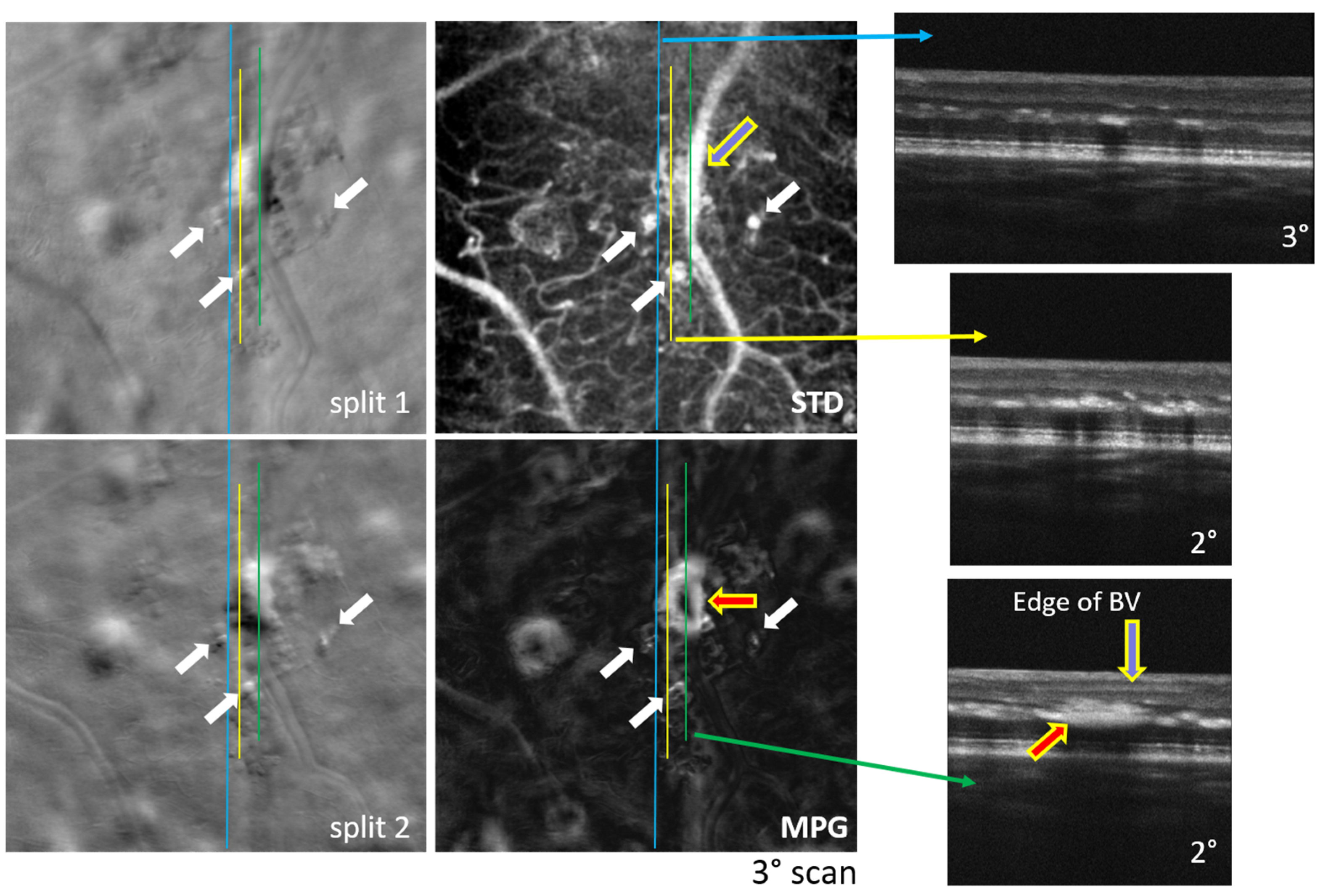
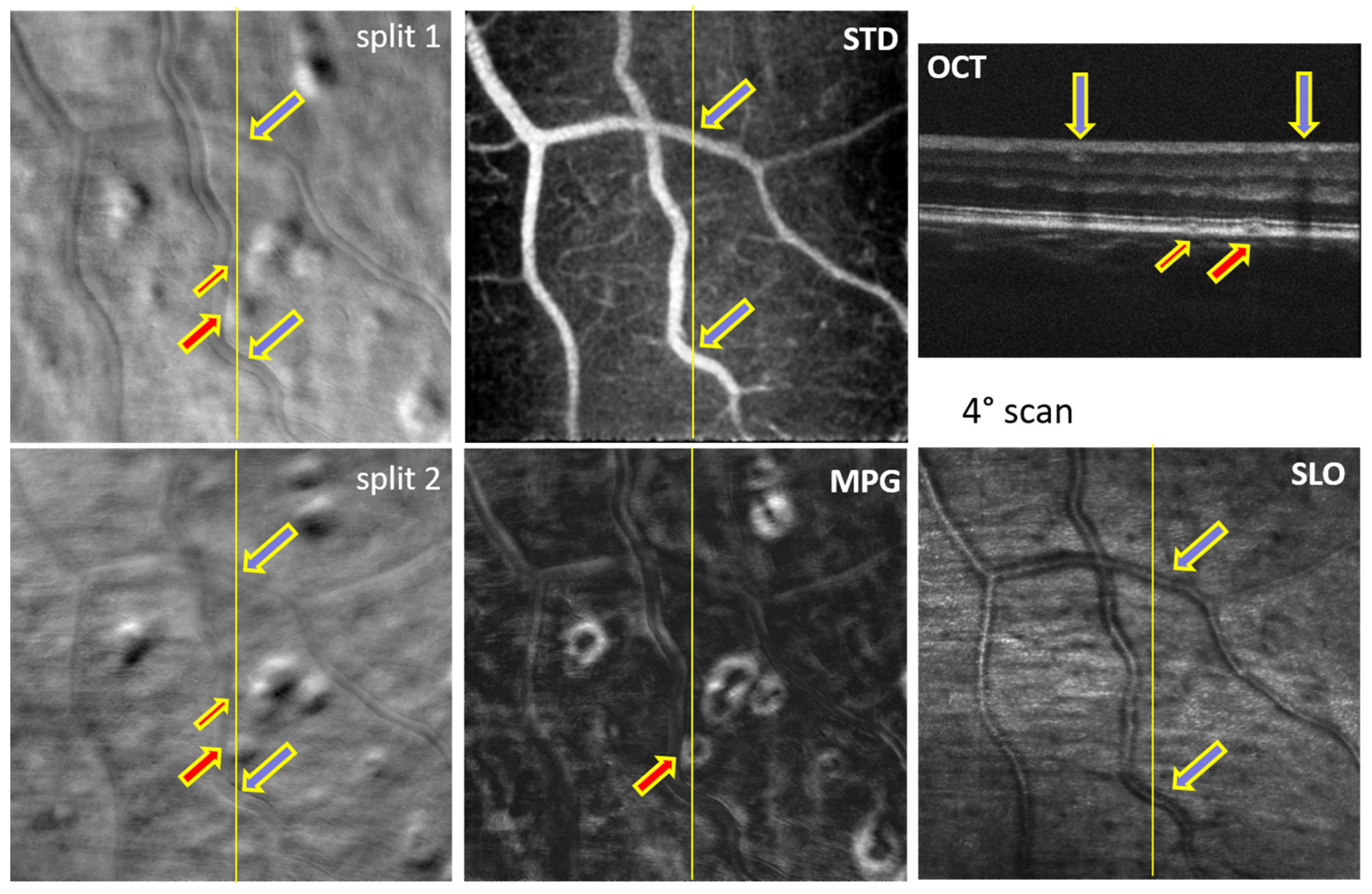
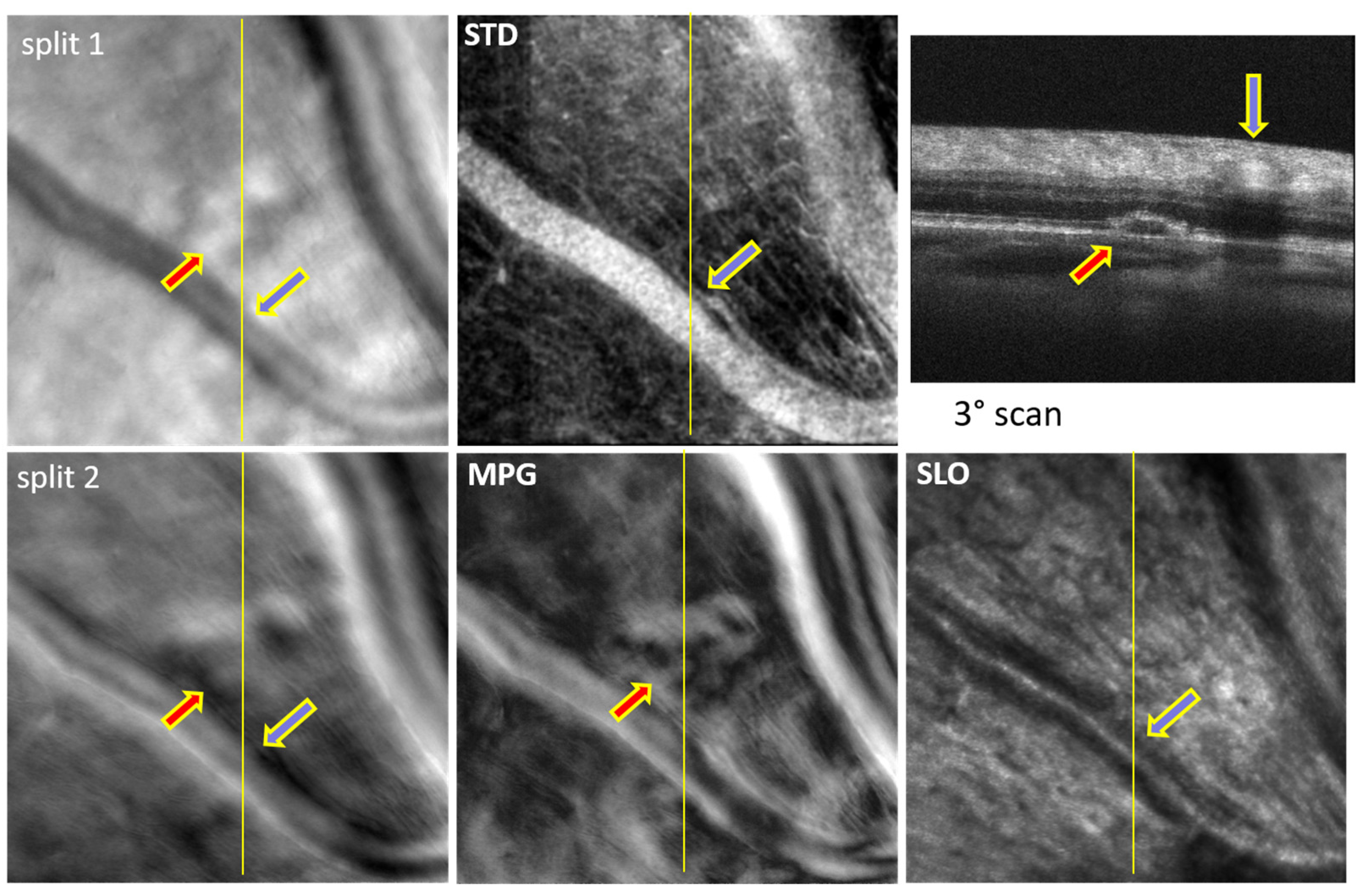
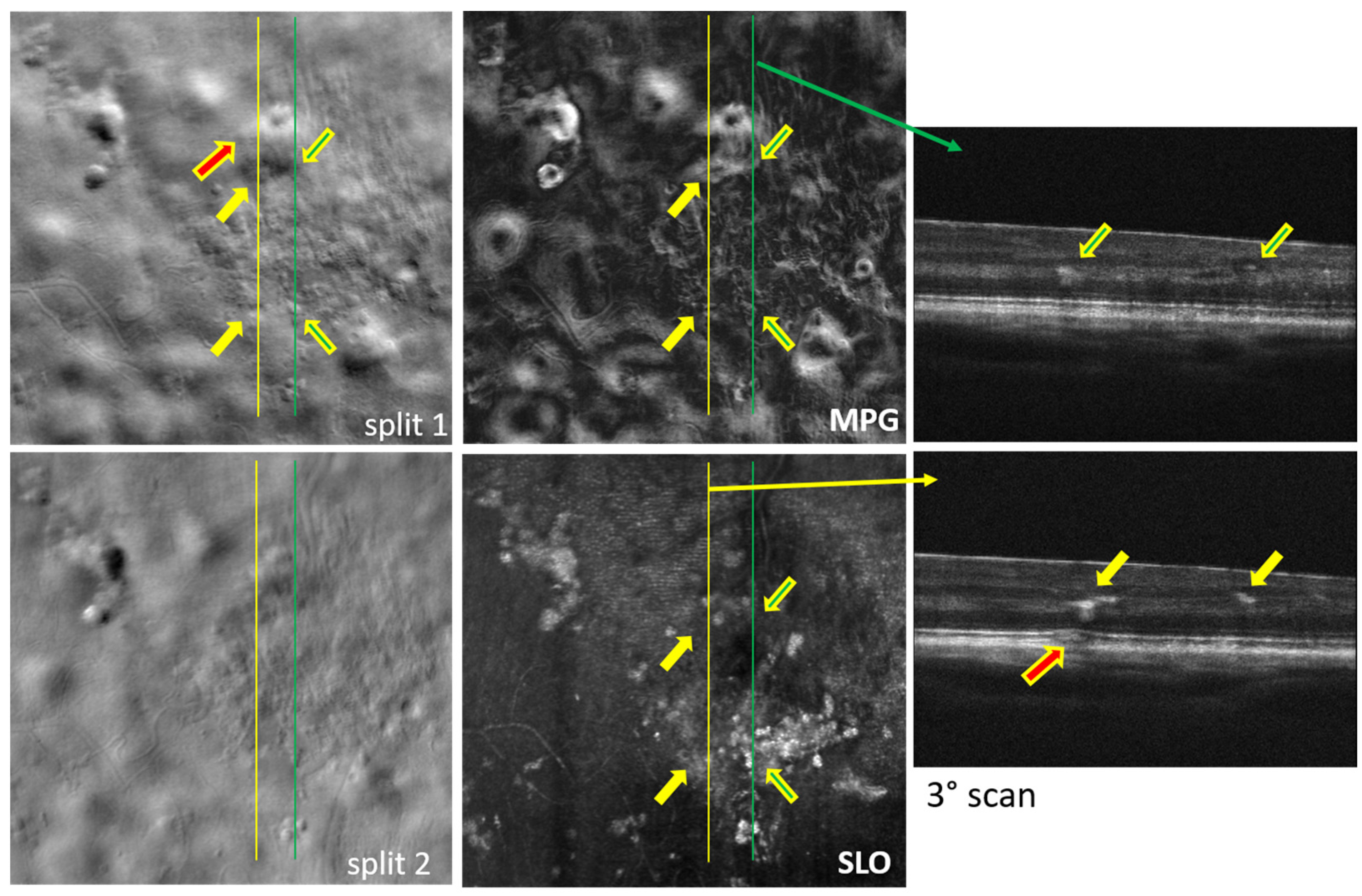
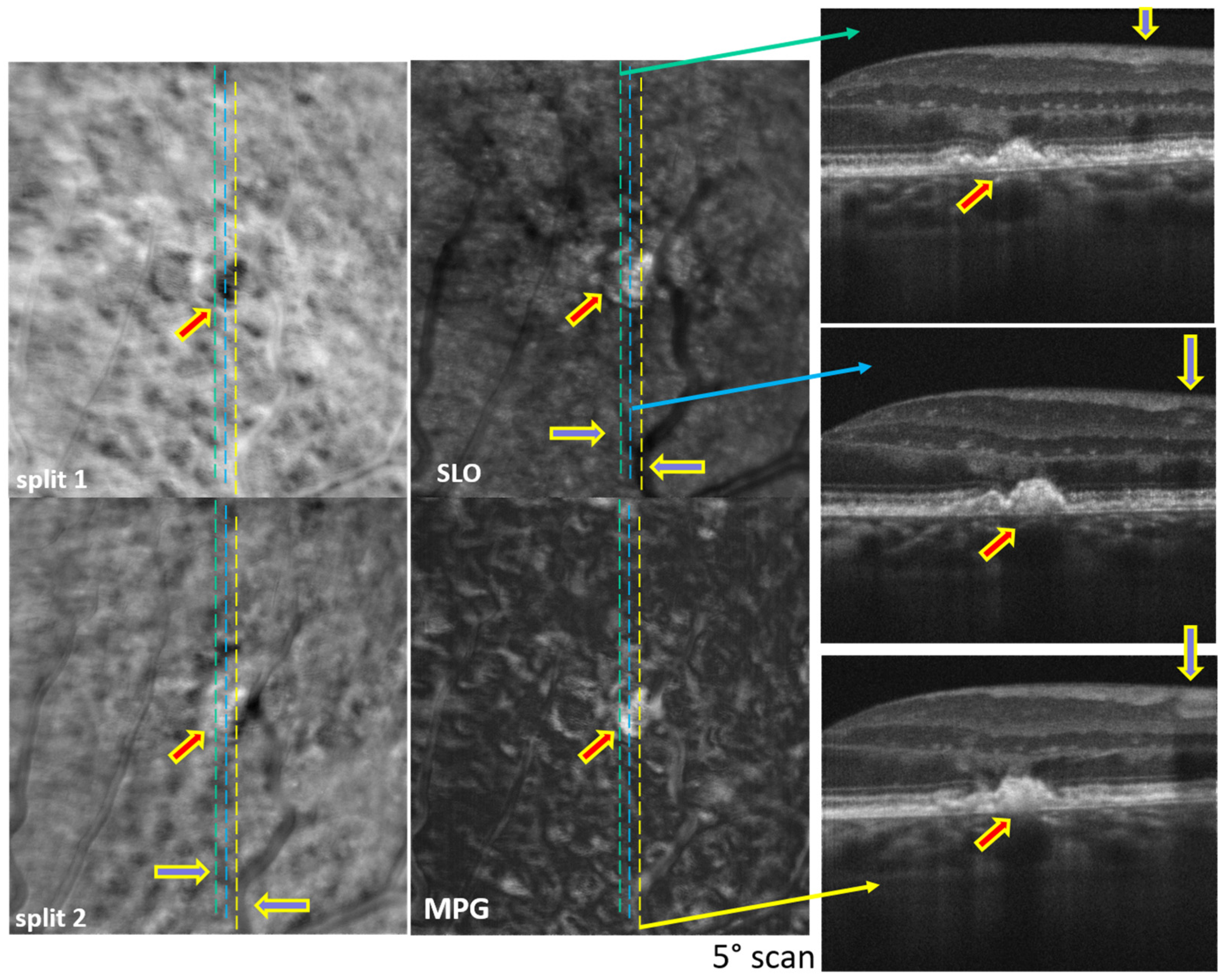
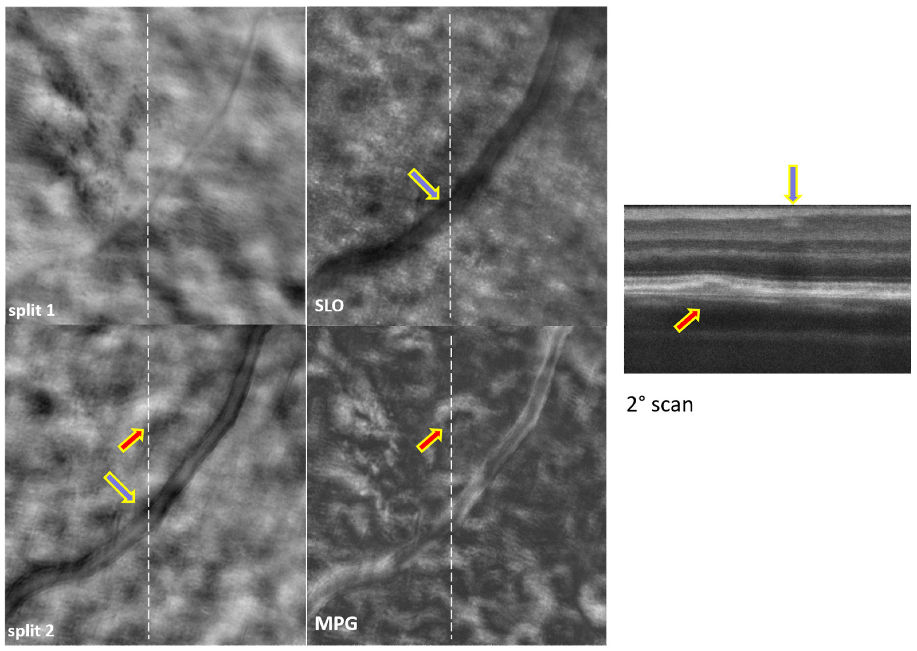
3. Results
4. Discussion
5. Conclusions
Supplementary Materials
Author Contributions
Funding
Institutional Review Board Statement
Informed Consent Statement
Data Availability Statement
Acknowledgments
Conflicts of Interest
References
- Chui, T.Y.P.; VanNasdale, D.A.; Burns, S.A. The use of forward scatter to improve retinal vascular imaging with an adaptive optics scanning laser ophthalmoscope. Biomed. Opt. Express 2012, 3, 2537–2549. [Google Scholar] [CrossRef]
- Scoles, D.; Sulai, Y.N.; Langlo, C.S.; Fishman, G.A.; Curcio, C.A.; Carroll, J.; Dubra, A. In Vivo Imaging of Human Cone Photoreceptor Inner Segments. Investig. Ophthalmol. Vis. Sci. 2014, 55, 4244–4251. [Google Scholar] [CrossRef]
- Amos, W.B.; Reichelt, S.; Cattermole, D.M.; Laufer, J. Re-evaluation of differential phase contrast (DPC) in a scanning laser microscope using a split detector as an alternative to differential interference contrast (DIC) optics. J. Microsc. 2003, 210, 166–175. [Google Scholar] [CrossRef]
- Hamilton, D.K.; Sheppard, C.J.R. Differential phase contrast in scanning optical microscopy. J. Microsc. 1984, 133, 27–39. [Google Scholar] [CrossRef]
- Hamilton, D.K.; Sheppard, C.J.R.; Wilson, T. Improved imaging of phase gradients in scanning optical microscopy. J. Microsc. 1984, 135, 275–286. [Google Scholar] [CrossRef]
- Mehta, S.B.; Sheppard, C.J.R. Quantitative phase-gradient imaging at high resolution with asymmetric illumination-based differential phase contrast. Opt. Lett. 2009, 34, 1924–1926. [Google Scholar] [CrossRef]
- Ferguson, R.D.; Mujat, M.; Maguluri, G.; Lu, Y.; Iftimia, N. Multi-fiber simultaneous offset-aperture AOSLO imaging in the inner retina. Investig. Ophthalmol. Vis. Sci. 2018, 59, 4640. [Google Scholar]
- Mozaffari, S.; Jaedicke, V.; Larocca, F.; Tiruveedhula, P.; Roorda, A. Versatile multi-detector scheme for adaptive optics scanning laser ophthalmoscopy. Biomed. Opt. Express 2018, 9, 5477–5488. [Google Scholar] [CrossRef]
- Rossi, E.A.; Granger, C.E.; Sharma, R.; Yang, Q.; Saito, K.; Schwarz, C.; Walters, S.; Nozato, K.; Zhang, J.; Kawakami, T.; et al. Imaging individual neurons in the retinal ganglion cell layer of the living eye. Proc. Natl. Acad. Sci. USA 2017, 114, 586–591. [Google Scholar] [CrossRef]
- Sredar, N.; Kowalski, B.; Razeen, M.M.; Steven, S.; Dubra, A. Non-confocal quad-detection adaptive optics scanning light ophthalmoscopy of the photoreceptor mosaic. Investig. Ophthalmol. Vis. Sci. 2018, 59, 4632. [Google Scholar]
- Mujat, M.; Sampani, K.; Patel, A.H.; Sun, J.K.; Iftimia, N. Cellular-Level Analysis of Retinal Blood Vessel Walls Based on Phase Gradient Images. Diagnostics 2023, 13, 3399. [Google Scholar] [CrossRef]
- Newman, E.A. Functional Hyperemia and Mechanisms of Neurovascular Coupling in the Retinal Vasculature. J. Cereb. Blood Flow Metab. 2013, 33, 1685–1695. [Google Scholar] [CrossRef]
- Garhöfer, G.; Chua, J.; Tan, B.; Wong, D.; Schmidl, D.; Schmetterer, L. Retinal Neurovascular Coupling in Diabetes. J. Clin. Med. 2020, 9, 2829. [Google Scholar] [CrossRef]
- Kotliar, K.; Hauser, C.; Ortner, M.; Muggenthaler, C.; Diehl-Schmid, J.; Angermann, S.; Hapfelmeier, A.; Schmaderer, C.; Grimmer, T. Altered neurovascular coupling as measured by optical imaging: A biomarker for Alzheimer’s disease. Sci. Rep. 2017, 7, 12906. [Google Scholar] [CrossRef] [PubMed]
- Huang, D.; Swanson, E.A.; Lin, C.P.; Schuman, J.S.; Stinson, W.G.; Chang, W.; Hee, M.R.; Flotte, T.; Gregory, K.; Puliafito, C.A.; et al. Optical Coherence Tomography. Science 1991, 254, 1178–1181. [Google Scholar] [CrossRef] [PubMed]
- Wojtkowski, M.; Leitgeb, R.; Kowalczyk, A.; Bajraszewski, T.; Fercher, A.F. In vivo human retinal imaging by Fourier domain optical coherence tomography. J. Biomed. Opt. 2002, 7, 457–463. [Google Scholar] [CrossRef] [PubMed]
- Liang, J.Z.; Williams, D.R.; Miller, D.T. Supernormal vision and high-resolution retinal imaging through adaptive optics. J. Opt. Soc. Am. A 1997, 14, 2884–2892. [Google Scholar] [CrossRef]
- Roorda, A.; Romero-Borja, F.; Donnelly, W.J.; Queener, H.; Hebert, T.J.; Campbell, M.C.W. Adaptive optics scanning laser ophthalmoscopy. Opt. Express 2002, 10, 405–412. [Google Scholar] [CrossRef]
- Zawadzki, R.J.; Jones, S.M.; Olivier, S.S.; Zhao, M.T.; Bower, B.A.; Izatt, J.A.; Choi, S.; Laut, S.; Werner, J.S. Adaptive-optics optical coherence tomography for high-resolution and high-speed 3D retinal in vivo imaging. Opt. Express 2005, 13, 8532–8546. [Google Scholar] [CrossRef]
- Mujat, M.; Ferguson, R.D.; Iftimia, N.; Hammer, D.X. Compact adaptive optics line scanning ophthalmoscope. Opt. Express 2009, 17, 10242–10258. [Google Scholar] [CrossRef]
- Ford, T.N.; Chu, K.K.; Mertz, J. Phase-gradient microscopy in thick tissue with oblique back-illumination. Nat. Methods 2012, 9, 1195–1197. [Google Scholar] [CrossRef] [PubMed]
- Tian, L.; Waller, L. Quantitative differential phase contrast imaging in an LED array microscope. Opt. Express 2015, 23, 11394–11403. [Google Scholar] [CrossRef]
- Mujat, M.; Akula, J.D.; Fulton, A.B.; Ferguson, R.D.; Iftimia, N. Non-Rigid Registration for High-Resolution Retinal Imaging. Diagnostics 2023, 13, 2285. [Google Scholar] [CrossRef] [PubMed]


Disclaimer/Publisher’s Note: The statements, opinions and data contained in all publications are solely those of the individual author(s) and contributor(s) and not of MDPI and/or the editor(s). MDPI and/or the editor(s) disclaim responsibility for any injury to people or property resulting from any ideas, methods, instructions or products referred to in the content. |
© 2024 by the authors. Licensee MDPI, Basel, Switzerland. This article is an open access article distributed under the terms and conditions of the Creative Commons Attribution (CC BY) license (https://creativecommons.org/licenses/by/4.0/).
Share and Cite
Mujat, M.; Sampani, K.; Patel, A.H.; Zambrano, R.; Sun, J.K.; Wollstein, G.; Ferguson, R.D.; Schuman, J.S.; Iftimia, N. Motion Contrast, Phase Gradient, and Simultaneous OCT Images Assist in the Interpretation of Dark-Field Images in Eyes with Retinal Pathology. Diagnostics 2024, 14, 184. https://doi.org/10.3390/diagnostics14020184
Mujat M, Sampani K, Patel AH, Zambrano R, Sun JK, Wollstein G, Ferguson RD, Schuman JS, Iftimia N. Motion Contrast, Phase Gradient, and Simultaneous OCT Images Assist in the Interpretation of Dark-Field Images in Eyes with Retinal Pathology. Diagnostics. 2024; 14(2):184. https://doi.org/10.3390/diagnostics14020184
Chicago/Turabian StyleMujat, Mircea, Konstantina Sampani, Ankit H. Patel, Ronald Zambrano, Jennifer K. Sun, Gadi Wollstein, R. Daniel Ferguson, Joel S. Schuman, and Nicusor Iftimia. 2024. "Motion Contrast, Phase Gradient, and Simultaneous OCT Images Assist in the Interpretation of Dark-Field Images in Eyes with Retinal Pathology" Diagnostics 14, no. 2: 184. https://doi.org/10.3390/diagnostics14020184
APA StyleMujat, M., Sampani, K., Patel, A. H., Zambrano, R., Sun, J. K., Wollstein, G., Ferguson, R. D., Schuman, J. S., & Iftimia, N. (2024). Motion Contrast, Phase Gradient, and Simultaneous OCT Images Assist in the Interpretation of Dark-Field Images in Eyes with Retinal Pathology. Diagnostics, 14(2), 184. https://doi.org/10.3390/diagnostics14020184