
Validation of Fixed Ultrasonography for Achilles Tendon Assessment: A Reliability Study

 ,
,  , , , , and
Abstract
1. Introduction
2. Materials and Methods
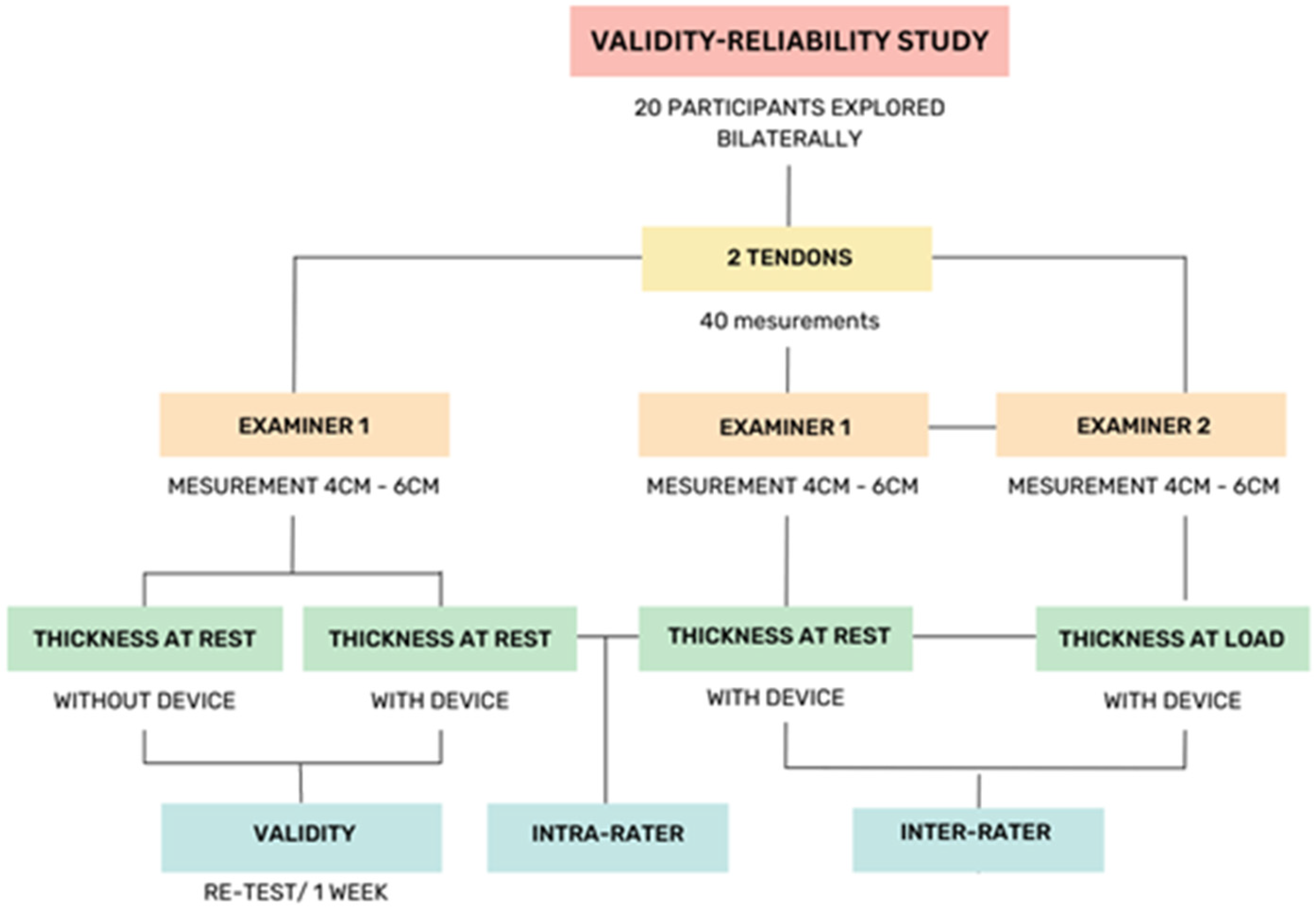
2.1. Design
2.2. Population
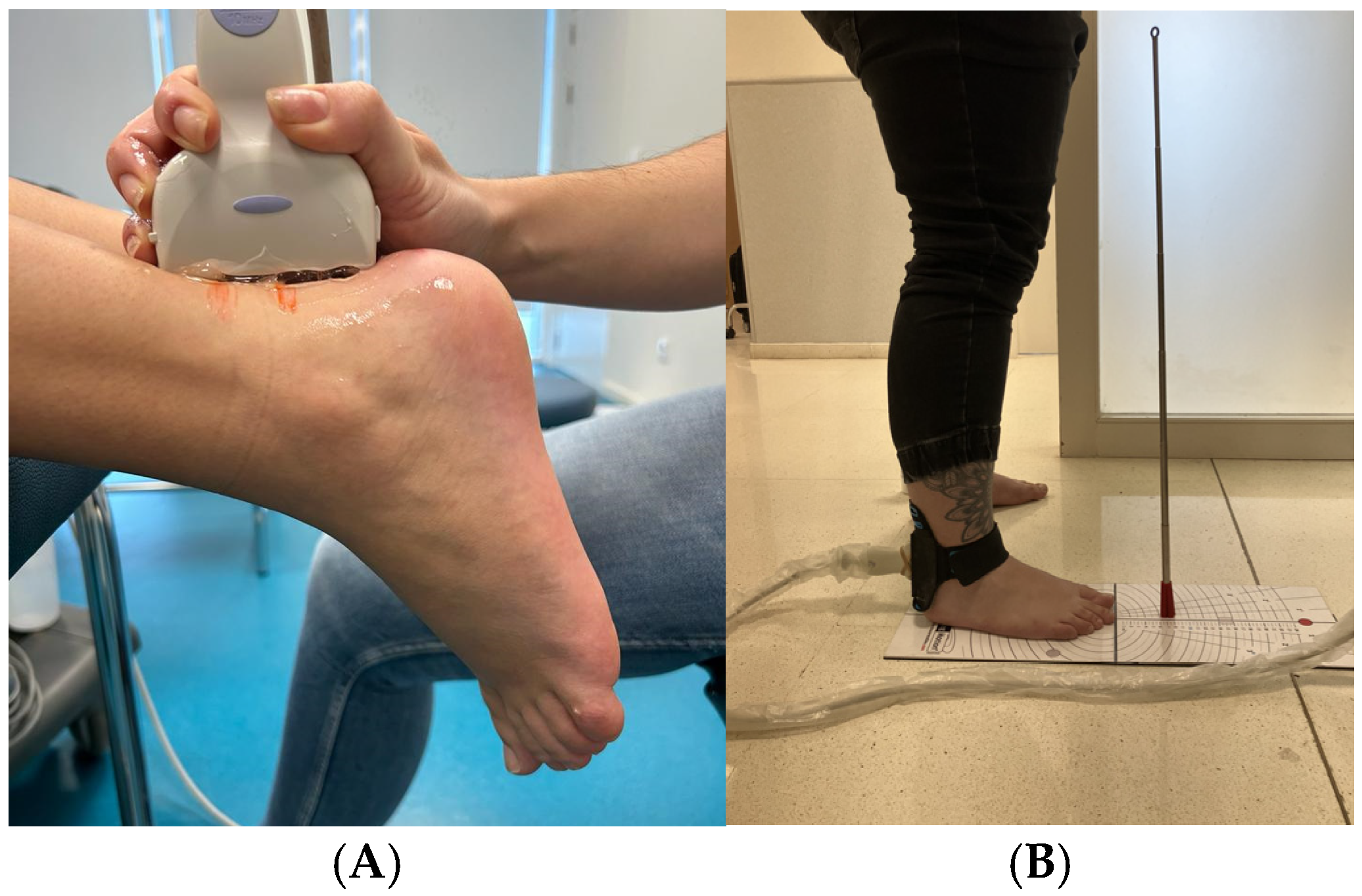
2.3. Procedure
2.4. Protocol Scanning
2.5. Statistical Analysis
3. Results
3.1. Sample Description
3.2. Ultrasound Data
3.3. Reliability
3.4. Inter-Rater Reliability
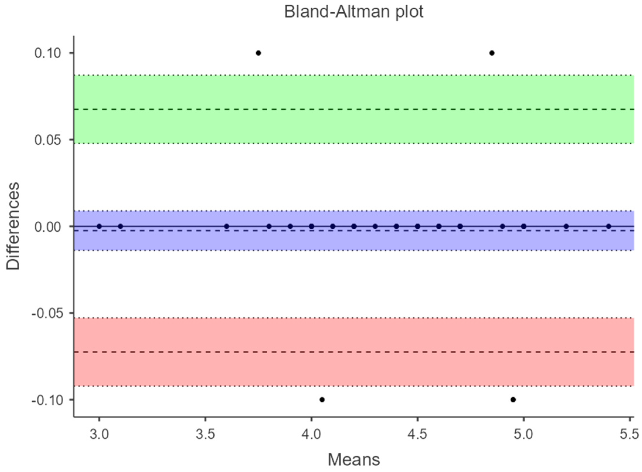
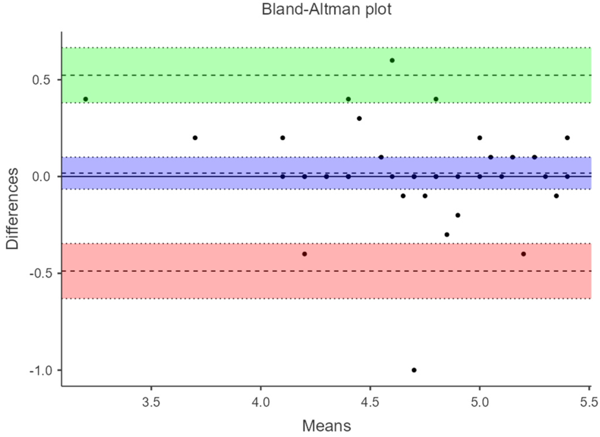
3.5. Bland–Altman Plots
4. Discussion
5. Conclusions
Author Contributions
Funding
Institutional Review Board Statement
Informed Consent Statement
Data Availability Statement
Conflicts of Interest
References
- French, C.N.; Walker, E.A.; Phillips, S.F.; Loeffert, J.R. Ultrasound in Sports Injuries. Clin. Sports Med. 2021, 40, 801–819. [Google Scholar] [CrossRef] [PubMed]
- Maffulli, N. Overuse tendon conditions: Time to change a confusing terminology. Arthrosc. J. Arthrosc. Relat. Surg. 1998, 14, 840–843. [Google Scholar] [CrossRef] [PubMed]
- Uquillas, C.A.; Guss, M.S.; Ryan, D.J.; Jazrawi, L.M.; Strauss, E.J. Everything Achilles: Knowledge Update and Current Concepts in Management. J. Bone Jt. Surg. 2015, 97, 1187–1195. [Google Scholar] [CrossRef] [PubMed]
- Hagoort, I.; Hortobágyi, T.; Vuillerme, N.; Lamoth, C.J.C.; Murgia, A. Age- and muscle-specific reliability of muscle architecture measurements assessed by two-dimensional panoramic ultrasound. Biomed. Eng. Online 2022, 21, 15. [Google Scholar] [CrossRef] [PubMed]
- Heres, H.M.; Sjoerdsma, M.; Schoots, T.; Rutten, M.C.M.; van de Vosse, F.N.; Lopata, R.G.P. Image acquisition stability of fixated musculoskeletal sonography in an exercise setting: A quantitative analysis and comparison with freehand acquisition. J. Med Ultrason. 2020, 47, 47–56. [Google Scholar] [CrossRef]
- Gao, J.; Rosander, A.; Rubin, J.M. Quantitative Assessment of Tendon Backscatter Anisotropy in B-Mode Ultrasound. Ultrasound Med. Biol. 2023, 49, 1408–1414. [Google Scholar] [CrossRef]
- Van Schie, H.T.; de Vos, R.J.; de Jonge, S.; Bakker, E.M.; Heijboer, M.P.; Verhaar, J.A.; Tol, J.L.; Weinans, H. Ultrasonographic tissue characterisation of human Achilles tendons: Quantification of tendon structure through a novel non-invasive approach. Br. J. Sports Med. 2010, 44, 1153–1159. [Google Scholar] [CrossRef]
- Daftary, A.; Adler, R.S. Sonographic Evaluation and Ultrasound-Guided Therapy of the Achilles Tendon. Ultrasound Q. 2009, 25, 103–110. [Google Scholar] [CrossRef]
- Alabau-Dasi, R.; Dominguez-Maldonado, G.; Gijon-Nogueron, G.; Ortega-Avila, A.B.; Delacroix, S. How susceptible are our Achilles Tendons? Sonoanatomical assessment. A cross-sectional study. J. Tissue Viability 2023, 32, 572–576. [Google Scholar] [CrossRef]
- Fukunaga, T.; Ichinose, Y.; Ito, M.; Kawakami, Y.; Fukashiro, S. Determination of fascicle length and pennation in a contracting human muscle in vivo. J. Appl. Physiol. 1997, 82, 354–358. [Google Scholar] [CrossRef]
- Färnqvist, K.; Pearson, S.; Malliaras, P. Adaptation of Tendon Structure and Function in Tendinopathy With Exercise and Its Relationship to Clinical Outcome. J. Sport Rehabil. 2020, 29, 107–115. [Google Scholar] [CrossRef] [PubMed]
- Rabusin, C.L.; Menz, H.B.; McClelland, J.A.; Evans, A.M.; Malliaras, P.; Docking, S.I.; Landorf, K.B.; Gerrard, J.M.; E. Munteanu, S. Efficacy of heel lifts versus calf muscle eccentric exercise for mid-portion Achilles tendinopathy (HEALTHY): A randomised trial. Br. J. Sports Med. 2021, 55, 486–492. [Google Scholar] [CrossRef]
- Shivapatham, G.; Richards, S.; Bamber, J.; Screen, H.; Morrissey, D. Ultrasound Measurement of Local Deformation in the Human Free Achilles Tendon Produced by Dynamic Muscle-Induced Loading: A Systematic Review. Ultrasound Med. Biol. 2023, 49, 1499–1509. [Google Scholar] [CrossRef]
- Fredberg, U.; Bolvig, L.; Andersen, N.T.; Stengaard-Pedersen, K. Ultrasonography in Evaluation of Achilles and Patella Tendon Thickness. Ultraschall Der Med.-Eur. J. Ultrasound 2007, 29, 60–65. [Google Scholar] [CrossRef]
- Hutchison, A.-M.; Evans, R.; Bodger, O.; Pallister, I.; Topliss, C.; Williams, P.; Vannet, N.; Morris, V.; Beard, D. What is the best clinical test for Achilles tendinopathy? Foot Ankle Surg. 2013, 19, 112–117. [Google Scholar] [CrossRef] [PubMed]
- Reiman, M.; Burgi, C.; Strube, E.; Prue, K.; Ray, K.; Elliott, A.; Goode, A. The Utility of Clinical Measures for the Diagnosis of Achilles Tendon Injuries: A Systematic Review With Meta-Analysis. J. Athl. Train. 2014, 49, 820–829. [Google Scholar] [CrossRef] [PubMed]
- Hopkins, W.G. Measures of Reliability in Sports Medicine and Science. Sports Med. 2000, 30, 1–15. [Google Scholar] [CrossRef] [PubMed]
- Bohm, S.; Mersmann, F.; Schroll, A.; Mäkitalo, N.; Arampatzis, A. Insufficient accuracy of the ultrasound-based determination of Achilles tendon cross-sectional area. J. Biomech. 2016, 49, 2932–2937. [Google Scholar] [CrossRef]
- Nadeau, M.; Desrochers, A.; Lamontagne, M.; Larivière, C.; Gagnon, D.H. Quantitative ultrasound imaging of Achilles tendon integrity in symptomatic and asymptomatic individuals: Reliability and minimal detectable change. J. Foot Ankle Res. 2016, 9, 30. [Google Scholar] [CrossRef]
- Bennell, K.; Talbot, R.; Wajswelner, H.; Techovanich, W.; Kelly, D.; Hall, A. Intra-rater and inter-rater reliability of a weight-bearing lunge measure of ankle dorsiflexion. Aust. J. Physiother. 1998, 44, 175–180. [Google Scholar] [CrossRef]
- Ríos-Díaz, J.; Martínez-Payá, J.J.; del Baño-Aledo, M.E.; de Groot-Ferrando, A.; Botía-Castillo, P.; Fernández-Rodríguez, D. Sonoelastography of Plantar Fascia: Reproducibility and Pattern Description in Healthy Subjects and Symptomatic Subjects. Ultrasound Med. Biol. 2015, 41, 2605–2613. [Google Scholar] [CrossRef] [PubMed]
- Wu, J.; Zhang, Y.-Z.; Gao, Y.; Luo, T.-Y. Assessment the reliability of ultrasonography in the imaging of the plantar fascia: A comparative study. BMC Med. Imaging 2019, 19, 62. [Google Scholar] [CrossRef] [PubMed]
- Barfod, K.W.; Riecke, A.F.; Anders, B.; Hansen, P.; Maier, J.F.; Døssing, S.; Troelsen, A. Validity and reliability of an ultrasound measurement of the free length of the Achilles tendon. Dan Med. J. 2018, 65, A5453. [Google Scholar]
- Alabau-Dasi, R.; Nieto-Gil, P.; Ortega-Avila, A.B.; Gijon-Nogueron, G. Variations in the Thickness of the Plantar Fascia After Training Based in Training Race. A Pilot Study. J. Foot Ankle Surg. 2022, 61, 1230–1234. [Google Scholar] [CrossRef]
- Ying, M.; Yeung, E.; Li, B.; Li, W.; Lui, M.; Tsoi, C.-W. Sonographic evaluation of the size of achilles tendon: The effect of exercise and dominance of the ankle. Ultrasound Med. Biol. 2003, 29, 637–642. [Google Scholar] [CrossRef]
- O’connor, P.J.; Grainger, A.J.; Morgan, S.R.; Smith, K.L.; Waterton, J.C.; Nash, A.F.P. Ultrasound assessment of tendons in asymptomatic volunteers: A study of reproducibility. Eur. Radiol. 2004, 14, 1968–1973. [Google Scholar] [CrossRef]
- Pang, B.S.F.; Ying, M. Sonographic Measurement of Achilles Tendons in Asymptomatic Subjects. J. Ultrasound Med. 2006, 25, 1291–1296. [Google Scholar] [CrossRef]
- Landis, J.R.; Koch, G.G. The Measurement of Observer Agreement for Categorical Data. Biometrics 1977, 33, 159–174. [Google Scholar] [CrossRef]
- Del Bano-Aledo, M.E.; Martinez-Paya, J.J.; Rios-Diaz, J.; Mejias-Suarez, S.; Serrano-Carmona, S.; de Groot-Ferrando, A. Ultrasound measures of tendon thickness: Intra-rater, Inter-rater and Inter-machine reliability. Muscle Ligaments Tendons J. 2017, 7, 192–199. [Google Scholar] [CrossRef]
- Wang, Y.-H.; Zhou, H.-H.; Nie, Z.; Cui, S. Prevalence of Achilles tendinopathy in physical exercise: A systematic review and meta-analysis. Sports Med. Health Sci. 2022, 4, 152–159. [Google Scholar] [CrossRef]
- Lewis, R.; Álvarez, C.B.G.; Rayman, M.; Lanham-New, S.; Woolf, A.; Mobasheri, A. Strategies for optimising musculoskeletal health in the 21st century. BMC Musculoskelet. Disord. 2019, 20, 164. [Google Scholar] [CrossRef] [PubMed]
- Nuri, L.; Obst, S.J.; Newsham-West, R.; Barrett, R.S. Three-dimensional morphology and volume of the free Achilles tendon at rest and under load in people with unilateral mid-portion Achilles tendinopathy. Exp. Physiol. 2018, 103, 358–369. [Google Scholar] [CrossRef] [PubMed]
- Bruno, F.; Arrigoni, F.; Palumbo, P.; Natella, R.; Splendiani, A.; Di Cesare, E.; Guglielmi, G.; Masciocchi, C.; Barile, A. Weight-bearing MR Imaging of Knee, Ankle and Foot. Semin. Musculoskelet. Radiol. 2019, 23, 594–602. [Google Scholar] [CrossRef] [PubMed]


| Gender (n) | Age (y) (Mean/SD) | Weight (k) (Mean/SD) | Height (cm) (Mean/SD) | BMI (Mean/SD) |
|---|---|---|---|---|
| Male (6) | 22.5 ± 3.12 | 79.8 ± 13.91 | 167 ±6.79 | 25.1 ± 2.99 |
| Female (14) | 22.6 ± 1.95 | 63.8 ± 6.94 | 178 ± 6.16 | 23 ± 2.77 |
| Total (20) | 22.55 ± 2.32 | 68.6 ± 12.51 | 170 ± 8.07 | 23.61 ± 2.97 |
| Intra-Rater | Inter-Rater | |||||||
|---|---|---|---|---|---|---|---|---|
| Variables | US Acquisition | Examiner 1 | Examiner 2 | Mean (SD) 95% CI | α Cronbach | * Mean Difference 95% LOA | ||
| Mean (SD) 95% CI | Mean (SD) 95% CI | ICC(1-1) (95% CI) | p-Value | |||||
| 4 cm At rest | manual acquisition | 4.50 (0.54) | 4.49 (0.60) | 0.91 (0.86–0.95) | <0.001 | 4.51 (0.528) | 0.998 | 0.005 (−0.104 to 0.094) |
| 6 cm At rest | manual acquisition | 4.63 (0.455) | 4.66 (0.611) | 0.895 (0.861–0.945) | <0.001 | 4.63 (0.450) | 0.996 | 0.02 (−0.0905 to 0.13) |
| 4 cm At rest | fixed ultrasonography | 4.58 (0.556) | 4.66 (0.579) | 0.926 (0.882–0.957) | <0.001 | 4.58 (0.543) | 0.997 | 0.000 (−0.109 to 0.109) |
| 6 cm At rest | fixed ultrasonography | 4.67 (0.461) | 4.66 (0.510) | 0.909 (0.861–0.945) | <0.001 | 4.67 (0.460) | 0.997 | 0.01 (−0.0872 to 0.1072) |
| 4 cm At load/DF | fixed ultrasonography | 4.24 (0.624) | 4.36 (0.614) | 0.894 (0.826–0.94) | <0.001 | 4.24 (0.626) | 1.000 | −0.005 (−0.0483 to 0.0383) |
| 6 cm At load/DF | fixed ultrasonography | 4.39 (0.531) | 4.36 (0.631) | 0.876 (0.802–0.928) | <0.001 | 4.39 (0.533) | 0.999 | −0.0025 (−0.0725 to 0.0675) |
Disclaimer/Publisher’s Note: The statements, opinions and data contained in all publications are solely those of the individual author(s) and contributor(s) and not of MDPI and/or the editor(s). MDPI and/or the editor(s) disclaim responsibility for any injury to people or property resulting from any ideas, methods, instructions or products referred to in the content. |
© 2024 by the authors. Licensee MDPI, Basel, Switzerland. This article is an open access article distributed under the terms and conditions of the Creative Commons Attribution (CC BY) license (https://creativecommons.org/licenses/by/4.0/).
Share and Cite
Alabau-Dasi, R.; Dominguez-Maldonado, G.; Ortega-Avila, A.B.; Gordillo-Fernandez, L.M.; Ortiz-Romero, M.; Melchor-Rodriguez, J.M.; Gijon-Nogueron, G. Validation of Fixed Ultrasonography for Achilles Tendon Assessment: A Reliability Study. Diagnostics 2024, 14, 2221. https://doi.org/10.3390/diagnostics14192221
Alabau-Dasi R, Dominguez-Maldonado G, Ortega-Avila AB, Gordillo-Fernandez LM, Ortiz-Romero M, Melchor-Rodriguez JM, Gijon-Nogueron G. Validation of Fixed Ultrasonography for Achilles Tendon Assessment: A Reliability Study. Diagnostics. 2024; 14(19):2221. https://doi.org/10.3390/diagnostics14192221
Chicago/Turabian StyleAlabau-Dasi, Raquel, Gabriel Dominguez-Maldonado, Ana Belen Ortega-Avila, Luis M. Gordillo-Fernandez, Mercedes Ortiz-Romero, Juan Manuel Melchor-Rodriguez, and Gabriel Gijon-Nogueron. 2024. "Validation of Fixed Ultrasonography for Achilles Tendon Assessment: A Reliability Study" Diagnostics 14, no. 19: 2221. https://doi.org/10.3390/diagnostics14192221
APA StyleAlabau-Dasi, R., Dominguez-Maldonado, G., Ortega-Avila, A. B., Gordillo-Fernandez, L. M., Ortiz-Romero, M., Melchor-Rodriguez, J. M., & Gijon-Nogueron, G. (2024). Validation of Fixed Ultrasonography for Achilles Tendon Assessment: A Reliability Study. Diagnostics, 14(19), 2221. https://doi.org/10.3390/diagnostics14192221