


Incidental Findings Following Dental Implant Procedures in the Mandible: A New Post-Processing CBCT Software Analysis

 , , and
Abstract
1. Introduction
2. Materials and Methods
- Positioning of dental implants: 0: implant located in the anterior right region of the mandible (right central incisor to right canine); 1: implant located in the anterior left region of the mandible (left central incisor to left canine); 2: implant located in the posterior right region of the mandible (right first premolar to right second molar); and 3: implant located in the posterior left region of the mandible (left first premolar to left second molar).
- Length of dental implants: 0: implant length less than 9 mm; 1: implant length between 9 mm and 14 mm; and 2: implant length greater than 14 mm.
- Anatomical relationship between the implant and mandibular canal: 0: implant above the upper limit of the mandibular canal by 1–2 mm; 1: implant in contact with the mandibular canal; and 2: implant within the mandibular canal (1 mm or more)
- Damage to adjacent teeth to the implants: 0: no damage to adjacent teeth; 1: damage to the tooth located anteriorly to the implant; and 2: damage to the tooth located posteriorly to the implant.
- Implant fracture: 0: no fracture in the implant; and 1: presence of fracture in the implant
- Bone support for the implant: 0: absence of bone support for the implant; and 1: presence of bone support for the implant.
3. Results
4. Discussion
5. Conclusions
Author Contributions
Funding
Institutional Review Board Statement
Informed Consent Statement
Data Availability Statement
Conflicts of Interest
References
- Dwingadi, E.; Soeroso, Y.; Lessang, R.; Priaminiarti, M. Evaluation of alveolar bone on dental implant treatment using cone beam computed tomography. Pesqui. Bras. Em Odontopediatria E Clínica Integr. 2019, 19, e4917. [Google Scholar] [CrossRef]
- Nulty, A. A literature review on prosthetically designed guided implant placement and the factors influencing dental implant success. Br. Dent. J. 2024, 236, 169–180. [Google Scholar] [CrossRef] [PubMed]
- Silva, K.S.; Nascimento, M.; de Souza, B.M.; Posch, A.T. Fatores que influênciam o planejamento de implantes dentários osseointegráveis. Braz. J. Implantol. Health Sci. 2022, 4, 17–34. [Google Scholar] [CrossRef]
- Clark, D.; Barbu, H.; Lorean, A.; Mijiritsky, E.; Levin, L. Incidental findings of implant complications on postimplantation CBCTs: A cross-sectional study. Clin. Implant. Dent. Relat. Res. 2017, 19, 776–782. [Google Scholar] [CrossRef] [PubMed]
- Gupta, A.; Rathee, S.; Agarwal, J.; Pachar, R.B. Measurement of Crestal Cortical Bone Thickness at Implant Site: A Cone Beam Computed Tomography Study. J. Contemp. Dent. Pract. 2017, 18, 785–789. [Google Scholar] [CrossRef]
- Hong, D.G.K.; Oh, J.H. Recent advances in dental implants. Maxillofac. Plast. Reconstr. Surg. 2017, 39, 33. [Google Scholar] [CrossRef]
- Thoma, D.S.; Sailer, I.; Ioannidis, A.; Zwahlen, M.; Makarov, N.; Pjetursson, B.E. A systematic review of the survival and complication rates of resin- bonded fixed dental prostheses after a mean observation period of at least 5 years. Clin. Oral. Implant. Res. 2017, 28, 1421–1432. [Google Scholar] [CrossRef]
- Wolff, C.; Mucke, T.; Wagenpfeil, S.; Kanatas, A.; Bissinger, O.; Deppe, H. Do CBCT scans alter surgical treatment plans? Comparison of preoperative surgical diagnosis using panoramic versus cone-beam CT images. J. Craniomaxillofac. Surg. 2016, 44, 1700–1705. [Google Scholar] [CrossRef]
- Silva, J.A.; de Alencar, A.H.; da Rocha, S.S.; Lopes, L.G.; Estrela, C. Three-dimensional image contribution for evaluation of operative procedural errors in endodontic therapy and dental implants. Braz. Dent. J. 2012, 23, 127–134. [Google Scholar] [CrossRef]
- Gaeta-Araujo, H.; Oliveira-Santos, N.; Mancini, A.X.M.; Oliveira, M.L.; Oliveira-Santos, C. Retrospective assessment of dental implant-related perforations of relevant anatomical structures and inadequate spacing between implants/teeth using cone-beam computed tomography. Clin. Oral Investig. 2020, 24, 3281–3288. [Google Scholar] [CrossRef]
- Kullar, A.S.; Miller, C.S. Are There Contraindications for Placing Dental Implants? Dent. Clin. N. Am. 2019, 63, 345–362. [Google Scholar] [CrossRef]
- Ribas, B.R.; Nascimento, E.H.L.; Freitas, D.Q.; Pontual, A.D.A.; Pontual, M.; Perez, D.E.C.; Ramos-Perez, F.M.M. Positioning errors of dental implants and their associations with adjacent structures and anatomical variations: A CBCT-based study. Imaging Sci. Dent. 2020, 50, 281–290. [Google Scholar] [CrossRef] [PubMed]
- Sun, Y.; Hu, S.; Xie, Z.; Zhou, Y. Relevant factors of posterior mandible lingual plate perforation during immediate implant placement: A virtual implant placement study using CBCT. BMC Oral Health 2023, 23, 76. [Google Scholar] [CrossRef]
- de Souza, L.A.; Souza Picorelli Assis, N.M.; Ribeiro, R.A.; Pires Carvalho, A.C.; Devito, K.L. Assessment of mandibular posterior regional landmarks using cone-beam computed tomography in dental implant surgery. Ann. Anat. 2016, 205, 53–59. [Google Scholar] [CrossRef] [PubMed]
- Sheridan, R.A.; Chiang, Y.C.; Decker, A.M.; Sutthiboonyapan, P.; Chan, H.L.; Wang, H.L. The Effect of Implant-Induced Artifacts on Interpreting Adjacent Bone Structures on Cone-Beam Computed Tomography Scans. Implant. Dent. 2018, 27, 10–14. [Google Scholar] [CrossRef] [PubMed]
- Kanewoff, E.; Alhallak, R.; de Carvalho Machado, V.; Chrcanovic, B.R. Immediate implant placement in the anterior mandible: A cone beam computed tomography study. BMC Oral Health 2024, 24, 393. [Google Scholar] [CrossRef]
- Safi, Y.; Amid, R.; Zadbin, F.; Ghazizadeh Ahsaie, M.; Mortazavi, H. The occurrence of dental implant malpositioning and related factors: A cross-sectional cone-beam computed tomography survey. Imaging Sci. Dent. 2021, 51, 251–260. [Google Scholar] [CrossRef]
- Nikkerdar, N.; Golshah, A.; Mahmoodivesali, R.; Falah-Kooshki, S. Assessment of Implant-Related Anatomical Landmarks in the Mandibular Interforaminal Region in an Iranian Population Using Cone-Beam Computed Tomography. Contemp. Clin. Dent. 2022, 13, 125–134. [Google Scholar] [CrossRef] [PubMed]
- Bueno, M.R.; Estrela, C.; Azevedo, B.C.; Diogenes, A. Development of a New Cone-Beam Computed Tomography Software for Endodontic Diagnosis. Braz. Dent. J. 2018, 29, 517–529. [Google Scholar] [CrossRef]
- Genc, T.; Duruel, O.; Kutlu, H.B.; Dursun, E.; Karabulut, E.; Tozum, T.F. Evaluation of anatomical structures and variations in the maxilla and the mandible before dental implant treatment. Dent. Med. Probl. 2018, 55, 233–240. [Google Scholar] [CrossRef]
- Leite, G.M.; Lana, J.P.; de Carvalho Machado, V.; Manzi, F.R.; Souza, P.E.; Horta, M.C. Anatomic variations and lesions of the mandibular canal detected by cone beam computed tomography. Surg. Radiol. Anat. 2014, 36, 795–804. [Google Scholar] [CrossRef] [PubMed]
- Machado, A.H.; Fardim, K.A.C.; de Souza, C.F.; Sotto-Maior, B.S.; Assis, N.; Devito, K.L. Effect of anatomical region on the formation of metal artefacts produced by dental implants in cone beam computed tomographic images. Dentomaxillofac. Radiol. 2018, 47, 20170281. [Google Scholar] [CrossRef]
- Bueno, M.R.; Azevedo, B.C.; Estrela, C.R.A.; Sousa-Neto, M.D.; Estrela, C. Method to Identify Accessory Root Canals using a New CBCT Software. Braz. Dent. J. 2021, 32, 28–35. [Google Scholar] [CrossRef] [PubMed]
- Estrela, C.; Costa, M.V.C.; Bueno, M.R.; Rabelo, L.E.G.; Decurcio, D.A.; Silva, J.A.; Estrela, C.R.A. Potential of a New Cone-Beam CT Software for Blooming Artifact Reduction. Braz. Dent. J. 2020, 31, 582–588. [Google Scholar] [CrossRef] [PubMed]
- Alghamdi, H.S.; Jansen, J.A. The development and future of dental implants. Dent. Mater. J. 2020, 39, 167–172. [Google Scholar] [CrossRef]
- Bueno, M.R.; Estrela, C.; Azevedo, B.C.; Cintra Junqueira, J.L. Root Canal Shape of Human Permanent Teeth Determined by New Cone-Beam Computed Tomographic Software. J. Endod. 2020, 46, 1662–1674. [Google Scholar] [CrossRef]
- Abdinian, M.; Yaghini, J.; Jazi, L. Comparison of intraoral digital radiography and cone-beam computed tomography in the measurement of periodontal bone defects. Dent. Med. Probl. 2020, 57, 269–273. [Google Scholar] [CrossRef]
- Nascimento, E.H.L.; Oliveira, M.L.; Freitas, D.Q. Incidental findings of implant complications on postimplantation CBCTs: A cross-sectional study-Methodological issues. Clin. Implant. Dent. Relat. Res. 2019, 21, 11–12. [Google Scholar] [CrossRef]
- Porto, O.C.L.; Silva, B.S.F.; Silva, J.A.; Estrela, C.R.A.; Alencar, A.H.G.; Bueno, M.D.R.; Estrela, C. CBCT assessment of bone thickness in maxillary and mandibular teeth: An anatomic study. J. Appl. Oral Sci. 2020, 28, e20190148. [Google Scholar] [CrossRef]
- Romanos, G.; Mulham, J.; Morrow, N.; Farber, A.H.; Mahdian, M. Neurological Risks During Implant Placement in the Anterior Maxilla and Mandible: A Literature Review. J. Oral Implantol. 2023, 49, 428–435. [Google Scholar] [CrossRef]
- Chackartchi, T.; Romanos, G.E.; Parkanyi, L.; Schwarz, F.; Sculean, A. Reducing errors in guided implant surgery to optimize treatment outcomes. Periodontol. 2000 2022, 88, 64–72. [Google Scholar] [CrossRef] [PubMed]
- Flügge, T.; Kramer, J.; Nelson, K.; Nahles, S.; Kernen, F. Digital implantology-a review of virtual planning software for guided implant surgery. Part II: Prosthetic set-up and virtual implant planning. BMC Oral Health 2022, 22, 23. [Google Scholar] [CrossRef]
- Parnia, F.; Fard, E.M.; Mahboub, F.; Hafezeqoran, A.; Gavgani, F.E. Tomographic volume evaluation of submandibular fossa in patients requiring dental implants. Oral Surg. Oral Med. Oral Pathol. Oral Radiol. Endod. 2010, 109, e32–e36. [Google Scholar] [CrossRef] [PubMed]


| Characteristics | n (%) | 95% IC |
|---|---|---|
| Sex | ||
| Female | 104 (48.6) | 41.7–55.5 |
| Male | 110 (51.4) | 44.5–58.3 |
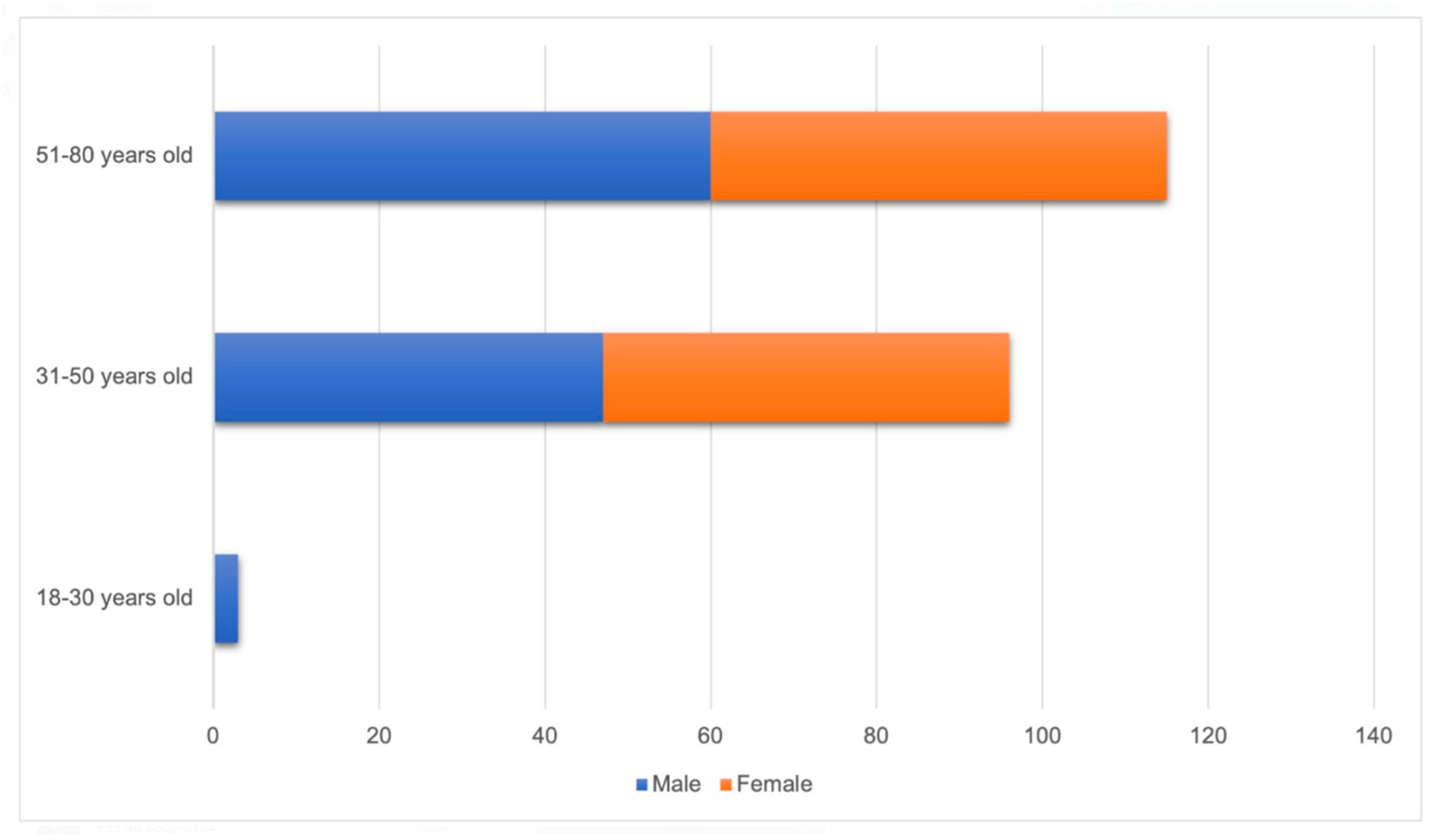
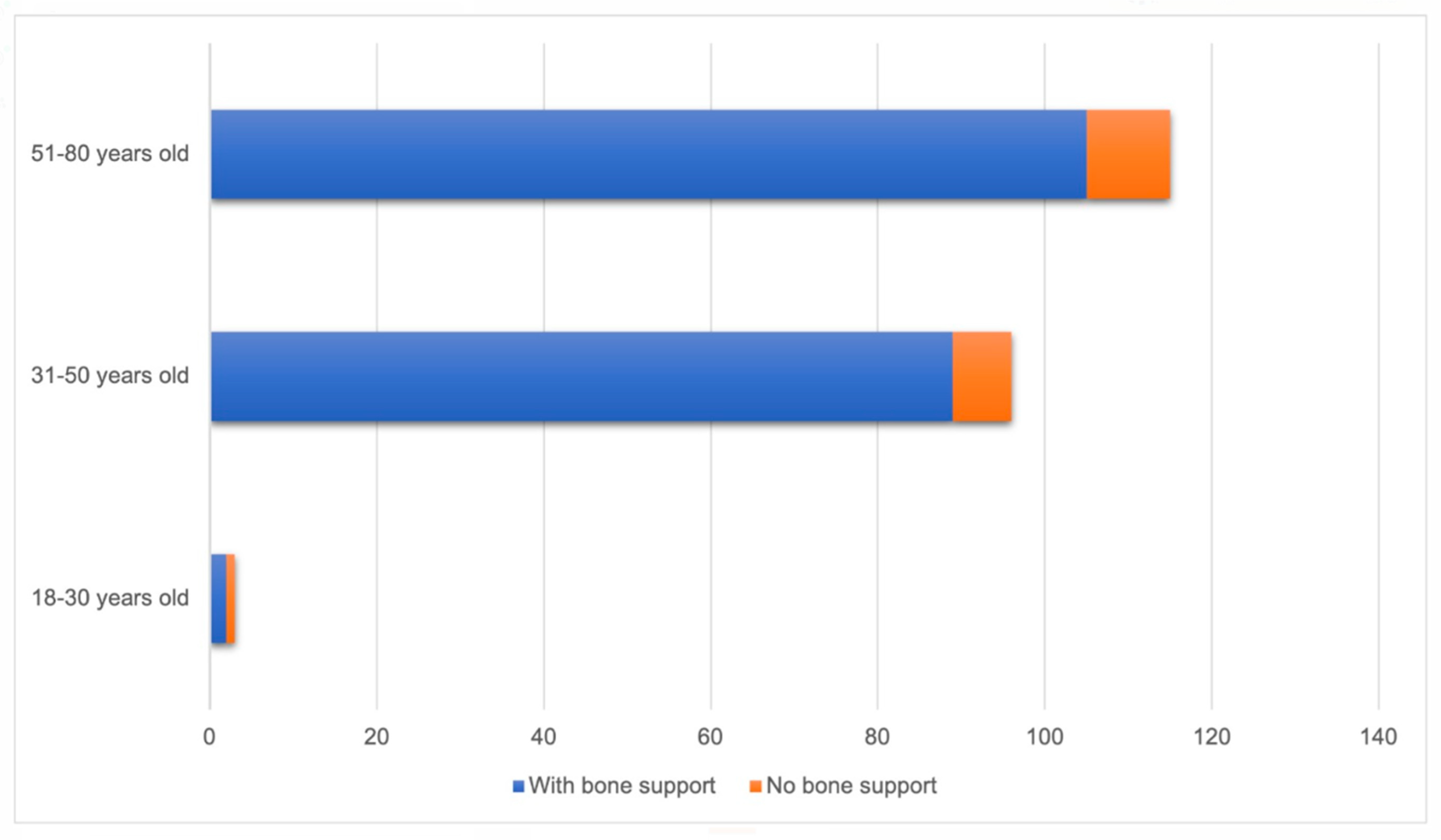
| Age | ||
| 18 to 30 years old | 3 (1.4) | 0.3–4.0 |
| 31 to 50 years old | 96 (44.8) | 38.1–51.8 |
| 51 to 80 years old | 115 (53.8) | 46.8–60.6 |
| Implant position | ||
| Implant located in the anterior region of the right mandible | 9 (4.2) | 1.9–7.8 |
| Implant located in the anterior region of the left mandible | 5 (2.3) | 0.8–5.4 |
| Implant located in the posterior region of the right mandible | 98 (45.8) | 39.0–52.7 |
| Implant located in the posterior region of the left mandible | 102 (47.7) | 40.8–54.6 |
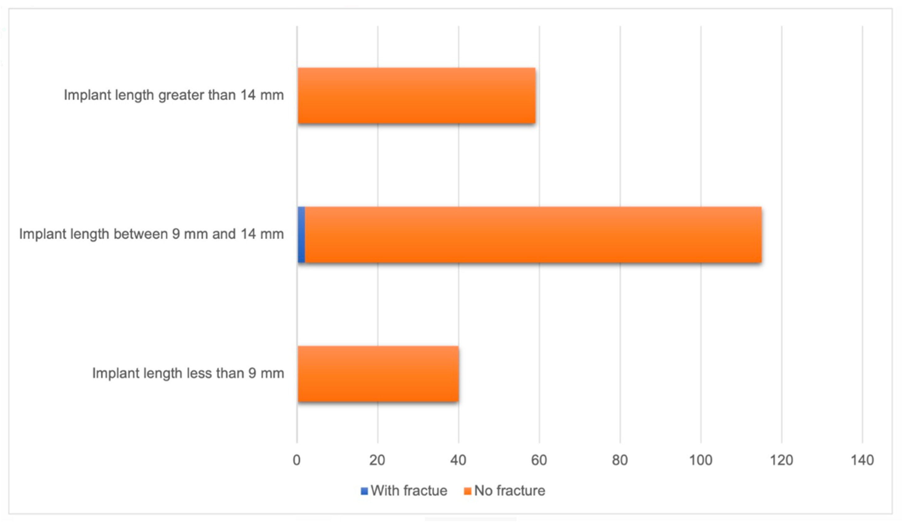
| Implant length | ||
| Implant length less than 9 mm | 40 (18.7) | 13.7–24.6 |
| Implant length between 9 mm and 14 mm | 115 (53.7) | 46.8–60.6 |
| Implant length greater than 14 mm | 59 (27.6) | 21.7–34.1 |
| Anatomical relationship between implant and mandibular canal | ||
| Implant short of the mandibular canal (1 to 2 mm) | 196 (91.6) | 87.0–94.9 |
| Implant in contact with the mandibular canal | 18 (8.4) | 5.1–13.0 |
| Implant within the mandibular canal (1 mm or more) | 0 (0) | 0–0 |
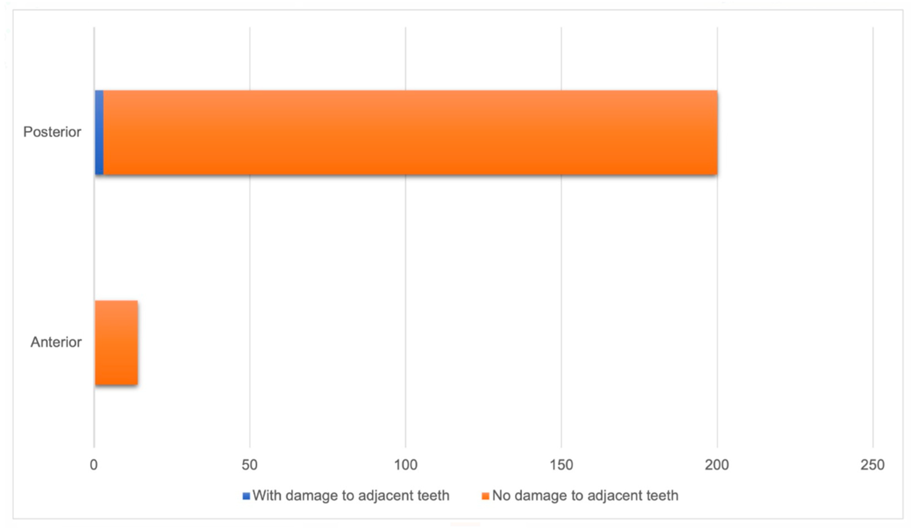
| Occurrence of damage to adjacent teeth | ||
| No damage to adjacent teeth | 211 (98.6) | 96.0–99.7 |
| Damage to the tooth located anterior to the implant | 0 (0) | 0.1–3.3 |
| Damage to the tooth located posterior to the implant | 3 (1.4) | 0.01–2.6 |
| Occurrence of implant fracture | ||
| No fracture in the implant | 212 (99.1) | 96.7–99.9 |
| Fracture in the implant | 2 (0.9) | 0.1–3.3 |
| Bone support for the implant | ||
| Absence of bone support for the implant * | 18 (8.4) | 5.1–13.0 |
| Presence of bone support for the implant | 196 (91.6) | 87.0–94.9 |
Disclaimer/Publisher’s Note: The statements, opinions and data contained in all publications are solely those of the individual author(s) and contributor(s) and not of MDPI and/or the editor(s). MDPI and/or the editor(s) disclaim responsibility for any injury to people or property resulting from any ideas, methods, instructions or products referred to in the content. |
© 2024 by the authors. Licensee MDPI, Basel, Switzerland. This article is an open access article distributed under the terms and conditions of the Creative Commons Attribution (CC BY) license (https://creativecommons.org/licenses/by/4.0/).
Share and Cite
Garrote, M.d.S.; Alencar, A.H.G.d.; Estrela, C.R.d.A.; Estrela, L.R.d.A.; Bueno, M.R.; Guedes, O.A.; Estrela, C. Incidental Findings Following Dental Implant Procedures in the Mandible: A New Post-Processing CBCT Software Analysis. Diagnostics 2024, 14, 1908. https://doi.org/10.3390/diagnostics14171908
Garrote MdS, Alencar AHGd, Estrela CRdA, Estrela LRdA, Bueno MR, Guedes OA, Estrela C. Incidental Findings Following Dental Implant Procedures in the Mandible: A New Post-Processing CBCT Software Analysis. Diagnostics. 2024; 14(17):1908. https://doi.org/10.3390/diagnostics14171908
Chicago/Turabian StyleGarrote, Marcel da Silva, Ana Helena Gonçalves de Alencar, Cyntia Rodrigues de Araújo Estrela, Lucas Rodrigues de Araújo Estrela, Mike Reis Bueno, Orlando Aguirre Guedes, and Carlos Estrela. 2024. "Incidental Findings Following Dental Implant Procedures in the Mandible: A New Post-Processing CBCT Software Analysis" Diagnostics 14, no. 17: 1908. https://doi.org/10.3390/diagnostics14171908
APA StyleGarrote, M. d. S., Alencar, A. H. G. d., Estrela, C. R. d. A., Estrela, L. R. d. A., Bueno, M. R., Guedes, O. A., & Estrela, C. (2024). Incidental Findings Following Dental Implant Procedures in the Mandible: A New Post-Processing CBCT Software Analysis. Diagnostics, 14(17), 1908. https://doi.org/10.3390/diagnostics14171908