Article

The Association between the Body Mass Index, Chronic Obstructive Pulmonary Disease and SUV of the Non-Tumorous Lung in the Pretreatment [18F]FDG-PET/CT of Patients with Lung Cancer

1
Department of Medicine V, LMU University Hospital, 80336 Munich, Germany
2
German Center for Lung Research (DZL CPC-M), 81377 Munich, Germany
3
Department of Dermatology and Allergy, LMU University Hospital, 80336 Munich, Germany
4
Department of Radiology, LMU University Hospital, 80336 Munich, Germany
5
Department of Nuclear Medicine, LMU University Hospital, 80336 Munich, Germany
6
Munich Cluster for Systems Neurology (SyNergy), 81377 Munich, Germany
7
German Center for Neurodegenerative Diseases (DZNE), 81377 Munich, Germany
8
Department of Dermatology, University Hospital Erlangen, Comprehensive Cancer Center Erlangen—European Metropolitan Region Nürnberg, CCC Alliance WERA, 91054 Erlangen, Germany
*
Author to whom correspondence should be addressed.
Diagnostics 2024, 14(11), 1139; https://doi.org/10.3390/diagnostics14111139
Submission received: 6 April 2024 / Revised: 26 May 2024 / Accepted: 29 May 2024 / Published: 30 May 2024
(This article belongs to the Special Issue Prognostic and Predictive Biomarkers of Lung Cancer)

Abstract

:
Background: A debate persists on the prognostic value of the pre-therapeutic standardized uptake value (SUV) of non-tumorous lung tissue for the risk assessment of therapy-related pneumonitis, with most studies lacking significant correlation. However, the influence of patient comorbidities on the pre-therapeutic lung SUV has not yet been systematically evaluated. Thus, we aimed to elucidate the association between comorbidities, biological variables and lung SUVs in pre-therapeutic [18F]FDG-PET/CT. Methods: In this retrospective study, the pre-therapeutic SUV in [18F]FDG-PET/CT was measured in non-tumorous areas of both lobes of the lung. SUVMEAN, SUVMAX and SUV95 were compared to a multitude of patient characteristics and comorbidities with Spearman’s correlation analysis, followed by a Bonferroni correction and multilinear regression. Results: In total, 240 patients with lung cancer were analyzed. An elevated BMI was significantly associated with increased SUVMAX (β = 0.037, p < 0.001), SUVMEAN (β = 0.017, p < 0.001) and SUV95 (β = 0.028, p < 0.001). Patients with chronic obstructive pulmonary disease (COPD) showed a significantly decreased SUVMAX (β = −0.156, p = 0.001), SUVMEAN (β = −0.107, p < 0.001) and SUV95 (β = −0.134, p < 0.001). Multiple other comorbidities did not show a significant correlation with the SUV of the non-tumorous lung. Conclusions: Failure to consider the influence of BMI and COPD on the pre-therapeutic SUV measurements may lead to an erroneous interpretation of the pre-therapeutic SUV and subsequent treatment decisions in patients with lung cancer.

1. Introduction

Lung cancer is one of the most common and deadly malignancies worldwide. Its global annual incidence is still increasing [1,2]. Therapy with immune checkpoint inhibitors (ICI) dramatically improved survival in patients with advanced-stage disease at first diagnosis [1,3,4,5,6,7]. However, severe immunotherapy-related adverse effects (irAEs) can occur during immune checkpoint inhibitor therapy and up to now are unpredictable. Approximately one out of every ten patients develops immune checkpoint inhibitor-associated pneumonitis (CIP) [8,9]. CIP has a major effect on the lung function, quality of life and overall survival of patients [9]. Higher-stage CIP can be life-threatening and often leads to a permanent discontinuation of immune checkpoint inhibitor therapy [10,11]. Hence, it is imperative to discern which patients are predisposed to developing CIP and which are not.
Recently, several studies investigated the pre-therapeutic standardized uptake value (SUV) of non-tumorous lung tissue in [18F]-fluorodeoxyglucose positron emission tomography/computed tomography (FDG-PET/CT) as a potential predictive biomarker for the risk of developing radiation therapy-associated and immune checkpoint inhibitor associated pneumonitis [12,13,14,15,16,17,18,19,20,21]. In individuals undergoing radiation therapy, a significant partial correlation exists between an increased pre-radiotherapeutic PET-measured glucose uptake and an elevated risk of radiation-induced pneumonitis [12,15]. However, most studies focusing on the predictability of immunotherapy-associated pneumonitis failed to establish a significant correlation between pre-therapeutic FDG uptake and the risk of therapy-related pneumonitis [16,17,18,19,20,21]. In the studies referenced, the impact of patients’ comorbidities on the SUV was not incorporated into the analyses. However, patients with lung cancer often show smoking-related comorbidities like chronic obstructive pulmonary disease (COPD) [22,23]. In these patients, an increased partial volume of trapped air could potentially lead to a lower SUV. On the contrary, in patients with obesity, a higher amount of mediastinal and abdominal fat could potentially lead to a higher lung density due to a functional restriction [24] and therefore to an elevated SUV of the lung tissue. There are many more biological variables and comorbidities potentially influencing the SUV of the lung and complicating the interpretation (e.g., cardiac congestion, pleural effusion, varying PET/CT scanner).
To ensure a correct interpretation of the SUV in non-tumorous lung tissue as a potential predictor for adverse events, understanding the impact of patient comorbidities on the pre-therapeutic SUV is imperative. To our knowledge, in patients with lung cancer, the impact of comorbidities and other clinical variables on the SUV of the non-tumorous lung has not yet been systematically evaluated. Thus, we aimed to investigate the influence of comorbidities and biological variables on the pre-therapeutic SUV of non-tumorous lung tissue in FDG-PET/CT, with the ultimate goal of improving pre-therapeutic assessment and therapy planning.

2. Materials and Methods

2.1. Patient Population

All patients with lung cancer who were treated with an immune checkpoint inhibitor therapy at the Department of Medicine V at LMU Klinikum between 2014 and 2022 and who were staged with FDG-PET/CT before the start of ICI therapy/combination therapy of ICI with chemotherapy and/or radiation therapy were included. Patients who received their FDG-PET/CT after or more than one year before the start of immunotherapy were excluded. This study was conducted in accordance with the Declaration of Helsinki and approved by the ethics committee der LMU München (Project No. 474-16 UE).

2.2. FDG-PET/CT Imaging

Before injection of the radiotracer, patients fasted for 4–6 h. At the time of injection, blood glucose levels were below 150 mg/dL. The scans were acquired in mid-breath-hold. The SUV was then calculated using the following standard calculation [21].
SUV calculation:
S U V = S p h e r e   a c t i v i t y B q m l b o d y   w e i g h t k g i n j e c t e d   d o s e

2.3. Image Analysis

FDG-PET/CT images were evaluated using Visage®7, Visage Imaging, Inc. (San Diego, CA, USA; version 7.1.18). Four three-dimensional spheres with a diameter of 30 mm were placed into lung parenchyma, ensuring placement in regions not infiltrated by lung tumor. One sphere was placed in each of the upper and lower lobes of both the right and left lungs, respectively (Figure 1). Spheres were excluded when it was not possible to place them exclusively in non-tumorous lung tissue (e.g., due to tumor infiltration, metastasis, or lobectomy). For normalization, one sphere was placed in the lumen of the aorta and two additional spheres were placed in liver tissue. The SUV (SUVMEAN), maximum SUV (SUVMAX), and the standard deviation (SD) of the SUV (SUVSD) were measured for each sphere. Mean values were measured for the SUVMEAN and SUVMAX of all four spheres. Additionally, mean values were computed for the two upper spheres, the two lower spheres and the two left or right spheres depending on tumor side (TFL). Furthermore, for normalization ratios were calculated between the SUVMEAN values obtained from all four spheres and those from the two liver spheres, as well as the sphere in the mediastinal blood pool. Lastly, SUV95 was measured for all four spheres. SUV95 describes the 95th percentile of SUVMAX and was calculated with the following formula:
SUV95 measurement:
S U V 95 = S U V M E A N + ( q n o r m ( 0.95 ) S U V S D )

2.4. Patient Characteristics

The following variables with a potential impact on the SUV were collected: sex, age, BMI, tumor stage, tumor side, smoking status, pack years, previous lung operation, previous lung radiation, COPD, diabetes mellitus type II, asthma, coronary heart disease (CHD), pericardial effusion (PCE), pleural effusion (PE), pneumonia (no more than three months before PET) and blood hemoglobin (Hb) level. A semi-automatic tool was built to analyze the text of electronic patient files using keywords for those characteristics. Possible spelling errors were considered. When keywords were found, the text was read manually. All collected data were anonymized and stored in a Microsoft Excel database (Redmond, WA, USA; version 2108).

2.5. Statistical Analyses

Categorical patient characteristics and clinical variables were summarized using absolute and relative frequencies. Following the Lilliefors normality test, the mean and standard deviation were calculated for Gaussian-distributed numerical variables, while the median and interquartile range (IQR) were computed for non-Gaussian-distributed numerical variables. Kruskal–Wallis test was used to test if mean SUVMEAN, mean SUVMAX and SUV95 were significantly different when using different PET/CT scanners. To determine the influence of different variables on SUV, Mann–Whitney U test was used for binary variables, and Spearman’s correlation analysis was used for numerical variables, with each of them being followed by a Bonferroni correction of the significance level for multiple testing. Additionally, a multilinear regression was performed, and the resulting beta coefficients were visualized in a forest plot. The association between biological variables and comorbidities and the SUV was evaluated for all patients as well as for the largest subgroup of patients that were scanned on the same PET/CT scanner. Boxplots and a scatterplot were used for the visualization of the test results. All statistical analyses and plots were conducted using R-Studio (Boston, MA, USA; version 2023.09.1+494) and R (Vienna, Austria; version 4.3.2).

3. Results

3.1. Patient Characteristics

A total of 293 patients underwent ICI therapy or combination therapy of ICI therapy with chemotherapy (n = 164) and/or radiation therapy (n = 63) as a first-line or subsequent therapy during the observation period. In total, 53 patients were excluded because the FDG-PET/CT was acquired after the start of ICI therapy or more than one year before ICI therapy commenced. The data of the remaining 240 patients are displayed in Table 1. PET/CT data from fifteen different locations were used for this analysis. In 137/293 patients, the pre-therapeutic FDG-PET/CT was acquired using the same imaging PET/CT scanner (GE Healthcare Discovery PET/CT 690 at LMU Klinikum).

3.2. Association of Biological Variables and Semiquantitative PET Parameters

Mean SUVMEAN of the lung varied between 0.16 g/mL and 1.37 g/mL. Mean SUVMAX of the lung varied between 0.31 g/mL and 2.63 g/mL; mean SUV95 varied between 0.16 g/mL and 1.94 g/mL. Prior to the correlation analysis, the congruence of SUVMEAN, SUVMAX and SUV95 obtained from various PET/CT scanners was tested with the Kruskal–Wallis test (Table A1). While mean SUVMEAN and SUV95 was comparable across all PET/CT scanners (mean SUVMEAN: p = 0.193–0.356 and SUV95: p = 0.075–0.591), there were significant differences regarding the mean SUVMAX (p = 0.007–0.160) and lung/blood ratio (p = 0.004–0.017). Thus, all analyses were not only carried out using the entire cohort of 240 patients but also verified within the subset consisting of 137 patients who underwent scanning with the identical PET/CT scanner.
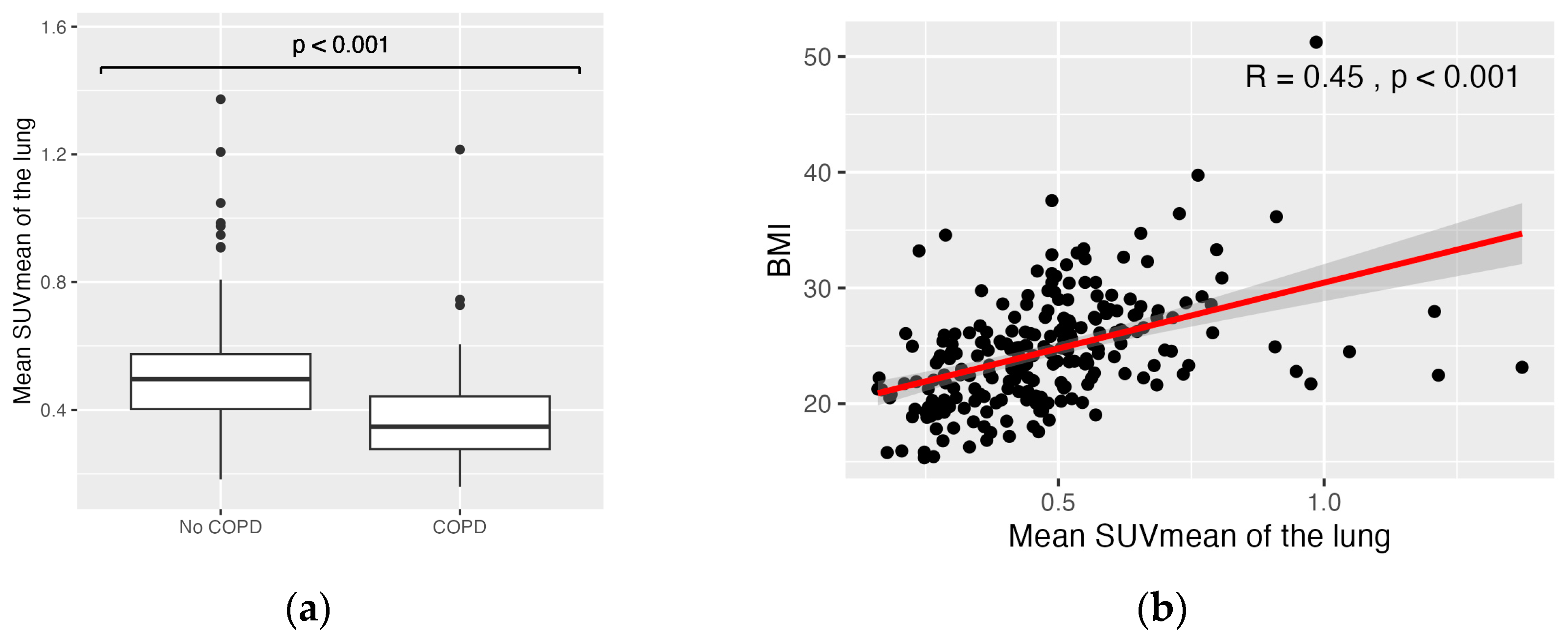
After the Bonferroni correction, BMI, smoking, pack years and COPD showed a significant correlation with the SUV (Table 2, Figure 2). The most robust correlation with tracer uptake was shown in the correlation analysis of the SUV of the whole lung and BMI (R = 0.32–0.58, p < 0.001). Furthermore, there were significantly decreased SUVs in patients with COPD (mean SUVMEAN: 0.38 g/mL vs. 0.51 g/mL, p < 0.001; mean SUVMAX: 0.77 g/mL vs. 0.97 g/mL, p < 0.001; mean SUV95: 0.54 g/mL vs. 0.71 g/mL, p < 0.001). The significant correlation of BMI and COPD with SUVMEAN and SUVMAX was confirmed in the subcohort of 137 patients who underwent scanning with the identical PET/CT scanner (Table A2). There was a variety of clinical parameters showing no significant correlation with the evaluated PET parameters: sex, age, tumor stage, lung operations, thorax radiotherapy, pleural effusion, pericardial effusion, diabetes mellitus type II, coronary heart disease and Hb did not show a significant effect on the SUV of non-tumorous lung-tissue. Normalization of the lung SUV to the SUV of the liver or the SUV of the mediastinal blood pool yielded equivalent results.
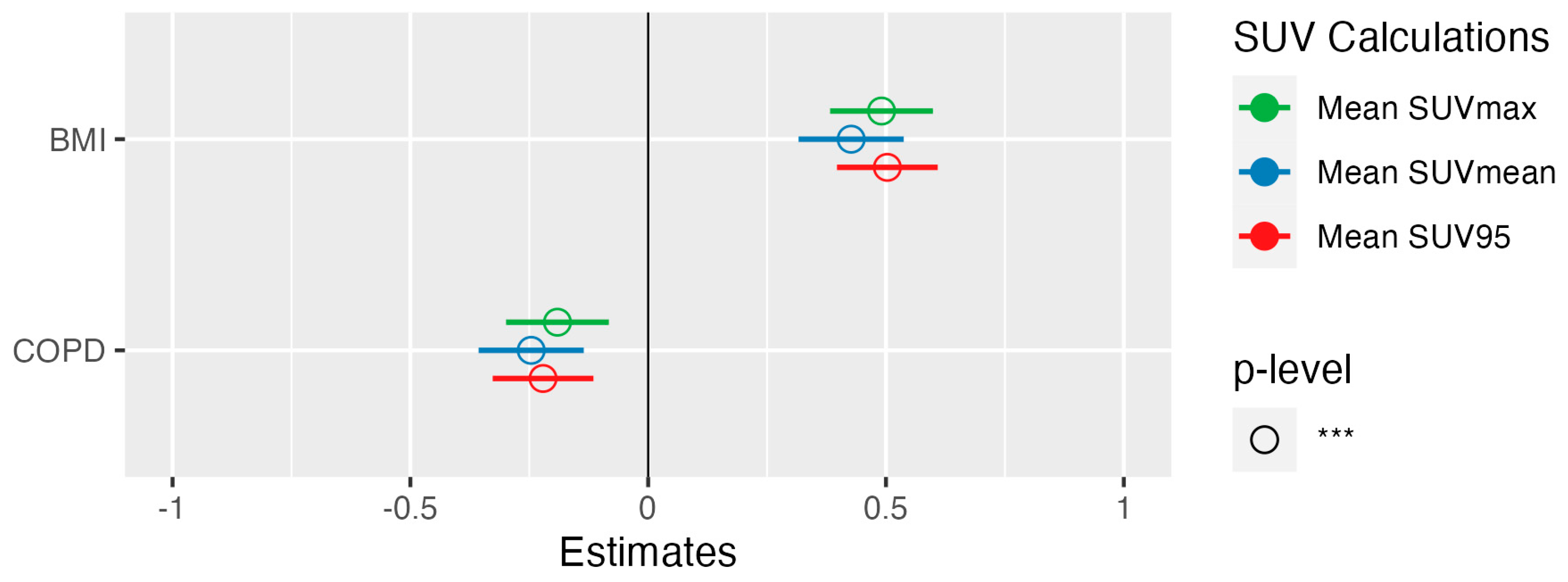
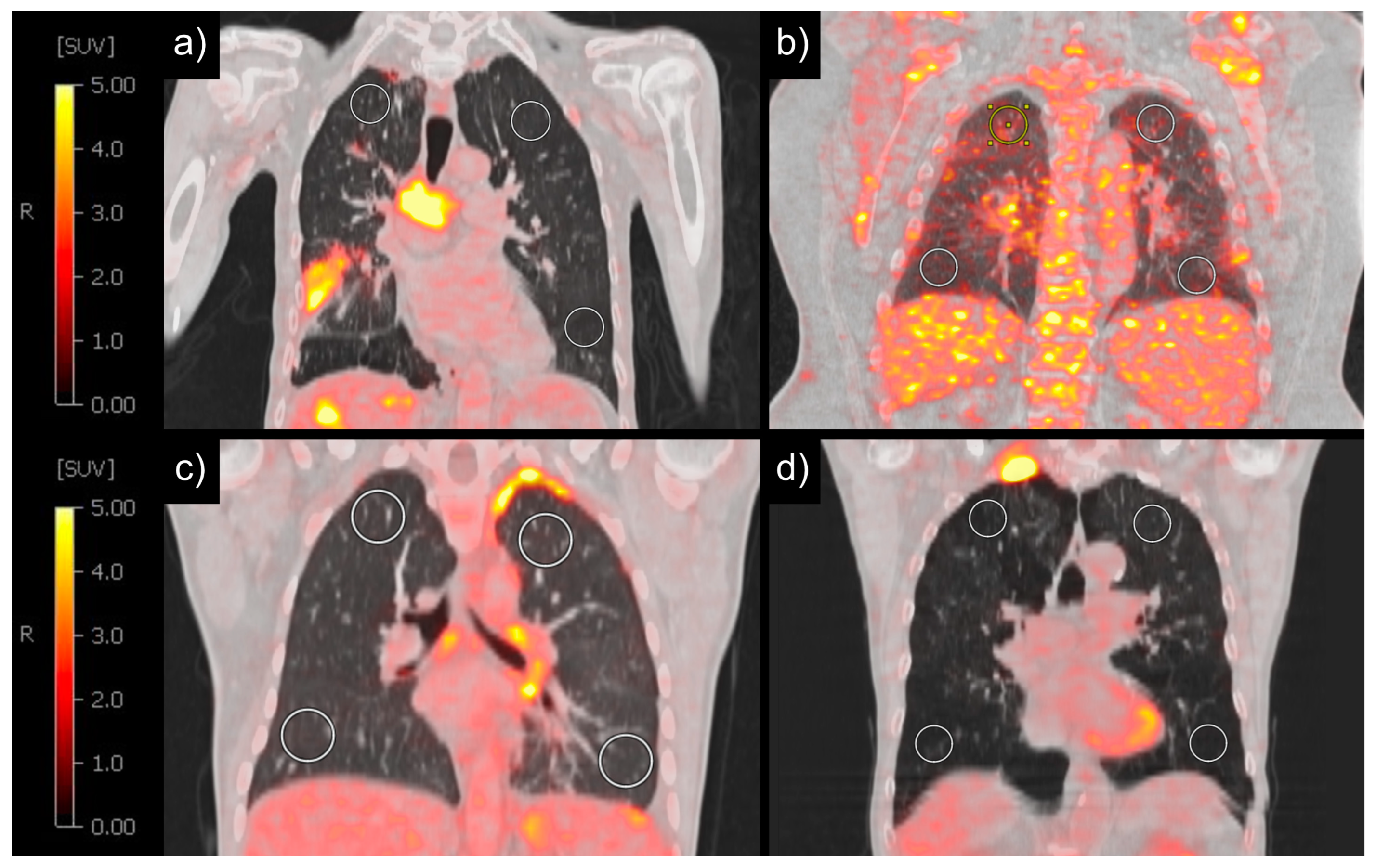
Multilinear regression was used to confirm the association between the previously significant clinical parameters in Table 2 and the PET parameters SUVMEAN, SUVMAX and SUV95. Only one of the smoking-related parameters (pack years and COPD) was used as an independent variable for multilinear regression, leaving BMI and COPD as independent variables. In multilinear regression, patients with increased BMI showed significantly increased mean SUVMAX (β = 0.037, p < 0.001), mean SUVMEAN (β = 0.017, p < 0.001) and SUV95 (β = 0.028, p < 0.001) (Figure 3, Figure 4a,b). Patients with COPD, on the other hand, showed a significantly decreased mean SUVMAX (β = −0.156, p = 0.001), mean SUVMEAN (β = −0.107, p < 0.001) and SUV95 (β = −0.134, p < 0.001) compared to the patients without COPD (Figure 3, Figure 4c,d). These results could be confirmed in the subcohort of 137 patients who underwent scanning with the identical PET/CT scanner (S3).

4. Discussion

Predictors for severe immune-related adverse events (irAEs) induced by immune checkpoint inhibitor therapy are desperately needed. Immune-related pneumonitis is the ICI-induced adverse event causing the most fatalities [10,11,25,26]. Thus, radiologic assessments from routine care carry a high potential to guide monitoring and treatment decisions if correlations with subsequent toxicity could be identified. The standardized uptake value (SUV) in FDG-PET/CT is gaining increasing interest, which is reflected by the wealth of research around the SUV as a predictor of treatment response and therapy-related adverse events (AEs). While most research has focused on the assessment of the SUV in tumor tissue as a prognostic marker, only few studies focused on the FDG uptake in healthy organs to identify patients with cancer at risk of AEs. Particularly in the context of immune-related adverse events (irAEs) associated with immune checkpoint inhibitor therapy, the SUV of non-tumorous tissue could provide a predictive advantage. The paramount aim is to identify, prior to treatment, patients with cancer prone to immunotherapy-associated adverse events through pre-therapeutic FDG-PET/CT. This would facilitate closer monitoring of susceptible patients, prompt therapy adjustments, and potentially enable a timelier integration of (inhalative) corticosteroids or other immune modulators into treatment protocols. Previous studies showed that an increased tracer uptake during immunotherapy reflected the presence of active immunotherapy related or radiation-therapy related adverse events [16,27,28]. Nevertheless, the majority of studies assessing the SUV before immunotherapy as a predictive biomarker for immune-related adverse events (irAEs) have yielded inconclusive results, failing to demonstrate a significant correlation between the pre-therapeutic SUV and the occurrence of ICI-induced irAEs [16,17,18,19]. Focusing on checkpoint inhibitor-related pneumonitis (CIP), only one study successfully predicted the probability of irAEs during therapy with pre-therapeutic PET, considering the CT and the combined PET/CT information in a radiomics score [21]. The SUV of the lung can be heavily influenced by confounding factors, including sex, body mass index (BMI) and diabetes [29,30,31]. Furthermore, comorbidities with an impact on tissue density such as lung emphysema could have a significant impact on the lung SUV and should therefore be considered when evaluating the pre-therapeutic FDG uptake in non-tumorous lung tissue. Notwithstanding the numerous studies examining the prognostic utility of SUV for the prediction of CIP in patients with lung cancer, a systematic exploration of the impact of comorbidities on the non-tumorous lung SUV within this patient cohort remains outstanding. Given this background, we aimed to elucidate the effect of biological variables and common comorbidities of patients with lung cancer on the pre-therapeutic SUV of the non-tumorous lung.
In our study, a higher BMI was consistently strongly associated with a higher SUVMEAN, SUVMAX and SUV95. When reviewing the literature, little information can be found on the influence of BMI on lung SUV [32,33]. A previous study investigating the effect of blood glucose levels on the SUV of healthy organs in over 5600 patients showed that an elevated BMI was significantly associated with an increased SUV of the lung [33]. Similarly, a small-scale study comprising 18 subjects demonstrated a significant association between a higher BMI and an increased SUV of the lung, implying the necessity for validation in a larger sample size [32]. The increase in mediastinal and intra-abdominal fat accompanying higher BMI levels likely contributes to elevated intra-thoracic pressure, a lower total lung capacity and an increased lung tissue density [34]. As the SUV is calculated based on measured radioactivity within a defined voxel, greater lung tissue density, with less air, results in higher SUV values. Conversely, reduced lung tissue density, with more air, leads to decreased SUV values. Additionally, a systemic chronic inflammatory response often associated with higher BMI levels might have contributed to the correlation between the BMI and the lung SUV [35,36,37]: an elevated BMI has been linked to increased levels of pro-inflammatory cytokines and adipokines, which could potentially enhance metabolic activity and inflammation within lung tissue, thereby influencing SUV measurements [35,36,37]. However, we also investigated a potential association of diabetes and the pre-therapeutic lung SUV, which is another comorbidity with a known influence on metabolic activity and is associated with chronic inflammatory activity. Our analysis did not reveal a significant correlation between diabetes status and lung SUV. Thus, although chronic inflammatory processes may indeed play a role in modifying metabolic activity within lung tissue, it is conceivable that factors influencing lung tissue density could exert a predominant influence on SUV measurements in this context. While a large study of over 5000 patients found that obesity paradoxically reduces the risk of pulmonary and other complications after minimally invasive thoracic surgery [38], in the context of checkpoint inhibitor therapy, multiple studies showed that an elevated BMI was linked to the occurrence of CIP [39,40,41]. In light of the substantial impact of the BMI on the lung SUV, we draw the conclusion that when investigating the relationship between the pre-therapeutic SUV and the risk of CIP, it is imperative to recognize that an elevated pre-therapeutic SUV may not exclusively reflect heightened inflammatory activity. Instead, pre-therapeutic SUVs are also influenced by increased tissue density and potentially altered metabolic activity associated with a higher BMI. Moreover, our findings highlight the potential to enhance the precision of discriminating between lung lesions through the inclusion of the BMI in the analysis. An SUV threshold of 2.5 has been the gold standard for discriminating between malignant and benign lung lesions [42,43]. Prior research suggests that the correlation between the tumor SUV of lung lesions and prognosis is stronger in normal weight individuals compared to their obese counterparts [44,45]. For patients with breast cancer, it was already shown that risk stratification using a tumor SUV could be ameliorated when using the BMI as a covariable independent of tumor subtype and clinical stage [46]. Consequently, there might be merit in considering the incorporation of patients’ BMI into the risk assessment framework for lung lesions.
Despite the acknowledged increase in inflammatory activity of the lungs in patients with COPD [47,48,49], our study consistently revealed a significantly lower SUV in the non-tumorous lung of patients with chronic obstructive pulmonary disease. COPD and smoking are linked to lung emphysema, manifesting as decreased tissue density [50] and potentially as a decreased density of glucose uptake. Our results therefore suggest a predominant effect of the decreased tissue density over the increased inflammatory activity of the lungs in patients with COPD. Previous studies showed an increased risk for CIP in patients with COPD and lung emphysema [51,52]. Considering that COPD is associated with a decreased SUV, a negative association between the SUV and the occurrence of pneumonitis would be expected. Based on our findings, we advocate for the distinct examination of SUV measurements within patient subgroups stratified by a COPD status and for the inclusion of COPD as a covariate in the analytical models.
It is recognized that for optimal patient care and comparability of PET scans, ideally, all imaging should be conducted using the same PET scanner [30]. However, upon comparing the standardized uptake values obtained from multiple PET/CT scanners, we observed that SUVMEAN and SUV95 were consistent across the diverse array of PET/CT scanners analyzed in this study. Nevertheless, disparities were evident in SUVMAX, probably resulting from varying reconstruction algorithms and post-processing methods, e.g., the application of different smoothing algorithms, potentially leading to an underestimation of SUVMAX [30,53]. To accommodate for possible confounders, we performed all statistical tests using the data of all included patients and with a subgroup that was scanned on the most frequently used PET/CT scanner (n = 137). These subgroup re-tests confirmed the results of the whole patient group, showing a highly significant impact of COPD and the BMI on lung SUV. In summary, our results suggest a sufficient comparability of SUVMEAN and SUV95 of imaging studies acquired with varying PET scanners. This finding could be useful in everyday clinical practice when patients come in for staging evaluations with FDG-PET/CT scans and have had PET imaging completed elsewhere before. In these patients, the 95th percentile of SUVMAX (SUV95) could be used instead of SUVMAX to assess disease progression. However, the comparability of the SUV95 of benign and malignant structures in FDG-PET/CT has to be confirmed in future studies.
The following limitations have to be considered when interpreting this study: First, the study cohort exclusively included patients receiving FDG-PET/CT before checkpoint inhibitor therapy. This might have led to a selection bias. Furthermore, this study is limited by its retrospective design. As the medical documentation was not primarily intended for this research, clinical information about patient comorbidities may have been incomplete.

5. Conclusions

Our study highlights a significant link between COPD, the BMI, and the pre-therapeutic lung SUV. In obese patients, a physiologically higher SUV in non-tumorous lung areas should be factored into the assessment of therapy-related pneumonitis risk. Moreover, in patients with a higher BMI, malignant lung lesions may show reduced contrast to surrounding non-tumorous lung tissue, potentially complicating diagnosis. Conversely, in patients with COPD, a lower SUV in non-tumorous lung regions may obscure inflammatory activity. In conclusion, acknowledging the influence of obesity and COPD is crucial for an accurate SUV interpretation. Future research should focus on tailored risk management of pre-therapeutic inflammatory activity and the assessment of malignancy risk of lung nodules in patients with obesity and COPD.

Supplementary Materials

The following supporting information can be downloaded at: https://www.mdpi.com/article/10.3390/diagnostics14111139/s1; Excel File S1: Data Used in Table 2 (Separate Sheets for p-values, Estimates, and Correlations); Excel File S2: Data Used in Table A2 (Separate Sheets for p-values, Estimates, and Correlations); Figure S3: Forest plot of patients scanned with GE Discovery 690.

Author Contributions

Conceptualization, L.W. and F.V.; methodology, L.W., J.W., T.P. and F.V.; formal analysis, L.W., J.W. and F.V.; data curation, L.W. and L.D.; writing—original draft preparation, L.W. and F.V.; writing—review and editing, J.W., L.D., L.H., F.K., M.I., C.S.-T., M.B., D.K.-G., A.T. and T.P.; visualization, L.W.; supervision, L.H., A.T. and T.P. All authors have read and agreed to the published version of the manuscript.

Funding

M.B. was funded by the Deutsche Forschungsgemeinschaft (DFG) under Germany’s Excellence Strategy within the framework of the Munich Cluster for Systems Neurology (EXC 2145 SyNergy—ID 390857198).

Institutional Review Board Statement

This study was conducted in accordance with the Declaration of Helsinki and approved by the Ethics Committee der LMU Munich with the project No. 474-16 UE.

Informed Consent Statement

Informed consent was obtained from all subjects involved in this study according to the Declaration of Helsinki.

Data Availability Statement

Research data and code can be found and downloaded at https://doi.org/10.5282/ubm/data.454 (accessed on 15 May 2024).

Acknowledgments

The authors thank A. Heißenberg for critically reading this manuscript.

Conflicts of Interest

M.B. received speaker honoraria from GE healthcare, Roche, and LMI, and is an advisor of LMI and MIAC. The authors declare no conflicts of interest.

Appendix A

Table A1. Comparing SUV measurements regarding PET-CT scanner.
SUV Measurementsp
SUVMAX
whole lung0.03 *
upper lung0.1603
lower lung0.0114 *
TFL0.0069 **
SUVMEAN
whole lung0.3562
upper lung0.2033
lower lung0.2462
TFL0.1932
SUV95
whole lung0.3664
upper lung0.5909
lower lung0.104
TFL0.0745
SUVMEAN lung/liver
whole lung0.2663
upper lung0.0534
lower lung0.1863
TFL0.0724
SUVMEAN lung/blood pool
whole lung0.0139 *
upper lung0.0044 **
lower lung0.0166 *
TFL0.0062 **
SUV = standardized uptake value, TFL = two left or right spheres depending on tumor side, adjusted alpha after Bonferroni correction: p-value < 0.0025, * = p-value < 0.05, ** = p-value < 0.01.

Appendix B

Table A2. Influence of patient characteristics on metabolic activity of lung tissue for patients scanned with GE Discovery 690 (n = 137).
VariableSexAgeBMISmokingPYCOPDStageOPRadPEPCEDMCHDHb
SUVMAX
  whole lung + -
  upper lung +
  lower lung +
  TFL +
SUVMEAN
  whole lung + -
  upper lung + --
  lower lung + -
  TFL + -
SUV95
  whole lung + -
  upper lung + --
  lower lung + -
  TFL +
SUVMEAN lung/liver
  whole lung + -
  upper lung + -
  lower lung + -
  TFL +
SUVMEAN lung/blood pool
  whole lung + -
  upper lung + -
  lower lung +
  TFL +
Diagnostics 14 01139 i002
Sex = male, BMI = body mass index, PY = pack years, COPD = chronic obstructive pulmonary disease, stage = tumor stage, OP = operation, Rad = thorax radiation, PE = pleural effusion, PCE = pericardial effusion, DM = diabetes mellitus type II, CHD = coronary heart disease, Hb = blood hemoglobin level, SUV = standardized uptake value, TFL = two left or right spheres depending on tumor side, adjusted alpha after Bonferroni correction: p-value < 0.0025. For additional details, please refer to the Supplementary Materials (S2).

References

  1. Leiter, A.; Veluswamy, R.R.; Wisnivesky, J.P. The global burden of lung cancer: Current status and future trends. Nat. Rev. Clin. Oncol. 2023, 20, 624–639. [Google Scholar] [CrossRef] [PubMed]
  2. Sung, H.; Ferlay, J.; Siegel, R.L.; Laversanne, M.; Soerjomataram, I.; Jemal, A.; Bray, F. Global cancer statistics 2020: GLOBOCAN estimates of incidence and mortality worldwide for 36 cancers in 185 countries. CA Cancer J. Clin. 2021, 71, 209–249. [Google Scholar] [CrossRef] [PubMed]
  3. Brahmer, J.; Reckamp, K.L.; Baas, P.; Crinò, L.; Eberhardt, W.E.; Poddubskaya, E.; Antonia, S.; Pluzanski, A.; Vokes, E.E.; Holgado, E. Nivolumab versus docetaxel in advanced squamous-cell non–small-cell lung cancer. N. Engl. J. Med. 2015, 373, 123–135. [Google Scholar] [CrossRef] [PubMed]
  4. Garon, E.B.; Rizvi, N.A.; Hui, R.; Leighl, N.; Balmanoukian, A.S.; Eder, J.P.; Patnaik, A.; Aggarwal, C.; Gubens, M.; Horn, L.; et al. Pembrolizumab for the treatment of non-small-cell lung cancer. N. Engl. J. Med. 2015, 372, 2018–2028. [Google Scholar] [CrossRef] [PubMed]
  5. Rittmeyer, A.; Barlesi, F.; Waterkamp, D.; Park, K.; Ciardiello, F.; Von Pawel, J.; Gadgeel, S.M.; Hida, T.; Kowalski, D.M.; Dols, M.C. Atezolizumab versus docetaxel in patients with previously treated non-small-cell lung cancer (OAK): A phase 3, open-label, multicentre randomised controlled trial. Lancet 2017, 389, 255–265. [Google Scholar] [CrossRef] [PubMed]
  6. Herbst, R.S.; Soria, J.-C.; Kowanetz, M.; Fine, G.D.; Hamid, O.; Gordon, M.S.; Sosman, J.A.; McDermott, D.F.; Powderly, J.D.; Gettinger, S.N. Predictive correlates of response to the anti-PD-L1 antibody MPDL3280A in cancer patients. Nature 2014, 515, 563–567. [Google Scholar] [CrossRef] [PubMed]
  7. Kinahan, P.E.; Fletcher, J.W. Positron emission tomography-computed tomography standardized uptake values in clinical practice and assessing response to therapy. In Seminars in Ultrasound, CT and MRI; WB Saunders: Philadelphia, PA, USA, 2010; pp. 496–505. [Google Scholar]
  8. Nishino, M.; Chambers, E.S.; Chong, C.R.; Ramaiya, N.H.; Gray, S.W.; Marcoux, J.P.; Hatabu, H.; Janne, P.A.; Hodi, F.S.; Awad, M.M. Anti-PD-1 Inhibitor-Related Pneumonitis in Non-Small Cell Lung Cancer. Cancer Immunol. Res. 2016, 4, 289–293. [Google Scholar] [CrossRef]
  9. Owen, D.H.; Wei, L.; Bertino, E.M.; Edd, T.; Villalona-Calero, M.A.; He, K.; Shields, P.G.; Carbone, D.P.; Otterson, G.A. Incidence, risk factors, and effect on survival of immune-related adverse events in patients with non–small-cell lung cancer. Clin. Lung Cancer 2018, 19, e893–e900. [Google Scholar] [CrossRef]
  10. Gandhi, L.; Rodriguez-Abreu, D.; Gadgeel, S.; Esteban, E.; Felip, E.; De Angelis, F.; Domine, M.; Clingan, P.; Hochmair, M.J.; Powell, S.F.; et al. Pembrolizumab plus Chemotherapy in Metastatic Non-Small-Cell Lung Cancer. N. Engl. J. Med. 2018, 378, 2078–2092. [Google Scholar] [CrossRef]
  11. Gettinger, S.N.; Horn, L.; Gandhi, L.; Spigel, D.R.; Antonia, S.J.; Rizvi, N.A.; Powderly, J.D.; Heist, R.S.; Carvajal, R.D.; Jackman, D.M. Overall survival and long-term safety of nivolumab (anti–programmed death 1 antibody, BMS-936558, ONO-4538) in patients with previously treated advanced non–small-cell lung cancer. J. Clin. Oncol. 2015, 33, 2004. [Google Scholar] [CrossRef]
  12. Castillo, R.; Pham, N.; Ansari, S.; Meshkov, D.; Castillo, S.; Li, M.; Olanrewaju, A.; Hobbs, B.; Castillo, E.; Guerrero, T. Pre-radiotherapy FDG PET predicts radiation pneumonitis in lung cancer. Radiat. Oncol. 2014, 9, 74. [Google Scholar] [CrossRef] [PubMed]
  13. Petit, S.F.; van Elmpt, W.J.; Oberije, C.J.; Vegt, E.; Dingemans, A.-M.C.; Lambin, P.; Dekker, A.L.; De Ruysscher, D. [18F] fluorodeoxyglucose uptake patterns in lung before radiotherapy identify areas more susceptible to radiation-induced lung toxicity in non-small-cell lung cancer patients. Int. J. Radiat. Oncol. Biol. Phys. 2011, 81, 698–705. [Google Scholar] [CrossRef] [PubMed]
  14. Zhang, Y.; Yu, Y.; Yu, J.; Fu, Z.; Liu, T.; Guo, S. (18)FDG PET-CT standardized uptake value for the prediction of radiation pneumonitis in patients with lung cancer receiving radiotherapy. Oncol. Lett. 2015, 10, 2909–2914. [Google Scholar] [CrossRef] [PubMed]
  15. Chaudhuri, A.A.; Binkley, M.S.; Rigdon, J.; Carter, J.N.; Aggarwal, S.; Dudley, S.A.; Qian, Y.; Kumar, K.A.; Hara, W.Y.; Gensheimer, M. Pre-treatment non-target lung FDG-PET uptake predicts symptomatic radiation pneumonitis following Stereotactic Ablative Radiotherapy (SABR). Radiother. Oncol. 2016, 119, 454–460. [Google Scholar] [CrossRef] [PubMed]
  16. Eshghi, N.; Garland, L.L.; Nia, E.; Betancourt, R.; Krupinski, E.; Kuo, P.H. 18F-FDG PET/CT can predict development of thyroiditis due to immunotherapy for lung cancer. J. Nucl. Med. Technol. 2018, 46, 260–264. [Google Scholar] [CrossRef]
  17. Ferrari, C.; Santo, G.; Merenda, N.; Branca, A.; Mammucci, P.; Pizzutilo, P.; Gadaleta, C.D.; Rubini, G. Immune checkpoint inhibitors in advanced NSCLC: [18F] FDG PET/CT as a troubleshooter in treatment response. Diagnostics 2021, 11, 1681. [Google Scholar] [CrossRef] [PubMed]
  18. Castello, A.; Rossi, S.; Toschi, L.; Lopci, E. Impact of antibiotic therapy and metabolic parameters in non-small cell lung cancer patients receiving checkpoint inhibitors. J. Clin. Med. 2021, 10, 1251. [Google Scholar] [CrossRef]
  19. Hribernik, N.; Huff, D.T.; Studen, A.; Zevnik, K.; Klanecek, Z.; Emamekhoo, H.; Skalic, K.; Jeraj, R.; Rebersek, M. Quantitative imaging biomarkers of immune-related adverse events in immune-checkpoint blockade-treated metastatic melanoma patients: A pilot study. Eur. J. Nucl. Med. Mol. Imaging 2022, 49, 1857–1869. [Google Scholar] [CrossRef] [PubMed]
  20. Anwar, H.; Sachpekidis, C.; Winkler, J.; Kopp-Schneider, A.; Haberkorn, U.; Hassel, J.C.; Dimitrakopoulou-Strauss, A. Absolute number of new lesions on 18 F-FDG PET/CT is more predictive of clinical response than SUV changes in metastatic melanoma patients receiving ipilimumab. Eur. J. Nucl. Med. Mol. Imaging 2018, 45, 376–383. [Google Scholar] [CrossRef]
  21. Mu, W.; Tunali, I.; Qi, J.; Schabath, M.B.; Gillies, R.J. Radiomics of 18F fluorodeoxyglucose PET/CT images predicts severe immune-related adverse events in patients with NSCLC. Radiol. Artif. Intell. 2020, 2, e190063. [Google Scholar] [CrossRef]
  22. Forey, B.A.; Thornton, A.J.; Lee, P.N. Systematic review with meta-analysis of the epidemiological evidence relating smoking to COPD, chronic bronchitis and emphysema. BMC Pulm. Med. 2011, 11, 1–61. [Google Scholar] [CrossRef]
  23. Durham, A.L.; Adcock, I.M. The relationship between COPD and lung cancer. Lung Cancer 2015, 90, 121–127. [Google Scholar] [CrossRef] [PubMed]
  24. Subramaniam, K.; Clark, A.R.; Hoffman, E.A.; Tawhai, M.H. Metrics of lung tissue heterogeneity depend on BMI but not age. J. Appl. Physiol. 2018, 125, 328–339. [Google Scholar] [CrossRef]
  25. Heinzerling, L.; de Toni, E.N.; Schett, G.; Hundorfean, G.; Zimmer, L. Checkpoint Inhibitors. Dtsch. Arztebl. Int. 2019, 116, 119–126. [Google Scholar] [CrossRef]
  26. Naidoo, J.; Wang, X.; Woo, K.M.; Iyriboz, T.; Halpenny, D.; Cunningham, J.; Chaft, J.E.; Segal, N.H.; Callahan, M.K.; Lesokhin, A.M.; et al. Pneumonitis in Patients Treated With Anti-Programmed Death-1/Programmed Death Ligand 1 Therapy. J. Clin. Oncol. 2017, 35, 709–717. [Google Scholar] [CrossRef]
  27. Demirev, A.K.; Kostadinova, I.D.; Gabrovski, I.R. 18F-FDG PET/CT in patients with parenchymal changes attributed to radiation pneumonitis. Mol. Imaging Radionucl. Ther. 2018, 27, 107. [Google Scholar] [CrossRef]
  28. Tatar, G.; Alçin, G.; Sengul Samanci, N.; Erol Fenercioglu, Ö.; Beyhan, E.; Cermik, T.F. Diagnostic impact of 18F-FDG PET/CT imaging on the detection of immune-related adverse events in patients treated with immunotherapy. Clin. Transl. Oncol. 2022, 24, 1903–1913. [Google Scholar] [CrossRef] [PubMed]
  29. Thie, J.A. Understanding the standardized uptake value, its methods, and implications for usage. J. Nucl. Med. 2004, 45, 1431–1434. [Google Scholar] [PubMed]
  30. Adams, M.C.; Turkington, T.G.; Wilson, J.M.; Wong, T.Z. A systematic review of the factors affecting accuracy of SUV measurements. Am. J. Roentgenol. 2010, 195, 310–320. [Google Scholar] [CrossRef]
  31. Zhuang, M.; García, D.V.; Kramer, G.M.; Frings, V.; Smit, E.; Dierckx, R.; Hoekstra, O.S.; Boellaard, R. Variability and repeatability of quantitative uptake metrics in 18F-FDG PET/CT of non–small cell lung cancer: Impact of segmentation method, uptake interval, and reconstruction protocol. J. Nucl. Med. 2019, 60, 600–607. [Google Scholar] [CrossRef]
  32. Well, D.; Meier, J.; Mahne, A.; Houseni, M.; Hernandez-Pampaloni, M.; Mong, A.; Mishra, S.; Zhuge, Y.; Souza, A.; Udupa, J.; et al. Structural and functional changes in lung with increasing body mass index (BMI). J. Nucl. Med. 2007, 48, 292P. [Google Scholar]
  33. Sprinz, C.; Zanon, M.; Altmayer, S.; Watte, G.; Irion, K.; Marchiori, E.; Hochhegger, B. Effects of blood glucose level on 18F fluorodeoxyglucose (18F-FDG) uptake for PET/CT in normal organs: An analysis on 5623 patients. Sci. Rep. 2018, 8, 2126. [Google Scholar] [CrossRef] [PubMed]
  34. Behazin, N.; Jones, S.B.; Cohen, R.I.; Loring, S.H. Respiratory restriction and elevated pleural and esophageal pressures in morbid obesity. J. Appl. Physiol. 2010, 108, 212–218. [Google Scholar] [CrossRef] [PubMed]
  35. Parasuaraman, G.; Ayyasamy, L.; Aune, D.; Sen, A.; Nagarajan, R.; Rajkumar, P.; Velusamy, S.; Manickam, P.; Sivaprakasam, S. The association between body mass index, abdominal fatness, and weight change and the risk of adult asthma: A systematic review and meta-analysis of cohort studies. Sci. Rep. 2023, 13, 7745. [Google Scholar] [CrossRef] [PubMed]
  36. Rodríguez-Hernández, H.; Simental-Mendía, L.E.; Rodríguez-Ramírez, G.; Reyes-Romero, M.A. Obesity and inflammation: Epidemiology, risk factors, and markers of inflammation. Int. J. Endocrinol. 2013, 2013, 678159. [Google Scholar] [CrossRef] [PubMed]
  37. Stienstra, R.; Tack, C.J.; Kanneganti, T.-D.; Joosten, L.A.; Netea, M.G. The inflammasome puts obesity in the danger zone. Cell Metab. 2012, 15, 10–18. [Google Scholar] [CrossRef] [PubMed]
  38. Parini, S.; Azzolina, D.; Massera, F.; Mastromarino, M.G.; Papalia, E.; Baietto, G.; Curcio, C.; Crisci, R.; Rena, O.; Alloisio, M.; et al. The Overweight Paradox: Impact of Body Mass Index on Patients Undergoing VATS Lobectomy or Segmentectomy. Semin. Thorac. Cardiovasc. Surg. 2023, 35, 164–176. [Google Scholar] [CrossRef] [PubMed]
  39. Eun, Y.; Kim, I.Y.; Sun, J.-M.; Lee, J.; Cha, H.-S.; Koh, E.-M.; Kim, H.; Lee, J. Risk factors for immune-related adverse events associated with anti-PD-1 pembrolizumab. Sci. Rep. 2019, 9, 14039. [Google Scholar] [CrossRef] [PubMed]
  40. Rogado, J.; Romero-Laorden, N.; Sanchez-Torres, J.M.; Ramos-Levi, A.M.; Pacheco-Barcia, V.; Ballesteros, A.I.; Arranz, R.; Lorenzo, A.; Gullon, P.; Garrido, A.; et al. Effect of excess weight and immune-related adverse events on the efficacy of cancer immunotherapy with anti-PD-1 antibodies. Oncoimmunology 2020, 9, 1751548. [Google Scholar] [CrossRef]
  41. Albitar, H.A.H.; Duma, N.; Leventakos, K.; Gallo De Moraes, A. Pulmonary Complications Secondary to Immune Checkpoint Inhibitors. Int. J. Chronic. Dis. 2020, 2020, 4928648. [Google Scholar] [CrossRef]
  42. Hickeson, M.; Yun, M.; Matthies, A.; Zhuang, H.; Adam, L.-E.; Lacorte, L.; Alavi, A. Use of a corrected standardized uptake value based on the lesion size on CT permits accurate characterization of lung nodules on FDG-PET. Eur. J. Nucl. Med. Mol. Imaging 2002, 29, 1639–1647. [Google Scholar] [CrossRef]
  43. Khalaf, M.; Abdel-Nabi, H.; Baker, J.; Shao, Y.; Lamonica, D.; Gona, J. Relation between nodule size and 18 F-FDG-PET SUV for malignant and benign pulmonary nodules. J. Hematol. Oncol. 2008, 1, 13. [Google Scholar] [CrossRef]
  44. Liu, J.; Dong, M.; Sun, X.; Li, W.; Xing, L.; Yu, J. Prognostic value of 18F-FDG PET/CT in surgical non-small cell lung cancer: A meta-analysis. PLoS ONE 2016, 11, e0146195. [Google Scholar] [CrossRef]
  45. Hyun, S.H.; Lee, K.H.; Choi, J.Y.; Kim, B.T.; Kim, J.; Zo, J.I.; Kim, H.; Kwon, O.J.; Ahn, H.K. Influence of Body Mass Index on the Prognostic Value of Tumor ¹⁸F-FDG Uptake in Stage I Non-Small Cell Lung Cancer. PLoS ONE 2015, 10, e0145020. [Google Scholar] [CrossRef]
  46. Hyun, S.H.; Ahn, H.K.; Lee, J.H.; Choi, J.Y.; Kim, B.T.; Park, Y.H.; Im, Y.H.; Lee, J.E.; Nam, S.J.; Lee, K.H. Body Mass Index with Tumor 18F-FDG Uptake Improves Risk Stratification in Patients with Breast Cancer. PLoS ONE 2016, 11, e0165814. [Google Scholar] [CrossRef]
  47. Rennard, S.I. Inflammation and repair processes in chronic obstructive pulmonary disease. Am. J. Respir. Crit. Care Med. 1999, 160, S12–S16. [Google Scholar] [CrossRef]
  48. Barnes, P.J. Immunology of asthma and chronic obstructive pulmonary disease. Nat. Rev. Immunol. 2008, 8, 183–192. [Google Scholar] [CrossRef]
  49. Brusselle, G.G.; Joos, G.F.; Bracke, K.R. New insights into the immunology of chronic obstructive pulmonary disease. Lancet 2011, 378, 1015–1026. [Google Scholar] [CrossRef]
  50. Guenard, H.; Diallo, M.H.; Laurent, F.; Vergeret, J. Lung density and lung mass in emphysema. Chest 1992, 102, 198–203. [Google Scholar] [CrossRef]
  51. Zhou, P.; Zhao, X.; Wang, G. Risk Factors for Immune Checkpoint Inhibitor-Related Pneumonitis in Cancer Patients: A Systemic Review and Meta-Analysis. Respiration 2022, 101, 1035–1050. [Google Scholar] [CrossRef]
  52. Zeng, Z.; Qu, J.; Yao, Y.; Xu, F.; Lu, S.; Zhang, P.; Yao, Y.; Li, N.; Zhou, J.; Wang, Y. Clinical outcomes and risk factor of immune checkpoint inhibitors-related pneumonitis in non-small cell lung cancer patients with chronic obstructive pulmonary disease. BMC Pulm. Med. 2022, 22, 458. [Google Scholar] [CrossRef] [PubMed]
  53. Yuji, H. Characteristics of Smoothing Filters to Achieve the Guideline Recommended Positron Emission Tomography Image without. Asia Ocean. J. Nucl. Med. Biol. 2018, 6, 15. [Google Scholar]
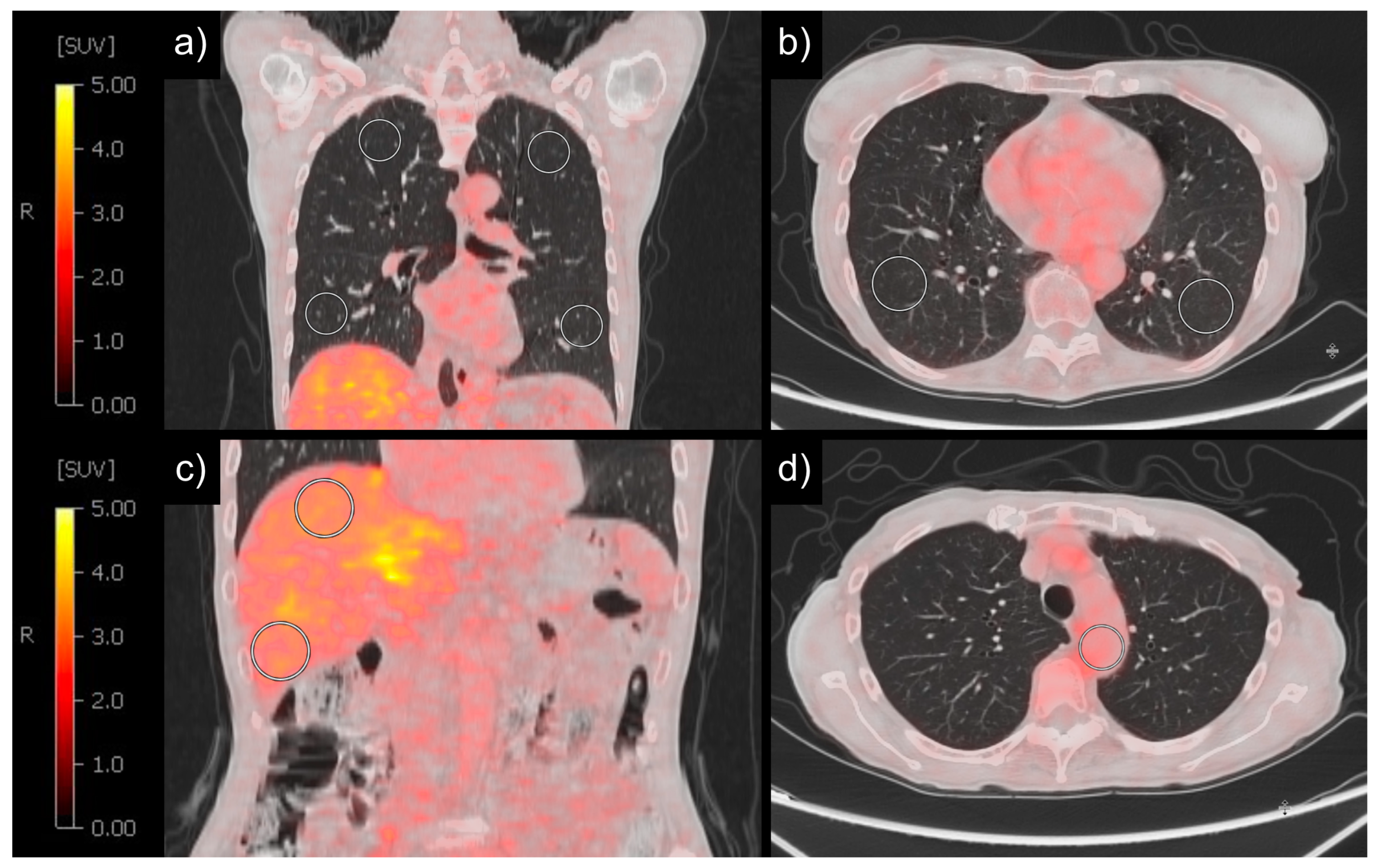
Figure 1. Example of PET/CT analysis with spheres. SUV measurement with spheres with 30 mm diameter in lung in coronal view (a), lung in axial view (b), and liver in coronal view (c). SUV measurement with one sphere and diameter depending on aorta size in blood localized in aorta in axial view (d).
Diagnostics 14 01139 g001
Figure 2. SUVMEAN of patients with and without COPD was significantly different (a); moderate correlation of SUVMEAN and BMI (b). COPD = chronic obstructive pulmonary disease, BMI = body mass index, SUV = standardized uptake value. The red line corresponds to the regression line.
Diagnostics 14 01139 g002
Figure 3. Forest plot of multilinear regression with SUV as response, and biological variables as independent variables. COPD = chronic obstructive pulmonary disease, BMI = body mass index, SUV = standardized uptake value, *** = p < 0.05.
Diagnostics 14 01139 g003
Figure 4. SUV measurements of the non-tumorous lung in four patients with a low BMI (15.3 kg/m2), resulting in a correspondingly reduced SUVMEAN (0.25 g/mL) (a); with a high BMI (51.2 kg/m2), resulting in a correspondingly higher SUVMEAN (0.99 g/mL) (b); without COPD (SUVMEAN = 0.47 g/mL) (c); and with COPD (SUVMEAN = 0.17 g/mL) (d). BMI = body mass index, SUV = standardized uptake value, COPD = chronic obstructive pulmonary disease.
Diagnostics 14 01139 g004
Table 1. Patient, tumor, PET/CT and treatment characteristics of the included 240 patients.
VariableMean/Median+/− SD/IQR
Physical characteristics
Age [y]68.014.25
BMI [kg/m≤]24.494.67
Smoking history
Pack years [y]34.3926.95
Blood count
Hb [g/dL]12.03.18
VariableNo.%
Physical characteristics
Biological sex
Female9137.9
Male14962.1
Tumor
Staging
I21.5
II4633.6
III8864.2
IV10.7
Histology
NSCLC adenocarcinoma13757.1
NSCLC squamous cell carcinoma6527.1
SCLC neuroendocrine carcinoma2410
Other lung cancer histology 1145.8
Clinical history
Thorax radiation before FDG-PET/CT6326.3
Lung operation239.6
Smoking history
Nicotine consumption19882.5
in female patients6975.8
in male patients12986.6
Comorbidities
COPD5723.8
Pericardial effusion156.3
Pleural effusion5723.8
Diabetes mellitus type II3615.1
Coronary heart disease3414.2
PET/CT Scanner
GE Discovery 69013757.1
Siemens Biograph 204217.5
Siemens Biograph 40197.9
Siemens Biograph 64145.8
Philips Guardian Body125
Other scanners166.7
1 Other lung cancer histology: NSCLC poorly differentiated lung carcinoma (4), NSCLC adenosquamous lung carcinoma (3), NSCLC neuroendocrine tumor (3), NSCLC mucoepidermoid carcinoma (2), NSCLC pleomorphic (1), NSCLC sarcomatoid carcinoma (1). Other PET/CT scanners: Siemens Biograph 128 (2), Siemens 1094 (6), Siemens SOMATOM (1), Philips Medical Systems GEMINI TF Big Bore (2), Philips Medical Systems Guardian Body (12), Philips Vereos (3), Philips Medical Systems GEMINI TF TO F16, (3), GE MEDICAL SYSTEMS Discovery 600 (1), CPS 1024 (1). BMI = body mass index, PET/CT = positron emission tomography/computed tomography, NSCLC = non-small cell lung cancer, SCLC = small cell lung cancer, COPD = chronic obstructive pulmonary disease.
Table 2. Influence of patient characteristics on metabolic activity of lung tissue. Spearman’s correlation analysis between standardized uptake values and clinical parameters. Adjusted alpha level after Bonferroni correction: p-value < 0.0025.
VariableSexAgeBMISmokingPYCOPDStageOPRadPEPCEDMCHDHb
SUVMAX
  whole lung + --
  upper lung + --
  lower lung + --
  TFL + -
SUVMEAN
  whole lung + --
  upper lung +---
  lower lung + --
  TFL + -
SUV95
  whole lung + --
  upper lung + --
  lower lung + --
  TFL + -
SUVMEAN lung/liver
  whole lung + --
  upper lung +---
  lower lung + -
  TFL + -
SUVMEAN lung/blood pool
  whole lung + --
  upper lung +---
  lower lung + -
  TFL + -
Diagnostics 14 01139 i001
Sex = male, BMI = body mass index, PY = pack years, COPD = chronic obstructive pulmonary disease, stage = tumor stage, OP = operation, Rad = thorax radiation, PE = pleural effusion, PCE = pericardial effusion, DM = diabetes mellitus type II, CHD = coronary heart disease, Hb = blood hemoglobin level, SUV = standardized uptake value, TFL = two left or right spheres depending on tumor side. For additional details, please refer to the Supplementary Materials (S1).
Disclaimer/Publisher’s Note: The statements, opinions and data contained in all publications are solely those of the individual author(s) and contributor(s) and not of MDPI and/or the editor(s). MDPI and/or the editor(s) disclaim responsibility for any injury to people or property resulting from any ideas, methods, instructions or products referred to in the content.