Brief Report

Captive Bolt Gun-Related Vascular Injury: A Single Center Experience
Department of Neurosurgery, University Medical Centre Ljubljana, 1000 Ljubljana, Slovenia
*
Author to whom correspondence should be addressed.
Diagnostics 2024, 14(10), 977; https://doi.org/10.3390/diagnostics14100977

Submission received: 25 March 2024 / Revised: 5 May 2024 / Accepted: 6 May 2024 / Published: 8 May 2024

Abstract

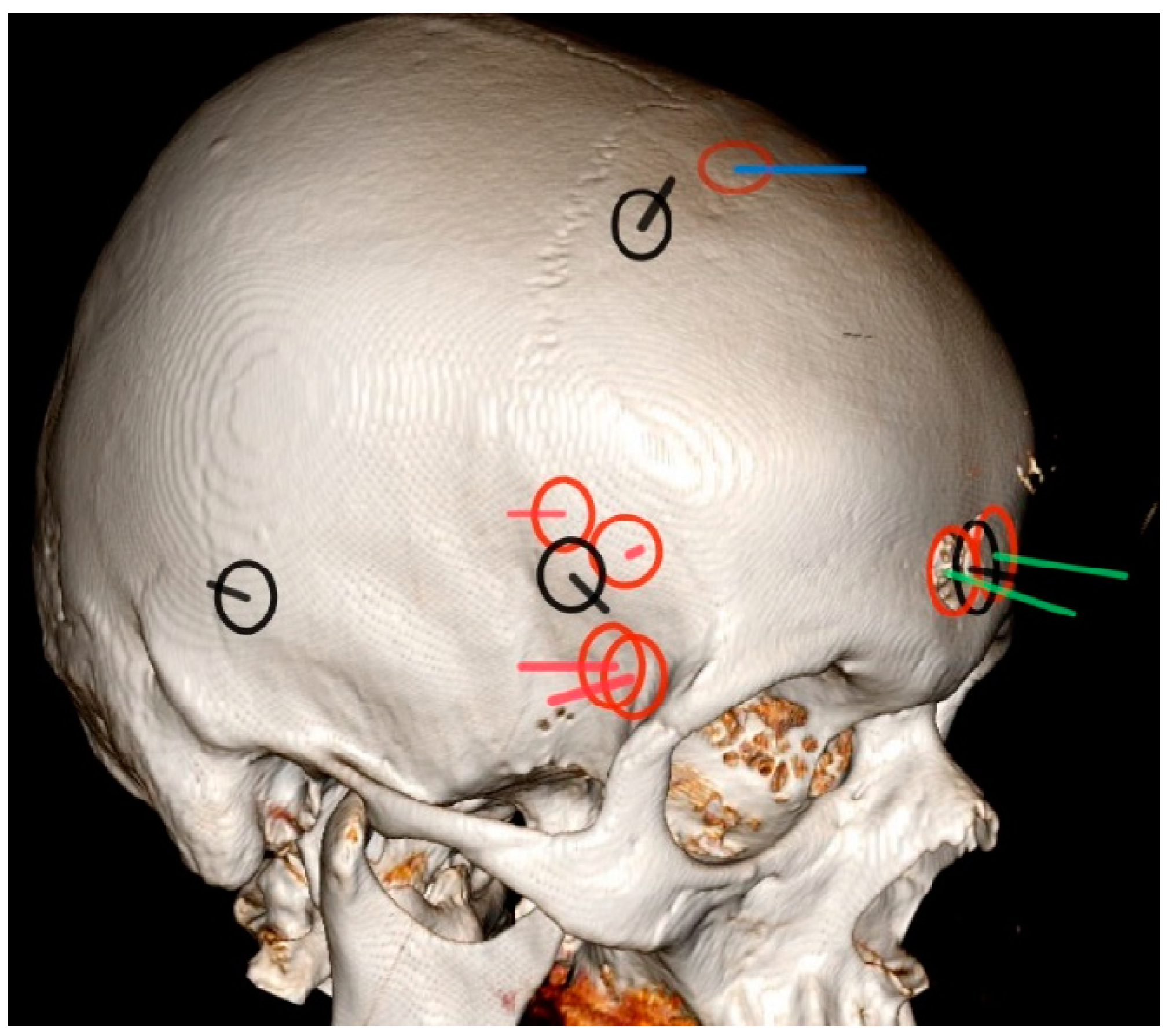
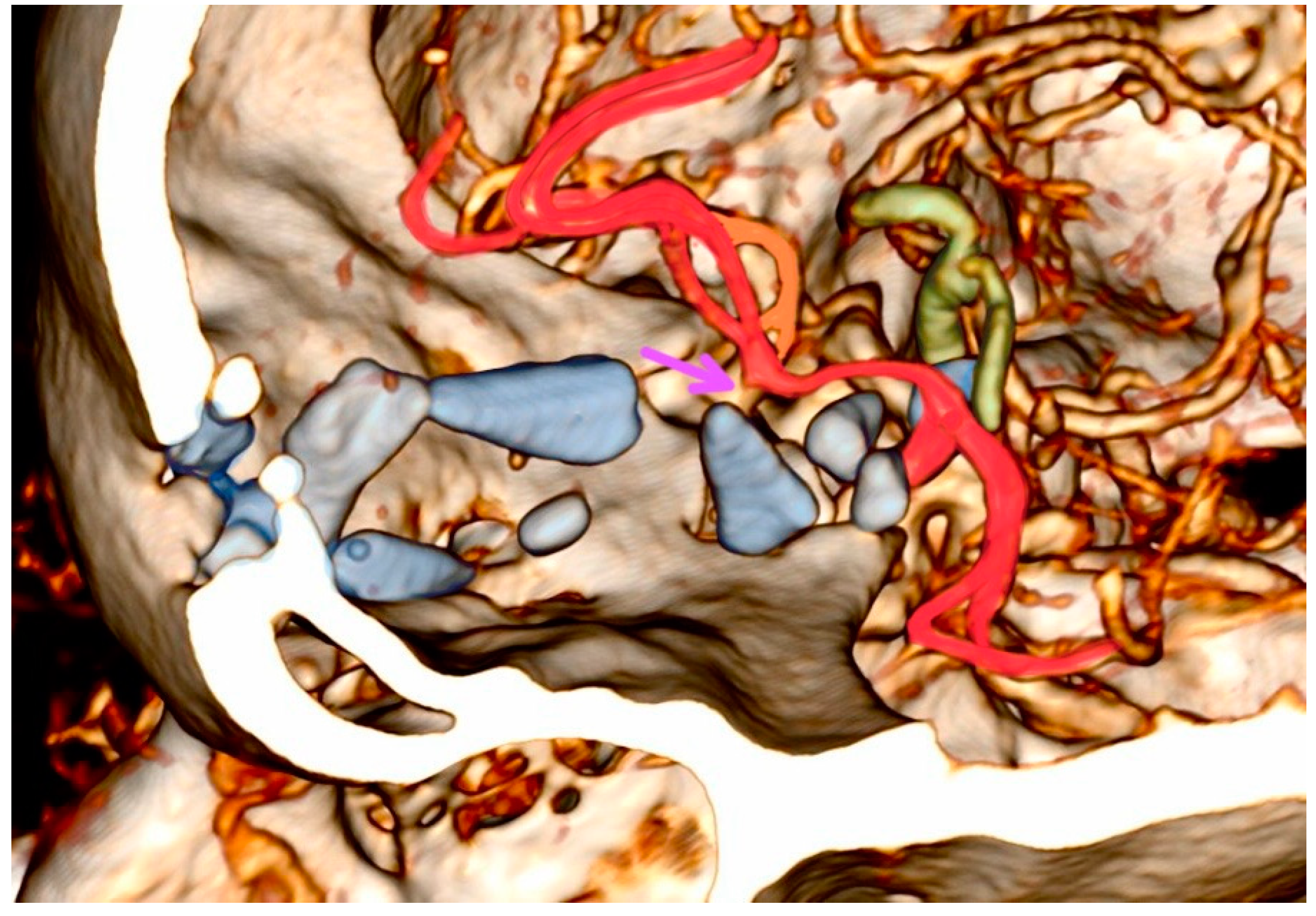
This article investigates the clinical and radiological characteristics of captive bolt gun head injuries, a rare form of low-velocity penetrating brain injury. Eleven consecutive patients were included in the study. Vascular injuries and the rate of infection were systematically analyzed. Radiological findings reveal common bolt trajectories in the anterior cranial fossa, with identified risk factors for a poor outcome including trajectory crossing midline, hematocephalus, and paranasal sinus involvement. Only one patient had a good outcome. Despite meticulous microsurgical techniques, this study highlights often unfavorable clinical outcomes in captive bolt gun injuries, with vascular injury identified as a potential contributing risk factor for a poor outcome. Knowledge of variant vascular tree anatomy and corresponding vascular territory is important. To avoid potential vascular injuries, a complete removal of bone fragments was not always performed and it did not increase the rate of infection, challenging the conventional wisdom advocating for the complete removal of bone fragments. These findings contribute novel insights into captive bolt gun-related injuries, paving the way for further research.
Keywords: captive bolt gun; vascular injury; low-velocity penetrating brain injury; skin–bone imprimatum; incomplete bone fragment debridement; frontopolar artery territory; anterior cranial fossa bolt trajectory captive bolt gun; vascular injury; low-velocity penetrating brain injury; skin–bone imprimatum; incomplete bone fragment debridement; frontopolar artery territory; anterior cranial fossa bolt trajectory