
Sonographic, Demographic, and Clinical Characteristics of Pre- and Postmenopausal Women with Endometrial Cancer; Results from a Post Hoc Analysis of the IETA4 (International Endometrial Tumor Analysis) Multicenter Cohort

 , , , , , , , , , , and
Abstract
:1. Introduction
2. Materials and Methods
2.1. Patients
2.2. Variables
2.3. Statistical Analyses
3. Results
3.1. Demographic and Anthropometric Characteristics
3.2. Histopathological Characteristics
3.3. Sonographic Characteristics
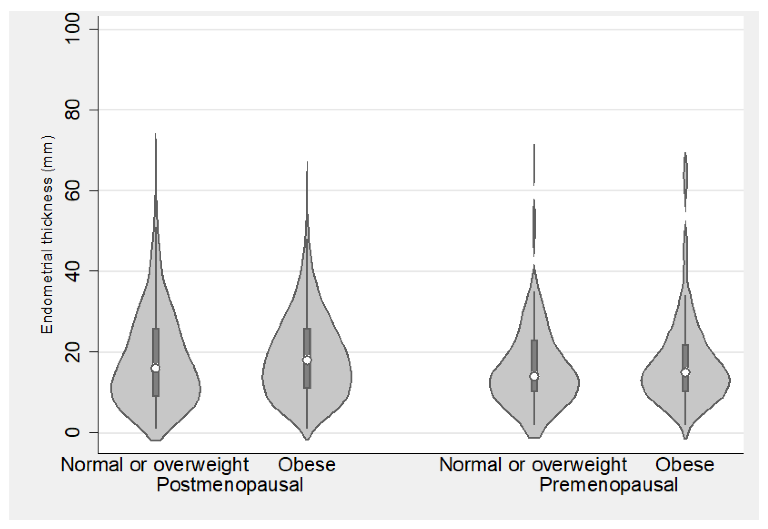
3.4. Endometrial Thickness in Relation to Anthropometric Characteristics
3.5. Sonographic and Anthropometric Characteristics in Relation to Risk Group
3.6. Results after Controlling the False Discovery Rate
4. Discussion
5. Conclusions
Supplementary Materials
Author Contributions
Funding
Institutional Review Board Statement
Informed Consent Statement
Data Availability Statement
Acknowledgments
Conflicts of Interest
References
- Amant, F.; Mirza, M.R.; Koskas, M.; Creutzberg, C.L. Cancer of the Corpus Uteri. Int. J. Gynecol. Obstet. 2015, 131, S96–S104. [Google Scholar] [CrossRef]
- Ferlay, J.; Soerjomataram, I.; Dikshit, R.; Eser, S.; Mathers, C.; Rebelo, M.; Parkin, D.M.; Forman, D.; Bray, F. Cancer Incidence and Mortality Worldwide: Sources, Methods and Major Patterns in GLOBOCAN 2012. Int. J. Cancer 2015, 136, E359–E386. [Google Scholar] [CrossRef] [PubMed]
- National Cancer Institute (NIH). Cancer Stat Facts: Endometrial Cancer. Available online: https://seer.cancer.gov/statfacts/html/corp.html (accessed on 30 October 2019).
- Cancer Research UK. Available online: https://www.cancerresearchuk.org/health-professional/cancer-statistics/statistics-by-cancer-type/uterine-cancer/incidence#heading-One (accessed on 30 October 2019).
- Socialstyrelsen, Statistikdatabas för Cancer. Available online: https://sdb.socialstyrelsen.se/if_can/val.aspx (accessed on 30 October 2019).
- Pennant, M.E.; Mehta, R.; Moody, P.; Hackett, G.; Prentice, A.; Sharp, S.J.; Lakshman, R. Premenopausal Abnormal Uterine Bleeding and Risk of Endometrial Cancer. BJOG 2017, 124, 404–411. [Google Scholar] [CrossRef] [PubMed]
- Clarke, M.A.; Long, B.J.; Del Mar Morillo, A.; Arbyn, M.; Bakkum-Gamez, J.N.; Wentzensen, N. Association of Endometrial Cancer Risk with Postmenopausal Bleeding in Women a Systematic Review and Meta-Analysis. JAMA Intern. Med. 2018, 178, 1201–1208. [Google Scholar] [CrossRef] [PubMed]
- Hernandez, A.V.; Pasupuleti, V.; Benites-Zapata, V.A.; Thota, P.; Deshpande, A.; Perez-Lopez, F.R. Insulin Resistance and Endometrial Cancer Risk: A Systematic Review and Meta-Analysis. Eur. J. Cancer 2015, 51, 2747–2758. [Google Scholar] [CrossRef] [PubMed]
- McPherson, C.P.; Sellers, T.A.; Potter, J.D.; Bostick, R.M.; Folsom, A.R. Reproductive Factors and Risk of Endometrial Cancer. The Iowa Women’s Health Study. Am. J. Epidemiol. 1996, 143, 1195–1202. [Google Scholar] [CrossRef] [PubMed]
- Soliman, P.T.; Oh, J.C.; Schmeler, K.M.; Sun, C.C.; Slomovitz, B.M.; Gershenson, D.M.; Burke, T.W.; Lu, K.H. Risk Factors for Young Premenopausal Women With Endometrial Cancer. Obstet. Gynecol. 2005, 105, 575–580. [Google Scholar] [CrossRef]
- Wise, M.R.; Jordan, V.; Lagas, A.; Showell, M.; Wong, N.; Lensen, S.; Farquhar, C.M. Obesity and Endometrial Hyperplasia and Cancer in Premenopausal Women: A Systematic Review. Am. J. Obstet. Gynecol. 2016, 214, 689.e1–689.e17. [Google Scholar] [CrossRef]
- Schmeler, K.M.; Soliman, P.T.; Sun, C.C.; Slomovitz, B.M.; Gershenson, D.M.; Lu, K.H. Endometrial Cancer in Young, Normal-Weight Women. Gynecol. Oncol. 2005, 99, 388–392. [Google Scholar] [CrossRef]
- Lee, N.K.; Cheung, M.K.; Shin, J.Y.; Husain, A.; Teng, N.N.; Berek, J.S.; Kapp, D.S.; Osann, K.; Chan, J.K. Prognostic Factors for Uterine Cancer in Reproductive-Aged Women. Obstet. Gynecol. 2007, 109, 655–662. [Google Scholar] [CrossRef]
- Pellerin, G.P.; Finan, M.A. Endometrial Cancer in Women 45 Years of Age or Younger: A Clinicopathological Analysis. Am. J. Obstet. Gynecol. 2005, 193, 1640–1644. [Google Scholar] [CrossRef]
- Colombo, N.; Creutzberg, C.; Amant, F.; Bosse, T.; González-Martín, A.; Ledermann, J.; Marth, C.; Nout, R.; Querleu, D.; Mirza, M.R.; et al. ESMO-ESGO-ESTRO Consensus Conference on Endometrial Cancer: Diagnosis, Treatment and Follow-Up. Ann. Oncol. 2016, 27, 16–41. [Google Scholar] [CrossRef]
- Verbakel, J.Y.; Mascilini, F.; Wynants, L.; Fischerova, D.; Testa, A.C.; Franchi, D.; Frühauf, F.; Cibula, D.; Lindqvist, P.G.; Fruscio, R.; et al. Validation of Ultrasound Strategies to Assess Tumor Extension and to Predict High-Risk Endometrial Cancer in Women from the Prospective IETA (International Endometrial Tumor Analysis)-4 Cohort. Ultrasound Obstet. Gynecol. 2020, 55, 115–124. [Google Scholar] [CrossRef] [PubMed]
- Epstein, E.; Fischerova, D.; Valentin, L.; Testa, A.C.; Franchi, D.; Sladkevicius, P.; Frühauf, F.; Lindqvist, P.G.; Mascilini, F.; Fruscio, R.; et al. Ultrasound Characteristics of Endometrial Cancer as Defined by International Endometrial Tumor Analysis (IETA) Consensus Nomenclature: Prospective Multicenter Study. Ultrasound Obstet. Gynecol. 2018, 51, 818–828. [Google Scholar] [CrossRef] [PubMed]
- Eriksson, L.S.E.; Epstein, E.; Testa, A.C.; Fischerova, D.; Valentin, L.; Sladkevicius, P.; Franchi, D.; Frühauf, F.; Fruscio, R.; Haak, L.A.; et al. Ultrasound-Based Risk Model for Preoperative Prediction of Lymph-Node Metastases in Women with Endometrial Cancer: Model-Development Study. Ultrasound Obstet. Gynecol. 2020, 56, 443–452. [Google Scholar] [CrossRef] [PubMed]
- Green, R.W.; Valentin, L.; Alcazar, J.L.; Chiappa, V.; Erdodi, B.; Franchi, D.; Frühauf, F.; Fruscio, R.; Guerriero, S.; Graupera, B.; et al. Endometrial Cancer Off-Line Staging Using Two-Dimensional Transvaginal Ultrasound and Three-Dimensional Volume Contrast Imaging: Intermethod Agreement, Interrater Reliability and Diagnostic Accuracy. Gynecol. Oncol. 2018, 150, 438–445. [Google Scholar] [CrossRef] [PubMed]
- Leone, F.P.G.; Timmerman, D.; Bourne, T.; Valentin, L.; Epstein, E.; Goldstein, S.R.; Marret, H.; Parsons, A.K.; Gull, B.; Istre, O.; et al. Terms, Definitions and Measurements to Describe the Sonographic Features of the Endometrium and Intrauterine Lesions: A Consensus Opinion from the International Endometrial Tumor Analysis (IETA) Group. Ultrasound Obstet. Gynecol. 2010, 35, 103–112. [Google Scholar] [CrossRef] [PubMed]
- Installé, A.J.F.; Van Den Bosch, T.; De Moor, B.; Timmerman, D. Clinical Data Miner: An Electronic Case Report Form System with Integrated Data Preprocessing and Machine-Learning Libraries Supporting Clinical Diagnostic Model Research. J. Med. Internet Res. 2014, 16, e28. [Google Scholar] [CrossRef] [PubMed]
- Pecorelli, S. Revised FIGO Staging for Carcinoma of the Vulva, Cervix, and Endometrium. Int. J. Gynecol. Obstet. 2009, 105, 103–104. [Google Scholar] [CrossRef]
- Kurman, R.J.; Carcangiu, M.L.; Herrington, S.C.; Young, R.H. WHO Classification of Tumours of Female Reproductive Organs, 4th ed.; International Agency for Research on Cancer: Lyon, France, 2014; ISBN 978-92-832-4487-5.
- Alberti, K.G.M.M.; Eckel, R.H.; Grundy, S.M.; Zimmet, P.Z.; Cleeman, J.I.; Donato, K.A.; Fruchart, J.C.; James, W.P.T.; Loria, C.M.; Smith, S.C. Harmonizing the Metabolic Syndrome: A Joint Interim Statement of the International Diabetes Federation Task Force on Epidemiology and Prevention; National Heart, Lung, and Blood Institute; American Heart Association; World Heart Federation; International. Circulation 2009, 120, 1640–1645. [Google Scholar] [CrossRef]
- Benjamini, Y.; Yekutieli, D. The Control of the False Discovery Rate in Multiple Testing under Dependency. Ann. Stat. 2001, 29, 1165–1188. [Google Scholar] [CrossRef]
- Newson, R. Multiple—Test Procedures and Smile Plots. Stata J. Promot. Commun. Stat. Stata 2003, 3, 109–132. [Google Scholar] [CrossRef]
- Gaus, W. Interpretation of Statistical Significance-Exploratory Versus Confirmative Testing in Clinical Trials, Epidemiological Studies, Meta-Analyses and Toxicological Screening (Using Ginkgo Biloba as an Example). Clin. Exp. Pharmacol. 2015, 5, 1000182. [Google Scholar] [CrossRef]
- Breijer, M.C.; Peeters, J.A.H.; Opmeer, B.C.; Clark, T.J.; Verheijen, R.H.M.; Mol, B.W.J.; Timmermans, A. Capacity of Endometrial Thickness Measurement to Diagnose Endometrial Carcinoma in Asymptomatic Postmenopausal Women: A Systematic Review and Meta-Analysis. Ultrasound Obstet. Gynecol. 2012, 40, 621–629. [Google Scholar] [CrossRef] [PubMed]
- Su, D.; Li, L.; Zhong, M.; Xia, Y. Capacity of Endometrial Thickness Measurement to Diagnose Endometrial Carcinoma in Asymptomatic Postmenopausal Women: A Systematic Review and Meta-Analysis. Ann. Palliat. Med. 2021, 10, 10840–10848. [Google Scholar] [CrossRef] [PubMed]
- Tsuda, H.; Ito, Y.M.; Todo, Y.; Iba, T.; Tasaka, K.; Sutou, Y.; Hirai, K.; Dozono, K.; Dobashi, Y.; Manabe, M.; et al. Measurement of Endometrial Thickness in Premenopausal Women in Office Gynecology. Reprod. Med. Biol. 2018, 17, 29–35. [Google Scholar] [CrossRef]
- Gao, Y.; Dai, X.; Lee, A.C.; Wise, M.R.; Shen, F.; Chen, Q. Body Mass Index Is Negatively Associated with Endometrial Cancer Stage, Regardless of Subtype and Menopausal Status. J. Cancer 2018, 9, 4756–4761. [Google Scholar] [CrossRef] [PubMed]
- Arem, H.; Irwin, M.L. Obesity and Endometrial Cancer Survival: A Systematic Review. Int. J. Obes. 2013, 37, 634–639. [Google Scholar] [CrossRef]
- Nevadunsky, N.S.; Van Arsdale, A.; Strickler, H.D.; Moadel, A.; Kaur, G.; Levitt, J.; Girda, E.; Goldfinger, M.; Goldberg, G.L.; Einstein, M.H. Obesity and Age at Diagnosis of Endometrial Cancer. Obstet. Gynecol. 2014, 124, 300–306. [Google Scholar] [CrossRef]
- Van Arsdale, A.; Miller, D.T.; Kuo, D.Y.; Isani, S.; Sanchez, L.; Nevadunsky, N.S. Association of Obesity with Survival in Patients with Endometrial Cancer. Gynecol. Oncol. 2019, 154, 156–162. [Google Scholar] [CrossRef]
- Eurostat. Available online: https://ec.europa.eu/eurostat/data/database (accessed on 13 December 2019).
- Santoro, N.; Lasley, B.; McConnell, D.; Allsworth, J.; Crawford, S.; Gold, E.B.; Finkelstein, J.S.; Greendale, G.A.; Kelsey, J.; Korenman, S.; et al. Body Size and Ethnicity Are Associated with Menstrual Cycle Alterations in Women in the Early Menopausal Transition: The Study of Women’s Health across the Nation (SWAN) Daily Hormone Study. J. Clin. Endocrinol. Metab. 2004, 89, 2622–2631. [Google Scholar] [CrossRef] [PubMed]
- Benjamin, D.J.; Berger, J.O.; Johannesson, M.; Nosek, B.A.; Wagenmakers, E.J.; Berk, R.; Bollen, K.A.; Brembs, B.; Brown, L.; Camerer, C.; et al. Redefine Statistical Significance. Nat. Hum. Behav. 2018, 2, 6–10. [Google Scholar] [CrossRef] [PubMed]
- Benjamini, Y.; Zeevi, Y. It’s the Selection’s Fault—Not the p-Values’: A Comment on “The Role of p-Values in Judging the Strength of Evidence and Realistic Replication Expectations”. Stat. Biopharm. Res. 2020, 13, 22–25. [Google Scholar] [CrossRef]
- Amrhein, V.; Greenland, S.; McShane, B. Scientists Rise up against Statistical Significance. Nature 2019, 567, 305–307. [Google Scholar] [CrossRef]
- Dirnagl, U. The p Value Wars (Again). Eur. J. Nucl. Med. Mol. Imaging 2019, 46, 2421–2423. [Google Scholar] [CrossRef] [PubMed]
- Ioannidis, J.P.A. Retiring Statistical Significance Would Give Bias a Free Pass. Nature 2019, 567, 461. [Google Scholar] [CrossRef] [PubMed]


| Characteristic | Premenopausal Women | Postmenopausal Women | ||||
|---|---|---|---|---|---|---|
| Background | Median | IQR | Median | IQR | Uncorrected p-value | Significance after Benjamini–Yekutieli procedure |
| Age | 48 | 43–52 | 66 | 61–72 | NA | |
| Parity | N | % | N | % | <0.001 | significant |
| 0 | 54 | 34% | 230 | 17% | ||
| 1 | 40 | 25% | 275 | 20% | ||
| 2 | 52 | 32% | 588 | 43% | ||
| 3+ | 15 | 9% | 284 | 21% | ||
| Family history of cancer | N | % | N | % | ||
| Endometrial | 7 | 4% | 94 | 7% | 0.311 | not significant |
| Ovarian | 1 | 1% | 20 | 1% | 0.717 | not significant |
| Breast | 16 | 10% | 149 | 11% | 0.893 | not significant |
| Colon | 26 | 16% | 103 | 7% | 0.001 | significant |
| Other | 30 | 19% | 278 | 20% | 0.679 | not significant |
| Lifestyle | ||||||
| Smoking | 0.398 | not significant | ||||
| Never | 122 | 76% | 1047 | 76% | ||
| Former | 19 | 12% | 198 | 14% | ||
| Present | 20 | 12% | 132 | 10% | ||
| Exercise | 0.243 | not significant | ||||
| Never | 53 | 33% | 515 | 37% | ||
| >20 min, 1–2/w. | 46 | 29% | 436 | 32% | ||
| >20 min, ≥3/w. | 44 | 27% | 328 | 24% | ||
| Strenuous, 1/w. | 10 | 6% | 54 | 4% | ||
| Strenuous, ≥2/w. | 8 | 5% | 44 | 3% | ||
| Alcohol use | 0.193 | not significant | ||||
| Never | 61 | 38% | 596 | 43% | ||
| 0–6 units/w. | 89 | 55% | 691 | 50% | ||
| 7–12 units/w. | 9 | 6% | 85 | 6% | ||
| >12 units/w. | 2 | 1% | 5 | 0% | ||
| Anthropometric | ||||||
| Constitution | 0.024 | not significant | ||||
| Abdominal adiposity | 70 | 43% | 742 | 54% | ||
| Female adiposity | 32 | 20% | 259 | 19% | ||
| Lean | 59 | 37% | 376 | 27% | ||
| Median | IQR | Median | IQR | |||
| Weight (kg) | 77 | 65–93 | 75 | 65–89 | 0.671 | not significant |
| Height (cm) | 165 | 162–170 | 163 | 159–168 | NA | |
| BMI (kg/m2) | 27.6 | 23.1–34.6 | 28.4 | 24.4–33.2 | 0.290 | not significant |
| Waist circumference (cm) | 92 | 82–108 | 98 | 87–112 | 0.002 | significant |
| Bra cup size | N | % | N | % | 0.87 | not significant |
| AA-B | 55 | 34% | 443 | 32% | ||
| C-D | 87 | 54% | 756 | 55% | ||
| E-I | 19 | 12% | 178 | 13% | ||
| Co-morbidity | ||||||
| Current use of hormonal treatment | 18 | 11% | 160 | 12% | 1 | not significant |
| Type 2 diabetes | 11 | 7% | 235 | 17% | NA | |
| Hypertension | 35 | 22% | 730 | 53% | NA | |
| Bleeding history | ||||||
| Abnormal uterine bleeding | 140 | 87% | 1226 | 89% | 0.428 | not significant |
| Median | IQR | Median | IQR | |||
| Duration of abnormal uterine bleeding (months) | 5 | 3–12 | 3 | 2–6 | <0.001 | significant |
| Characteristic | Premenopausal Women | Postmenopausal Women | ||||
|---|---|---|---|---|---|---|
| N | % | N | % | Uncorrected p-value | Significance after Benjamini–Yekutieli procedure | |
| FIGO surgical stage | <0.001 ϕ | significant | ||||
| IA | 135 | 84% | 801 | 58% | ||
| IB | 9 | 6% | 315 | 23% | ||
| II | 7 | 4% | 79 | 6% | ||
| III | 10 | 6% | 159 | 12% | ||
| IV | 0 | 0% | 23 | 2% | ||
| Histologic subtype | ||||||
| Endometrioid | 158 | 98% | 1172 | 85% | <0.001 Ω | significant |
| Grade 1 | 91 | 58% | 512 | 44% | 0.0012 Ψ | significant |
| Grade 2 | 49 | 31% | 463 | 40% | ||
| Grade 3 | 18 | 11% | 197 | 17% | ||
| Non-endometrioid | 3 | 2% | 205 | 15% | NA | |
| Serous | 0 | 0% | 91 | 44% | ||
| Carcinosarcoma | 2 | 67% | 39 | 19% | ||
| Clear cell carcinoma | 0 | 0% | 33 | 16% | ||
| Mixed cell carcinoma | 1 | 33% | 35 | 17% | ||
| Undifferentiated | 0 | 0% | 7 | 3% | ||
| ESMO-ESGO-ESTRO risk group ε | <0.001 | significant | ||||
| Low | 126 | 78% | 636 | 46% | ||
| Intermediate/High | 35 | 22% | 741 | 54% | ||
| Finding | Premenopausal Women | Postmenopausal Women | ||||
|---|---|---|---|---|---|---|
| Sonographic findings, all women | N | % | N | % | Uncorrected p-value | Significance after Benjamini–Yekutieli procedure |
| Endometrium | 0.356 | not significant | ||||
| Measurable | 154 | 96% | 1274 | 93% | ||
| Not measurable | 4 | 2% | 45 | 3% | ||
| Not visible | 3 | 2% | 58 | 4% | ||
| Tumor | 0.001 | significant | ||||
| Defined | 118 | 73% | 1157 | 84% | ||
| Not defined | 43 | 27% | 220 | 16% | ||
| Myometrium | ||||||
| Fibroid present | 48 | 30% | 503 | 37% | 0.099 | not significant |
| Adenomyosis suspected | 25 | 16% | 96 | 7% | 0.001 | significant |
| Sonographic findings, in women w. visible endometrium only | ||||||
| Endometrial–myometrial border | <0.001 | significant | ||||
| Regular | 61 | 39% | 298 | 23% | ||
| Endometrial echogenicity | ||||||
| Uniform | 65 | 41% | 496 | 38% | 0.387 ϕ | not significant |
| Hyperechoic | 49 | 75% | 377 | 76% | ||
| Hypo-/iso-/three layered | 16 | 25% | 119 | 24% | ||
| Non-uniform | 93 | 59% | 823 | 62% | ||
| Homogenous w. cysts | 9 | 10% | 70 | 9% | ||
| Heterogenous without cysts | 77 | 83% | 661 | 80% | ||
| Heterogenous w. cysts | 7 | 8% | 92 | 11% | ||
| Bright edge sign | 0.007 | not significant | ||||
| Yes | 11 | 7% | 191 | 14% | ||
| Endometrial midline | <0.001 | significant | ||||
| Seen | 37 | 23% | 147 | 11% | ||
| Undefined/not seen | 121 | 77% | 1172 | 89% | ||
| Color score | 0.545 Ω | not significant | ||||
| No flow | 25 | 16% | 267 | 20% | ||
| Minimal flow | 32 | 20% | 246 | 19% | ||
| Moderate flow | 65 | 41% | 436 | 33% | ||
| Abundant flow | 36 | 23% | 370 | 28% | ||
| Vascular pattern | 0.439 Ψ | not significant | ||||
| No flow | 25 | 16% | 267 | 20% | ||
| Single +/− branching | 21 | 13% | 150 | 11% | ||
| Multiple vessels, focal | 26 | 16% | 214 | 16% | ||
| Multiple vessels, multifocal | 57 | 36% | 519 | 39% | ||
| Scattered vessels | 28 | 18% | 168 | 13% | ||
| Circular | 1 | 1% | 1 | 0% | ||
| Endometrial thickness (mm) | 14 | 10–23 | 17 | 10–26 | 0.132 | not significant |
| Tumor volume (cm3) | 7.3 | 2.4–23.3 | 7.9 | 2.7–21.1 | 0.786 ε | not significant |
| Finding | Premenopausal Women | Postmenopausal Women | ||||||||||
|---|---|---|---|---|---|---|---|---|---|---|---|---|
| Low risk | Intermediate/high risk | Low risk | Intermediate/high risk | |||||||||
| Sonographic findings, all women | N | % | N | % | Uncorrected p-value | Significance after Benjamini–Yekutieli procedure | N | % | N | % | Uncorrected p-value | Significance after Benjamini–Yekutieli procedure |
| Tumor | 0.196 | not significant | <0.001 | significant | ||||||||
| Defined | 89 | 71% | 29 | 83% | 509 | 80% | 648 | 87% | ||||
| Not defined | 37 | 29% | 6 | 17% | 127 | 20% | 93 | 93% | ||||
| BMI | 0.696 | not significant | <0.001 | significant | ||||||||
| <30 | 77 | 61% | 23 | 66% | 338 | 53% | 473 | 64% | ||||
| ≥30 | 49 | 39% | 12 | 34% | 298 | 47% | 268 | 36% | ||||
| Waist group | 1.000 | not significant | 0.001 | significant | ||||||||
| <80 cm | 49 | 39% | 13 | 37% | 146 | 23% | 229 | 31% | ||||
| ≥80 cm | 77 | 61% | 22 | 63% | 490 | 77% | 512 | 69% | ||||
| Sonographic findings, in women w. visible endometrium only | ||||||||||||
| Endometrial–myometrial border | <0.001 | significant | <0.001 | significant | ||||||||
| Regular | 58 | 46% | 3 | 9% | 207 | 33% | 91 | 13% | ||||
| Non-regular | 68 | 54% | 29 | 91% | 415 | 67% | 606 | 87% | ||||
| Endometrial echogenicity | 0.232 | not significant | <0.001 | significant | ||||||||
| Uniform | 55 | 44% | 10 | 31% | 266 | 43% | 230 | 33% | ||||
| Non-uniform | 71 | 56% | 22 | 69% | 356 | 57% | 467 | 67% | ||||
| Endometrial midline | 0.818 | not significant | 0.431 | not significant | ||||||||
| Seen | 29 | 23% | 8 | 25% | 74 | 12% | 73 | 10% | ||||
| Undefined/not seen | 97 | 77% | 24 | 75% | 548 | 88% | 624 | 90% | ||||
| Color score | 0.156 | not significant | <0.001 | significant | ||||||||
| No flow–minimal flow | 49 | 39% | 8 | 25% | 324 | 52% | 189 | 27% | ||||
| Moderate flow–abundant flow | 77 | 61% | 24 | 75% | 298 | 48% | 508 | 73% | ||||
| Vascular pattern | 0.216 | not significant | <0.001 | significant | ||||||||
| All others | 84% | 67% | 17 | 53% | 449 | 72% | 351 | 50% | ||||
| Multiple vessels, multifocal | 42 | 33% | 15 | 47% | 173 | 28% | 346 | 50% | ||||
Disclaimer/Publisher’s Note: The statements, opinions and data contained in all publications are solely those of the individual author(s) and contributor(s) and not of MDPI and/or the editor(s). MDPI and/or the editor(s) disclaim responsibility for any injury to people or property resulting from any ideas, methods, instructions or products referred to in the content. |
© 2023 by the authors. Licensee MDPI, Basel, Switzerland. This article is an open access article distributed under the terms and conditions of the Creative Commons Attribution (CC BY) license (https://creativecommons.org/licenses/by/4.0/).
Share and Cite
Green, R.W.; Fischerová, D.; Testa, A.C.; Franchi, D.; Frühauf, F.; Lindqvist, P.G.; di Legge, A.; Cibula, D.; Fruscio, R.; Haak, L.A.; et al. Sonographic, Demographic, and Clinical Characteristics of Pre- and Postmenopausal Women with Endometrial Cancer; Results from a Post Hoc Analysis of the IETA4 (International Endometrial Tumor Analysis) Multicenter Cohort. Diagnostics 2024, 14, 1. https://doi.org/10.3390/diagnostics14010001
Green RW, Fischerová D, Testa AC, Franchi D, Frühauf F, Lindqvist PG, di Legge A, Cibula D, Fruscio R, Haak LA, et al. Sonographic, Demographic, and Clinical Characteristics of Pre- and Postmenopausal Women with Endometrial Cancer; Results from a Post Hoc Analysis of the IETA4 (International Endometrial Tumor Analysis) Multicenter Cohort. Diagnostics. 2024; 14(1):1. https://doi.org/10.3390/diagnostics14010001
Chicago/Turabian StyleGreen, Rasmus W., Daniela Fischerová, Antonia C. Testa, Dorella Franchi, Filip Frühauf, Pelle G. Lindqvist, Alessia di Legge, David Cibula, Robert Fruscio, Lucia A. Haak, and et al. 2024. "Sonographic, Demographic, and Clinical Characteristics of Pre- and Postmenopausal Women with Endometrial Cancer; Results from a Post Hoc Analysis of the IETA4 (International Endometrial Tumor Analysis) Multicenter Cohort" Diagnostics 14, no. 1: 1. https://doi.org/10.3390/diagnostics14010001
APA StyleGreen, R. W., Fischerová, D., Testa, A. C., Franchi, D., Frühauf, F., Lindqvist, P. G., di Legge, A., Cibula, D., Fruscio, R., Haak, L. A., Opolskiene, G., Vidal Urbinati, A. M., Timmerman, D., Bourne, T., van den Bosch, T., & Epstein, E. (2024). Sonographic, Demographic, and Clinical Characteristics of Pre- and Postmenopausal Women with Endometrial Cancer; Results from a Post Hoc Analysis of the IETA4 (International Endometrial Tumor Analysis) Multicenter Cohort. Diagnostics, 14(1), 1. https://doi.org/10.3390/diagnostics14010001