Article

Evaluation of Signal Transducer and Activator of Transcription 3 (STAT-3) Protein Expression in Non-Hodgkin Lymphoma Cases in Hospital USM

by
Izyan Rifhana Muhamad
1,2,
Noorul Balqis Che Ibrahim
1,2 and
Faezahtul Arbaeyah Hussain
1,2,*
1
Department of Pathology, School of Medical Sciences, Universiti Sains Malaysia Health Campus, Kubang Kerian 16150, Kelantan, Malaysia
2
Hospital Universiti Sains Malaysia, Universiti Sains Malaysia, Kubang Kerian 16150, Kelantan, Malaysia
*
Author to whom correspondence should be addressed.
Diagnostics 2023, 13(9), 1649; https://doi.org/10.3390/diagnostics13091649
Submission received: 10 February 2023 / Revised: 2 May 2023 / Accepted: 3 May 2023 / Published: 7 May 2023
(This article belongs to the Special Issue Haematology: Diagnosis and Management)

Abstract

:
Background: Evolving targeted therapy on Janus Associated Kinase-Signal Transducer and Activator of Transcription (JAK-STAT) signaling pathway, especially pertaining to STAT-3 protein in non-Hodgkin lymphoma (NHL), provides new treatment strategies. STAT-3 protein also relates to the prognostication of NHL. Hence, we aimed to evaluate the expression of STAT-3 protein in NHL cases diagnosed in Hospital Universiti Sains Malaysia (USM). Methods: A retrospective cross sectional study using formalin fixed paraffin embedded (FFPE) tissue blocks of 95 NHL cases were obtained. STAT-3 immunostaining was performed and evaluated. The proportion and association between the expression of STAT-3 protein with subtypes of NHL were statistically analyzed. Results: The majority of the cases (78.9%) had positive STAT-3 protein expression. 64.2% were among aggressive B cell NHL, whilst 20.0% of them were diffuse large B cell lymphoma, a non-germinal center B subtype (DLBCL-NGCB). There is also an association between STAT-3 protein expression with DLBCL subtypes (p = 0.046). Conclusion: Our study demonstrated a remarkable expression of STAT-3 protein in NHL, in which DLBCL subtypes had significant association. A larger scale study with a combination of JAK protein evaluation should be undertaken in the future.

1. Introduction

The pathogenesis of lymphoma is relatively complex and may involve a variety of gene mutations in the multistep origin of this cancer development. Mutations that increase tyrosine kinase activity are one of the common aberrations that drive cell growth [1]. Tyrosine kinase is an enzyme that mediates intracellular signaling by phosphorylating proteins on tyrosine residues to activate the Janus Associated Kinase-Signal Transducer and Activator of Transcription (JAK/STAT) signaling pathway [2]. The evolution of drugs targeting the JAK-STAT intracellular signalling pathway are becoming more recognized as drugs that can contribute to the treatment of haematolymphoid malignancies. Although JAK-STAT signaling is more well-known in hematological malignancies, more studies have been explored regarding its role in the pathogenesis of solid tumors, including lymphoma [3]. Four recognizable Janus-associated kinase (JAK) families are JAK-1, JAK-2, JAK-3, and TYK-2 proteins, while signal transducer and activator of transcription (STAT) families consist of seven major proteins, which are STAT-1, STAT-2, STAT-3, STAT-4, STAT-5a, STAT-5b, and STAT-6 [4].
The JAK-STAT pathway is an intracellular signaling pathway for gene expression mediated by cytokines in the form of interferon, interleukin, and growth factors [3]. The cytokine receptors receive extracellular signals once the cytokine molecules bind on them, resulting in their aggregation. The JAK protein, which is associated with cytokine receptors intracellularly, will phosphorylate and cause activation of the tyrosine kinase domain, providing docking sites for the STAT protein [3]. Recruited STAT monomers by the JAK-mediated phosphorylation result in their dimerization and translocation from the cell cytoplasm across the nuclear membrane to the cell nucleus. STAT dimers enhance the transcription of specific genes in the nucleus [3]. Changes in gene expression, cell proliferation and differentiation with the prevention of apoptosis occur because of this intracellular complex cascade of biochemical events of the JAK-STAT pathway [3,4,5], as shown in Figure 1. The development of some cancers as a result of genetic mutations, amplifications, or polymorphisms causes aberrant activation of the JAK-STAT pathway and leads to its continuous activation [3]. Other mechanisms that lead to the continuous activation of the JAK-STAT signaling pathways include extracellular signals by excessive cytokine expression, such as interleukin-6 upregulation by tumor cells and tumor microenvironment [6,7,8]. Therefore, the role of the aberrant activation of JAK-STAT signaling contributes to carcinogenesis, either as a tumor intrinsic driver of cancer growth/metastasis or as a modulator of immune surveillance [6].
In numerous human malignancies, phosphorylated tyrosine kinase and STAT nuclear expressions are signs of JAK-STAT pathway activation [3]. JAK-1 is known to be the main activator for STAT-3 activity, and excessive STAT-3 activation is common in lymphoma [7,9]. In fact, aberrant STAT-3 signaling plays an important role in immune evasion, and it becomes an oncogenic driver in several types of B-cell lymphoma and, also, in most T-cell lymphomas [7]. Targeting JAK/STAT3 signaling inhibits tumor growth and enhances anti-tumor immune responses [7]. In addition, previous studies agree that STAT-3 functions as an oncogene. Thus, inhibiting STAT-3 activity is an effective method for treating cancer. Aberrant STAT-3 activation has also been linked to cancer hallmarks and poor patient outcomes. Based on the outcomes of clinical trials, several STAT-3 inhibitors were well tolerated, and they may increase the overall survival of cancer patients, including lymphoma cases [8]. Therefore, aiming for JAK/STAT pathways in lymphoma has potential clinical value, and is a promising therapeutic target for treating the disease [7,10]. Thus, in line with the emerging therapeutic intervention targeting the JAK-STAT intracellular signaling pathway, focusing on STAT-3 expression in NHL types may provide some prognostic information. This study aims to evaluate the expression of STAT-3 protein, and to determine its prevalence and its associated clinicopathologic characteristics in NHL cases in Hospital Universiti Sains Malaysia (USM).

2. Materials and Methods

This retrospective cross-sectional study was carried out in the Department of Pathology, Hospital Universiti Sains Malaysia (Hospital USM). The study period was between January 2015 and December 2020. The keyword “lymphoma” was used to search the laboratory information system database. A total of 286 cases were reported within that period. However, only 95 of these cases were selected for this study, which were the ones that fulfilled the inclusion and exclusion criteria. The inclusion criteria included formalin fixed paraffin embedded (FFPE) tissue blocks that have adequate tumoral tissue, newly diagnosed cases, and complete clinical information from the patients’ records. Referral cases from other centers were excluded from this study.
The 95 selected FFPE-tissue blocks were sectioned at 3 µm thickness. The STAT-3 primary antibody, recombinant rabbit monoclonal [EPR787Y] from ABCAM, was diluted at 1:200 dilution. Heat mediated antigen retrieval was performed using detection kit K8012 (DAKO) with high pH (pH9). Subsequently, all slides were placed in a manual stainer and incubated overnight at 4 °C. Endogenous peroxidase blocking, incubation with secondary antibody, and application of substrate chromogen and haematoxylin counterstaining in sequence were then applied. All samples were examined using an Olympus CX31 microscope at 400× magnification, with 10 high power field (HPF). STAT-3 staining can be seen in both the nuclear and the cytoplasm. In lymphoma cases, only those with nuclear staining were considered as positive (Figure 2) [11]. The scoring method (Table 1) was utilized [11].
Age, ethnicity, gender, the localization and size of tumors, B symptoms, lactate dehydrogenase (LDH) level, and the disease stage were included as clinicopathologic features in this study. Tumor histopathological characteristics were classified based on the WHO classification, revised 4th edition [12]. Data were analyzed using a Statistical Package for Social Sciences (SPSS) v. 27.0 software. Continuous data were expressed by mean and medium, while descriptive and categorical data were expressed by proportion, and Pearson chi-square and the Fisher-exact test with appropriate application. p < 0.05 were considered statistically significant.

3. Results

A total of 95 cases with NHL of various subtypes were selected, and the clinicopathologic characteristics are shown in Table 2 and Table 3. The mean age of this study was 54 years old. The ethnic was mainly among Malay (90.5%) ethnicity and males in gender (65.3%). Most of the patients had extra nodal site involvement (49.5%), a size of tumor less than 10 cm (63.2%), the presence of B symptoms (56.8%), a high lactate dehydrogenase (LDH) level (67.4%), and an advanced stage (III-IV) of disease (70.5%) during the first presentation. Many of them were diagnosed as aggressive B cell NHL (77.9%), largely contributed by DLBL subtypes (64.3% in total), and, specifically, DLBCL-NGCB subtypes (21.1%).
Amongst these 95 NHL cases, STAT-3 protein expressions were expressed in more than two-thirds (78.9%) of the cases, as shown in Table 4, using the scoring system with negative score in 20 cases, weak positive in 25 cases, and strong positive in 50 cases, respectively. Overall, STAT-3 protein expression was observed in 61 cases (64.2%) of aggressive B cell NHL, with 44 cases (88%) having a strong expression. The strong expressions were observed in DLBCL variants with 12 (24%) NOS, 15 (30%) NGCB, 6 (12%) GCB, 3 (6%) double expressions, and 1 (2%) triple expressions. The other aggressive type of B cell NHL is listed in Table 4, and had strong positive staining.
There was no significant association between STAT-3 protein expression with clinicopathologic characteristic age, ethnicity, and gender, nor the site and size of the tumors, B symptoms, LDH level, or disease staging (Table 5). Generally, there was no significant association, either, between STAT-3 protein expression with different NHL types based on cell lineage group and specific lymphoma subtypes. We categorized B cell NHL into an indolent subtype and an aggressive subtype. The later was further classified into two large groups: the DLBCL group and another aggressive B cell lymphoma group based on the WHO classification [12]. Among B cell NHL subtypes, we found that there was an association between DLBCL subtypes with STAT-3 protein expression (p = 0.046) (Table 6).

4. Discussion

This study involved 95 cases of NHL that have been diagnosed in a 5-year period in Hospital USM. The mean age of patients with NHL was 53 years old, which was almost similar to the median age (54 years old) among Asian populations, as reported in a previous study [13]. Younger age presentations across Asian populations were related to regional variation, endemic infection, and environmental exposure [14]. Lower prevalence of nodal type lymphoma was also hypothesized to be the contributing factor to the presentation of lymphoma in a younger population in Asian as compared to Western regions [13]. This was consistent with our study, whereby only a minority (31%) of patients had nodal site tumor involvement. Although the size of tumoral size was less than 10 cm (63.2%) in most of the cases, B symptoms (56.8%) experienced by the patients were postulated to be the reason why the patient came to seek the treatment in our center. Pyrexia of unknown origin (PUO) warranted the physician to do extensive clinical investigation, include a malignancy work up.
NHL is so far consistently more common among males of Malay ethnicity, and patients tend to have an advanced stage of the disease during the initial presentations, which were also observed in our study population, as reported by Ab Manan et al. [15]. Since the advanced stage of the disease was frequently observed among our patients, the baseline high LDH levels of ≥480 U/L (67.4%) reflecting the high tumor burden was expected.
In our study, the majority (78.9%) of NHL cases expressed STAT-3 protein. Comparing different NHL subtypes, aggressive B cell lymphomas (64.2%) were predominantly the STAT-3 protein expression, especially DLBCL subtypes (54.7%). Most NHL subtypes had no correlation with STAT-3 expression. However, when we specifically focused on B cell NHL subtypes, there was significant association between STAT-3 protein expression with DLBCL subtypes (p = 0.046).
Between B cell NHL, Ohgami et al. found the STAT-3 expression was highest among DLBL (36%) compared to other subtypes [16]. Furthermore, in several studies focusing only on DLBCL, 41% to 84% of cases showed STAT-3 expression in which high STAT-3 expression was observed in 25.7% to 46% of the cases [11,17,18,19]. While Wu et al. and Kwon et al. identified 32% and 83% of the DLBCL-NGCB subtype expressed STAT-3 protein, respectively, our study also demonstrated that the DLBCL-NGCB subtype was the highest (20%) among DLBCL subtypes [17,19]. Among DLBCL subtypes, a higher percentage of DLBCL-NGCB subtype (83%) was associated with STAT-3 expression [19]. Concerning STAT-3 expression in DLBCL, it was related to concurrent downregulation of BCL-6 and MYC, which leads to STAT-3 activation, especially those with the DLBCL-NGCB subtype [16,19]. The current study agrees with previous studies, as epidemiological data of DLBCL subtypes remain the major NHL subtypes throughout the world [13]. Therefore, there has been much interest in DLBCL subtypes, and they have been explored.
Apart from DLBCL, there was little difference between other cases of lymphoma subtypes that did not express STAT-3 and those that did. Interestingly, STAT-3 expressions were present in all three cases of mantle cell lymphomas (MCL) and three out of four primary mediastinal large B cell lymphoma (PMBL) cases. These were contradicted with the findings of previous studies of STAT-3 expression in PMBL or MCL [16]. Our study also found that most T/Natural Killer cells NHL expressed STAT-3, apart from Anaplastic Large cell lymphoma (ALCL) and Anaplastic Lymphoma Kinase-positive (ALK-positive). The latter barely accounted for one in three ALCL cases. However, there is still a lack of work on using IHC technique for comparison. Nevertheless, earlier molecular studies do show that none to a very small proportion of ALCL ALK-positive cases have STAT3 gene mutation [20,21]. In contrast, a wide range (6% to 72.4%) of Extra nodal Natural Killer/T cell lymphoma nasal type cases harbored the STAT3 gene mutation [7,20,22,23,24]. In fact, STAT-3 IHC was reliable as a surrogate marker in routine samples [11,17]. There was no previous study pertaining to the expression of STAT-3 protein in T-lymphoblastic leukemia/lymphoma for comparison.
No correlation has been found between STAT-3 expression and the clinicopathological parameters included in this study, which agreed with the studies completed by Wu et al. and also by Kwon et al. [17,19]. However, in the most recent studies, STAT-3 activity was also associated with advanced stage (stage III/IV), multiple extra-nodal sites involvement, the advanced age of the patient, the presence of B symptoms (presence of fever, weight loss, night sweat), bone marrow involvement, bulky lymph nodes, and a poor prognosis in DLBCL [11,18,25]. These studies were comparable to our study in terms of the sample size, the IHC technique, and the interpretation of the result. The difference was that our study compared the association of both non-expressed STAT-3 and expressed STAT-3 cases rather than comparing weak- and strong STAT-3 expressions. Therefore, there was discrepancy between the two groups of researchers.
There are several limitations to our study. Only a small sample size (95) of NHL cases were qualified in this study in a single institutional center. Furthermore, the initial purpose of this study was to investigate in detail the overall JAK-STAT signaling pathway that will contribute the general understanding of lymphomagenesis. However, due to time constraints, the limitation of the budget, and the difficulty of optimizing the JAK-2 primary antibody, only a part of the initial objective was successfully fulfilled. In future, we hope that a study akin to this can be undertaken on a larger scale, with a combination of JAK protein evaluation by collaborating with other institutions and companies in order to establish more significant findings.

5. Conclusions

Our study demonstrated more frequent expression of STAT-3 protein in NHL, in which DLBCL subtypes are statistically significant (p = 0.046). These observations may give additional local data to aid in the understanding of the involvement of this protein in NHLs.

Author Contributions

Conceptualization, F.A.H.; methodology, I.R.M.; validation, I.R.M. and F.A.H.; formal analysis, I.R.M. and F.A.H.; investigation, I.R.M.; resources, F.A.H.; data curation, I.R.M. and F.A.H.; writing—original draft preparation, I.R.M.; writing—review and editing, N.B.C.I. and F.A.H.; visualization, I.R.M., N.B.C.I. and F.A.H.; supervision, N.B.C.I. and F.A.H.; project administration, F.A.H.; funding acquisition, F.A.H. All authors have read and agreed to the published version of the manuscript.

Funding

This research was funded by Research University Grant 1001.PPSP.8012247 from Universiti Sains Malaysia.

Institutional Review Board Statement

The study was conducted in accordance with the Declaration of Helsinki, and approved by the Human Research Ethics Committees Universiti Sains Malaysia (USM), USM/JEPeM/21030209, approved on 4 April 2021 for studies involving humans.

Informed Consent Statement

Not applicable.

Data Availability Statement

Not applicable.

Acknowledgments

We would like to thank the administration of the Hospital Universiti Sains Malaysia, Universiti Sains Malaysia, Jalan Raja Perempuan Zainab 2, 16150, Kota Bharu, Kelantan, Malaysia for giving us the permission to conduct this research. We would also like to thank Normayazi Mohemed Ali, who has helped us with the technical part of the immunohistochemistry part of this work.

Conflicts of Interest

The authors declare no conflict of interest. The funders had no role in the design of the study; in the collection, analyses, or interpretation of data; in the writing of the manuscript; or in the decision to publish the results.

References

  1. Menet, C.J.; Van Rompaey, L.; Geney, R. Advances in the discovery of selective JAK inhibitors. Prog. Med. Chem. 2013, 52, 153–223. [Google Scholar]
  2. Kumar, V.; Abbas, A.; Aster, J. Robbins Basic Pathology, 10th ed.; Elsevier Inc.: Philadelphia, PA, USA, 2018; pp. 463–474. [Google Scholar]
  3. Hoffbrand, A.V.; Moss, P.A.H. Hoffbrand’s Essential Haematology, 7th ed.; Wiley Blackwell: Hoboken, NJ, USA, 2016; p. 634. [Google Scholar]
  4. Luo, N.; Balko, M. Role of JAK-STAT Pathway in Cancer Signaling. Predict. Biomark. Oncol. Appl. Precis. Med. 2018, 311–319. [Google Scholar]
  5. Scott, L.M.; Gandhi, M.K. Deregulated JAK/STAT signalling in lymphomagenesis, and its implications for the development of new targeted therapies. Blood Rev. 2015, 29, 405–415. [Google Scholar] [CrossRef]
  6. Brooks, A.J.; Putoczki, T. Jak-stat signalling pathway in cancer. Cancers 2020, 12, 1971. [Google Scholar] [CrossRef]
  7. Zhu, F.; Wang, K.B.; Rui, L. Stat3 activation and oncogenesis in lymphoma. Cancers 2020, 12, 19. [Google Scholar] [CrossRef]
  8. Wang, H.Q.; Man, Q.W.; Huo, F.Y.; Gao, X.; Lin, H.; Li, S.R.; Wang, J.; Su, F.C.; Cai, L.; Shi, Y.; et al. STAT3 pathway in cancers: Past, present, and future. MedComm 2022, 3, 1–29. [Google Scholar] [CrossRef] [PubMed]
  9. Rébé, C.; Végran, F.; Berger, H.; Ghiringhelli, F. STAT3 activation: A key factor in tumor immunoescape. Jak-Stat 2013, 2, e23010. [Google Scholar]
  10. Derenzini, E.; Younes, A. Targeting the JAK-STAT pathway in lymphoma: A focus on pacritinib. Drug Eval. 2013, 22, 775–785. [Google Scholar] [CrossRef] [PubMed]
  11. Harb, O.A.; Kaf, R.M.; Taha, H.F.; Ahmed, R.Z.; Mandour, D.; Al Attar, A.Z.; Fathy, A.; Almoregy, A.S.; Osman, G.; Gertallah, L.M. Prognostic values and clinical implications of programmed cell death-ligand 1 (PD-L1), fork head transcription factor P-1 (FOXP-1) and signal transducer and activator of transcription-3 (STAT-3) expression in diffuse large B-cell lymphoma (DLBCL); an immu. Surg. Exp. Pathol. 2019, 2, 15. [Google Scholar] [CrossRef]
  12. Swerdlow, S.H.; Campo, E.; Harris, N.L.; Jaffe, E.S.; Pileri, S.A.; Stein, H.; Thiele, J.; Vardiman, J.W. WHO Classification of Tumours of Haematopoietic and Lymphoid Tissues, 4th ed.; WHO Press: Geneva, Switzerland, 2008; Volume 108. [Google Scholar]
  13. Mozaheb, Z. Epidemiology of Lymphoid Malignancy in Asia. Epidemiol. Insights 2012, 16, 326–342. [Google Scholar]
  14. Miranda-Filho, A.; Piñeros, M.; Znaor, A.; Marcos-Gragera, R.; Steliarova-Foucher, E.; Bray, F. Global patterns and trends in the incidence of non-Hodgkin lymphoma. Cancer Causes Control. 2019, 30, 489–499. [Google Scholar] [CrossRef] [PubMed]
  15. Manan, A.A.; Basri, H.; Kaur, N.; Abd Rahman, S.Z.; Amir, P.N.; Ali, N.; Raman, S.; Bahtiar, B.; Mustafa Ramdzuan, N.S.; Syed Soffian, S.S.; et al. Malaysia National Cancer Registry Report (MNCRR) 2012–2016. Natl. Cancer Regist. 2019, 42–44. [Google Scholar]
  16. Ohgami, R.S.; Ma, L.; Monabati, A.; Zehnder, J.L.; Arber, D.A. STAT3 mutations are present in aggressive B-cell lymphomas including a subset of diffuse large B-cell lymphomas with CD30 expression. Haematologica 2014, 99, 105–107. [Google Scholar] [CrossRef] [PubMed]
  17. Wu, Z.; Song, Y.; Shi, Y.; Zhu, J. High nuclear expression of STAT3 is associated with unfavorable prognosis in diffuse large B-cell lymphoma. J. Hematol. Oncol. 2011, 4, 31. [Google Scholar] [PubMed]
  18. Ok, C.Y.; Chen, J.; Xu-Monette, Z.Y.; Tzankov, A.; Manyam, G.C.; Li, L.; Visco, C.; Montes, S.; Dybkaer, K.; Chiu, A.; et al. Clinical implications of phosphorylated STAT3 expression in de Novo diffuse large B-cell lymphoma. Clin. Cancer Res. 2014, 20, 5113–5123. [Google Scholar] [CrossRef] [PubMed]
  19. Kwon, H.J.; Yang, J.M.; Lee, J.O.; Lee, J.S.; Paik, J.H. Clinicopathologic implication of PD-L1 and phosphorylated STAT3 expression in diffuse large B cell lymphoma. J. Transl. Med. 2018, 1, 16. [Google Scholar] [CrossRef] [PubMed]
  20. Seffens, A.; Herrera, A.; Tegla, C.; Buus, T.B.; Hymes, K.B.; Ødum, N.; Geskin, L.J.; Koralov, S.B. STAT3 dysregulation in mature T and NK cell lymphomas. Cancers 2019, 11, 1711. [Google Scholar] [CrossRef] [PubMed]
  21. Crescenzo, R.; Abate, F.; Lasorsa, E.; Tabbo, F.; Gaudiano, M.; Chiesa, N.; Di Giacomo, F.; Spaccarotella, E.; Barbarossa, L.; Ercole, E.; et al. Convergent mutations and kinase fusions lead to oncogenic STAT3 activation in anaplastic large cell lymphoma. Cancer Cell 2015, 27, 516–532. [Google Scholar] [CrossRef]
  22. Liu, J.; Liang, L.; Li, D.; Nong, L.; Zheng, Y.; Huang, S.; Zhang, B.; Li, T. JAK3/STAT3 oncogenic pathway and PRDM1 expression stratify clinicopathologic features of extranodal NK/T-cell lymphoma, nasal type. Oncol. Rep. 2019, 41, 3219–3232. [Google Scholar] [CrossRef]
  23. Lee, S.; Park, H.Y.; Kang, S.Y.; Kim, S.J.; Hwang, J.; Lee, S.; Kwak, S.H.; Park, K.S.; Yoo, H.Y.; Kim, W.S.; et al. Genetic alterations of JAK/STAT cascade and histone modification in extranodal NK/T-cell lymphoma nasal type. Oncotarget 2015, 6, 17764–17776. [Google Scholar] [CrossRef]
  24. Song, T.L.; Nairismägi, M.L.; Laurensia, Y.; Lim, J.Q.; Tan, J.; Li, Z.M.; Pang, W.L.; Kizhakeyil, A.; Wijaya, G.C.; Huang, D.C.; et al. Oncogenic activation of the STAT3 pathway drives PD-L1 expression in natural killer/T-cell lymphoma. Blood 2018, 132, 1146–1158. [Google Scholar] [CrossRef] [PubMed]
  25. Lu, L.; Zhu, F.; Zhang, M.; Li, Y.; Drennan, A.C.; Kimpara, S.; Rumball, I.; Selzer, C.; Cameron, H.; Kellicut, A.; et al. Gene regulation and suppression of type i interferon signaling by STAT3 in diffuse large B cell lymphoma. Proc. Natl. Acad. Sci. USA 2018, 115, E498–E505. [Google Scholar] [CrossRef] [PubMed]
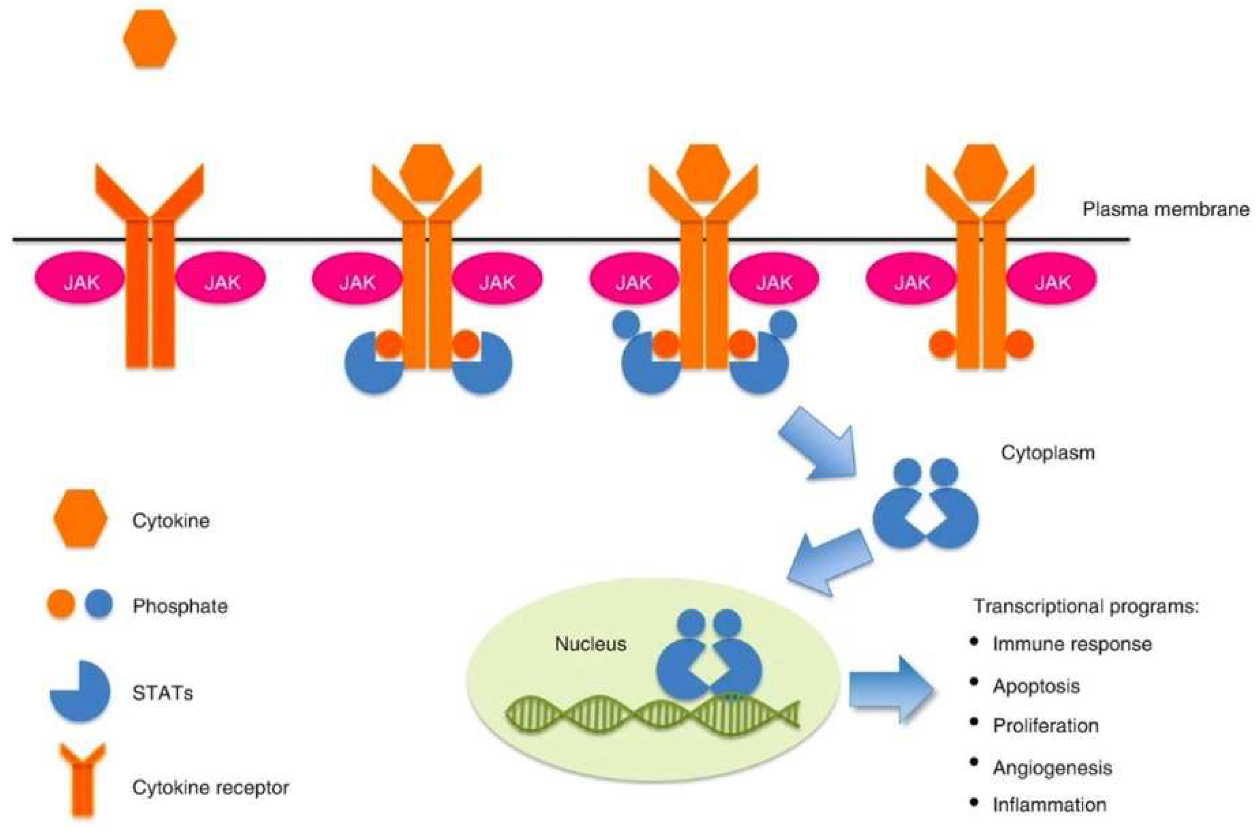
Figure 1. JAK-STAT signaling pathway [3].
Diagnostics 13 01649 g001
Figure 2. Expression of STAT-3 immunostaining: (A) Weak nuclear staining, (B) Moderate nuclear staining, and (C) Strong nuclear staining (400× magnification).
Diagnostics 13 01649 g002
Table 1. Scoring system for STAT3 stain [11].
ScoreExtent of Markers ExpressionIntensity of Positive Score
0No positive cells/HPFNil
1<10% positive cells/HPFWeak
210–30% positive cells/HPFModerate
3>30% positive cells/HPFStrong
Score = Intensity × Extent. (Values: 0 to 9, >3 is high expression; <3 is low expression; 0 is negative). HPF: high power field.
Table 2. Demographic and clinicopathological characteristics of patients (n = 95).
n(%)
Age Group
(years)
≤6054 (mean)51(53.7)
>60 44(46.3)
Ethnic groupMalay 86(90.5)
Non-Malay
- Chinese 7(7.4)
- Indian 1(1.1)
- Others 1(1.1)
GenderFemale 33(34.7)
Male 62(65.3)
SiteNodal 30(31.6)
Extra nodal 47(49.5)
Nodal and extra nodal 18(18.9)
Tumour size (cm)<107 (median)60(63.2)
≥10 35(36.8)
B symptomsAbsent 41(43.2)
Present 54(56.8)
LDH level (U/L)<480 (Normal)720 (median)31(32.6)
≥480 (Elevated) 64(67.4)
Stage of diseaseStage I–II (Early) 28(29.5)
Stage III–IV (Advanced) 67(70.5)
Table 3. NHL histopathological subtypes (n = 95).
n(%)
Type T/Natural Killer cells7(7.4)
B cells88(92.6)
SubtypesIndolent B cell14(14.7)
Follicular lymphoma4(4.2)
Mantle cell lymphoma3(3.2)
Extra nodal Marginal Zone Lymphoma3(3.2)
Lymphoplasmacytic lymphoma1(1.1)
Primary cutaneous follicular centre lymphoma1(1.1)
SLL2(2.1)
Aggressive B cell
DLBCL group
74
61
(77.9)
(64.3)
- DLBCL-NGCB20(21.1)
- DLBCL-GCB15(15.8)
- DLBCL NOS17(17.9)
- DLBCL double expressions7(7.4)
- DLBCL triple expressions2(2.1)
Others aggressive B cell13(14.0)
- PMBL4(4.2)
- Primary DLBCL of CNS3(3.2)
- EBV-positive DLBCL, NOS1(1.1)
- T cell/histiocyte- rich B cell lymphoma1(1.1)
- B-cell lymphoma, unclassifiable, with features intermediate between diffuse large B-cell lymphoma and classic Hodgkin lymphoma1(1.1)
- Plasmablastic lymphoma1(1.1)
- B-lymphoblastic leukaemia/lymphoma1(1.1)
- Burkitt Lymphoma1(1.1)
T cell NHL7(7.4)
T-lymphoblastic leukaemia/lymphoma3(3.2)
ALCL, ALK-positive3(3.2)
Extra nodal Natural Killer/T cell lymphoma, nasal type1(1.1)
Abbreviation: DLBCL: Diffuse large B cell lymphoma; DLBCL-NGCB: Diffuse large B cell lymphoma, Non-Germinal Centre B subtypes; DLBCL-GCB: Diffuse large B cell lymphoma, Germinal Centre B subtypes; NOS: Not Otherwise Specified; CNS: Central Nervous System; PMBL: Primary Mediastinal Large B Cell Lymphoma.
Table 4. The expression of STAT-3 protein in NHL histopathological subtypes (n = 95).
Subtypes STAT-3 Protein Expression
NegativePositive
n (%)n (%)
B cell NHLIndolent B cell5 (5.3)9 (9.5)
Follicular lymphoma2 (2.1)2 (2.1)
Mantle cell lymphoma0 (0.0)3 (3.2)
Extra nodal Marginal Zone Lymphoma1 (1.1)2 (2.1)
Lymphoplasmacytic lymphoma1 (1.1)0 (0.0)
Primary cutaneous follicular centre lymphoma0 (0.0)1 (1.1)
SLL1 (1.1)1 (1.1)
Aggressive B cell13 (13.7)61 (64.2)
1. DLBCL group9 (9.5)52 (54.7)
- DLBCL-NGCB1 (1.1)19 (20.0)
- DLBCL-GCB4 (4.2)11 (11.6)
- DLBCL NOS2 (2.1)15 (15.8)
- DLBCL double expressions2 (2.1)5 (5.3)
- DLBCL triple expressions0 (0.0)2 (2.1)
2. Others aggressive B cell4 (4.2)9 (9.5)
- PMBL1 (1.1)3 (3.2)
- Primary DLBCL of CNS1 (1.1)2 (2.1)
- EBV-positive DLBCL, NOS0 (0.0)1 (1.1)
- T cell/histiocyte- rich B cell lymphoma0 (0.0)1 (1.1)
- B-cell lymphoma, unclassifiable, with features intermediate between diffuse large B-cell lymphoma and classic Hodgkin lymphoma0 (0.0)1 (1.1)
- Plasmablastic lymphoma1 (1.1)0 (0.0)
- B-lymphoblastic leukaemia/lymphoma1 (1.1)0 (0.0)
- Burkitt Lymphoma0 (0.0)1 (1.1)
T cell NHLT cell2 (2.1)5 (5.3)
T-lymphoblastic leukaemia/lymphoma0 (0.0)3 (3.2)
ALCL, ALK-positive2 (2.1)1 (1.1)
Extra nodal Natural Killer/T cell lymphoma, nasal type0 (0.0)1 (1.1)
Total95 (100.0)20 (21.1)75 (78.9)
Abbreviation: DLBCL: Diffuse large B cell lymphoma; DLBCL-NGCB: Diffuse large B cell lymphoma, Non-Germinal Centre B subtypes; DLBCL-GCB: Diffuse large B cell lymphoma, Germinal Centre B subtypes; NOS: Not Otherwise Specified; CNS: Central Nervous System; PMBL: Primary Mediastinal Large B Cell Lymphoma.
Table 5. The association between clinicopathologic characteristics with STAT-3 protein expression in NHL (n = 95).
STAT-3 Protein Expression
NegativePositivep Value
n(%)n(%)
Age group (years)≤6011(55.0)40(53.3)0.894 a
>609(45.0)35(46.7)
Ethnic groupMalay18(90.0)68(90.7)>0.950 b
Non-Malay2(10.0)7(9.3)
GenderFemale7(35.0)26(34.7)0.978 a
Male13(65.0)49(65.3)
SiteNodal3(15.0)27(36.0)0.196 a
Extra nodal12(60.0)35(46.7)
Nodal and extra nodal5(25.0)13(17.3)
Tumour size (cm)<1010(50.0)50(66.7)0.170 a
≥1010(50.0)25(33.3)
B symptomsAbsent11(55.0)30(40.0)0.229 a
Present9(45.0)45(60.0)
LDH level (U/L)<480 (Normal)6(30.0)25(33.3)0.778 a
≥480 (Elevated)14(70.0)50(66.7)
Stage of diseaseStage I-II (Early)7(35.0)21(28.0)0.542 a
Stage III-IV (Advanced)13(65.0)54(72.0)
a Pearson Chi Square test. b Fisher’s exact test.
Table 6. The association between NHL type/subtypes with STAT-3 protein expression (n = 95).
STAT-3 Protein Expression
NegativePositivep Value
n(%)n(%)
TypeT/Natural Killer cells2(10.0)5(6.7)0.636 b
B18(90.0)70(93.3)
SubtypeIndolent B cell5(25.0)9(12.0)0.214 b
Aggressive B cell13(65.0)61(81.3)
T/Natural Killer cells2(10.0)5(6.7)
B cell NHLDLBCL group9(50)52(74.3)0.046 a
Others B cell NHL9(50)18(25.7)
a Pearson Chi Square test. b Fisher’s exact test.
Disclaimer/Publisher’s Note: The statements, opinions and data contained in all publications are solely those of the individual author(s) and contributor(s) and not of MDPI and/or the editor(s). MDPI and/or the editor(s) disclaim responsibility for any injury to people or property resulting from any ideas, methods, instructions or products referred to in the content.