


Usefulness of Chromogenic Media with Fluconazole Supplementation for Presumptive Identification of Candida auris

 , , ,
, , ,  , , and
Abstract
1. Introduction
2. Materials and Methods
3. Results
4. Discussion
Author Contributions
Funding
Institutional Review Board Statement
Informed Consent Statement
Data Availability Statement
Acknowledgments
Conflicts of Interest
References
- Satoh, K.; Makimura, K.; Hasumi, Y.; Nishiyama, Y.; Uchida, K.; Yamaguchi, H. Candida aurissp. nov., a novel ascomycetous yeast isolated from the external ear canal of an inpatient in a Japanese hospital. Microbiol. Immunol. 2009, 53, 41–44. [Google Scholar] [CrossRef]
- Jeffery-Smith, A.; Taori, S.K.; Schelenz, S.; Jeffery, K.; Johnson, E.M.; Borman, A.; Manuel, R.; Brown, C.S. Candida auris: A review of the literature. Clin. Microbiol. Rev. 2017, 31, e00029-17. [Google Scholar] [CrossRef]
- Ruiz-Gaitán, A.; Moret, A.M.; Tasias-Pitarch, M.; Aleixandre-López, A.I.; Martínez-Morel, H.; Calabuig, E.; Salavert-Lletí, M.; Ramírez, P.; López-Hontangas, J.L.; Hagen, F.; et al. An outbreak due to Candida auris with prolonged colonisation and candidaemia in a tertiary care European hospital. Mycoses 2018, 61, 498–505. [Google Scholar] [CrossRef] [PubMed]
- Welsh, R.M.; Bentz, M.L.; Shams, A.; Houston, H.; Lyons, A.; Rose, L.J.; Litvintseva, A.P. Survival, Persistence, and Isolation of the Emerging Multidrug-Resistant Pathogenic Yeast Candida auris on a Plastic Health Care Surface. J. Clin. Microbiol. 2017, 55, 2996–3005. [Google Scholar] [CrossRef] [PubMed]
- Garcia-Bustos, V.; Cabanero-Navalon, M.D.; Ruiz-Saurí, A.; Ruiz-Gaitán, A.C.; Salavert, M.; Tormo, M.Á.; Pemán, J. What Do We Know about Candida auris? State of the Art, Knowledge Gaps, and Future Directions. Microorganisms 2021, 9, 2177. [Google Scholar] [CrossRef] [PubMed]
- Centers for Disease Control. Antibiotic Resistance Threats in the United States, 2019; CDC: Atlanta, Georgia, 2019. [Google Scholar]
- Wang, X.; Bing, J.; Zheng, Q.; Zhang, F.; Liu, J.; Yue, H.; Tao, L.; Du, H.; Wang, Y.; Wang, H.; et al. The first isolate of Candida auris in China: Clinical and biological aspects. Emerg. Microbes Infect. 2018, 7, 1–9. [Google Scholar] [CrossRef]
- Bidaud, A.; Chowdhary, A.; Dannaoui, E. Candida auris: An emerging drug resistant yeast—A mini-review. J. Med. Mycol. 2018, 28, 568–573. [Google Scholar] [CrossRef] [PubMed]
- Prakash, A.; Sharma, C.; Singh, A.; Singh, P.K.; Kumar, A.; Hagen, F.; Govender, N.; Colombo, A.; Meis, J.; Chowdhary, A. Evidence of genotypic diversity among Candida auris isolates by multilocus sequence typing, matrix-assisted laser desorption ionization time-of-flight mass spectrometry and amplified fragment length polymorphism. Clin. Microbiol. Infect. 2016, 22, 277.e1–277.e9. [Google Scholar] [CrossRef]
- Perry, J.D. A Decade of Development of Chromogenic Culture Media for Clinical Microbiology in an Era of Molecular Diagnostics. Clin. Microbiol. Rev. Am. Soc. Microbiol. 2017, 30, 449–479. [Google Scholar] [CrossRef]
- Fasciana, T.; Cortegiani, A.; Ippolito, M.; Giarratano, A.; Di Quattro, O.; Lipari, D.; Graceffa, D.; Giammanco, A. Candida auris: An Overview of How to Screen, Detect, Test and Control This Emerging Pathogen. Antibiotics 2020, 9, 778. [Google Scholar] [CrossRef]
- Das, S.; Singh, S.; Tawde, Y.; Chakrabarti, A.; Rudramurthy, S.M.; Kaur, H.; Shankarnarayan, S.A.; Ghosh, A. A Selective Medium for Isolation and Detection of Candida auris, an Emerging Pathogen. J. Clin. Microbiol. 2021, 59, e00326-20. [Google Scholar] [CrossRef] [PubMed]
- Orenga, S.; James, A.L.; Manafi, M.; Perry, J.D.; Pincus, D.H. Enzymatic substrates in microbiology. J. Microbiol. Methods 2009, 79, 139–155. [Google Scholar] [CrossRef] [PubMed]
- De Angelis, G.; Menchinelli, G.; Torelli, R.; De Carolis, E.; Posteraro, P.; Sanguinetti, M.; Posteraro, B. Different detection capabilities by mycological media for Candida isolates from mono- or dual-species cultures. PLoS ONE 2020, 15, e0226467. [Google Scholar] [CrossRef]
- Tamura, T.; Alshahni, M.M.; Makimura, K. Evaluation of CHROMagarTM Candida Plus chromogenic agar for the presumptive identification of Candida auris. Microbiol. Immunol. 2022, 66, 292–298. [Google Scholar] [CrossRef] [PubMed]
- Borman, A.M.; Fraser, M.; Johnson, E.M. CHROMagarTM Candida Plus: A novel chromogenic agar that permits the rapid identification of Candida auris. Med. Mycol. 2020, 11, 1–6. [Google Scholar] [CrossRef]
- Mulet Bayona, J.V.; Salvador García, C.; Tormo Palop, N.; Gimeno Cardona, C. Evaluation of a novel chromogenic medium for Candida spp. identification and comparison with CHROMagarTM Candida for the detection of Candida auris in surveillance samples. Diagn. Microbiol. Infect. Dis. 2020, 98, 115168. [Google Scholar] [CrossRef]
- Bayona, J.V.M.; García, C.S.; Palop, N.T.; Martín, A.V.; Padrón, C.G.; Rodríguez, J.C.; Pemán, J.; Cardona, C.G. Novel Chromogenic Medium CHROMagarTM Candida Plus for Detection of Candida auris and Other Candida Species from Surveillance and Environmental Samples: A Multicenter Study. J. Fungi 2022, 8, 281. [Google Scholar] [CrossRef]
- Bellemain, E.; Carlsen, T.; Brochmann, C.; Coissac, E.; Taberlet, P.; Kauserud, H. ITS as an environmental DNA barcode for fungi: An in silico approach reveals potential PCR biases. BMC Microbiol. 2010, 10, 189. [Google Scholar] [CrossRef]
- Chowdhary, A.; Sharma, C.; Meis, J.F. Candida auris: A rapidly emerging cause of hospital-acquired multidrug-resistant fungal infections globally. PLoS Pathog. 2017, 13, e1006290. [Google Scholar] [CrossRef]
- de Jong, A.W.; Dieleman, C.; Carbia, M.; Tap, R.M.; Hagen, F. Performance of Two Novel Chromogenic Media for the Identification of Multidrug-Resistant Candida auris Compared with other Commercially Available Formulations. J. Clin. Microbiol. 2021, 59, e03220-20. [Google Scholar] [CrossRef]
- Kumar, A.; Sachu, A.; Mohan, K.; Vinod, V.; Dinesh, K.; Karim, S. Simple low cost differentiation of Candida auris from Candida haemulonii complex using CHROMagar Candida medium supplemented with Pal’s medium. Rev. Iberoam Micol. 2017, 34, 109–111. [Google Scholar] [CrossRef] [PubMed]
- Sasoni, N.; Maidana, M.; Latorre-Rapela, M.G.; Morales-Lopez, S.; Berrio, I.; Gamarra, S.; Garcia-Effron, G. Candida auris and some Candida parapsilosis strains exhibit similar characteristics on CHROMagarTMCandida Plus. Med. Mycol. 2022, 60, myac062. [Google Scholar] [CrossRef] [PubMed]
- Sharma, C.; Kumar, N.; Meis, J.F.; Pandey, R.; Chowdhary, A. Draft Genome Sequence of a Fluconazole-Resistant Candida auris Strain from a Candidemia Patient in India. Genome Announc. 2015, 3, e00722-15. [Google Scholar] [CrossRef] [PubMed]
- Chowdhary, A.; Sharma, C.; Duggal, S.; Agarwal, K.; Prakash, A.; Singh, P.K.; Jain, S.; Kathuria, S.; Randhawa, H.S.; Hagen, F.; et al. New Clonal Strain of Candida auris, Delhi, India. Emerg. Infect. Dis. 2013, 19, 1670–1673. [Google Scholar] [CrossRef]
- Berkow, E.L.; Lockhart, S.R. Fluconazole resistance in Candida species: A current perspective. Infect. Drug Resist. 2017, 10, 237–245. [Google Scholar] [CrossRef]
- Lockhart, S.R.; Etienne, K.A.; Vallabhaneni, S.; Farooqi, J.; Chowdhary, A.; Govender, N.P.; Colombo, A.L.; Calvo, B.; Cuomo, C.A.; Desjardins, C.A.; et al. Simultaneous Emergence of Multidrug-Resistant Candida auris on 3 Continents Confirmed by Whole-Genome Sequencing and Epidemiological Analyses. Clin. Infect. Dis. 2017, 64, 134–140. [Google Scholar] [CrossRef]
- Pino-Calm, B.; de Artola, D.G.M.; Gil-Campesino, H.; Alcoba-Flórez, J. Restriction fragment length polymorphism de las regiones ITS1-ITS2 como método para identificar Candida auris. Rev. Iberoam Micol. 2018, 35, 167–168. [Google Scholar] [CrossRef]
- Bao, J.R.; Master, R.N.; Azad, K.N.; Schwab, D.A.; Clark, R.B.; Jones, R.S.; Moore, E.C.; Shier, K.L. Rapid, Accurate Identification of Candida auris by Using a Novel Matrix-Assisted Laser Desorption Ionization–Time of Flight Mass Spectrometry (MALDI-TOF MS) Database (Library). J. Clin. Microbiol. 2018, 56, e01700-17. [Google Scholar] [CrossRef]
- Mahmoudi, S.; Afshari, S.A.K.; Gharehbolagh, S.A.; Mirhendi, H.; Makimura, K. Methods for identification of Candida auris, the yeast of global public health concern: A review. J. Med. Mycol. 2019, 29, 174–179. [Google Scholar] [CrossRef]


| Mixed Culture/Organism | Recovered Colonies (n) | |||
|---|---|---|---|---|
| CHROMagarTM Candida Plus | HiCromeTM Candida | CHROMagarTM Candida | CHROMagar Candida Plus Fluconazole | |
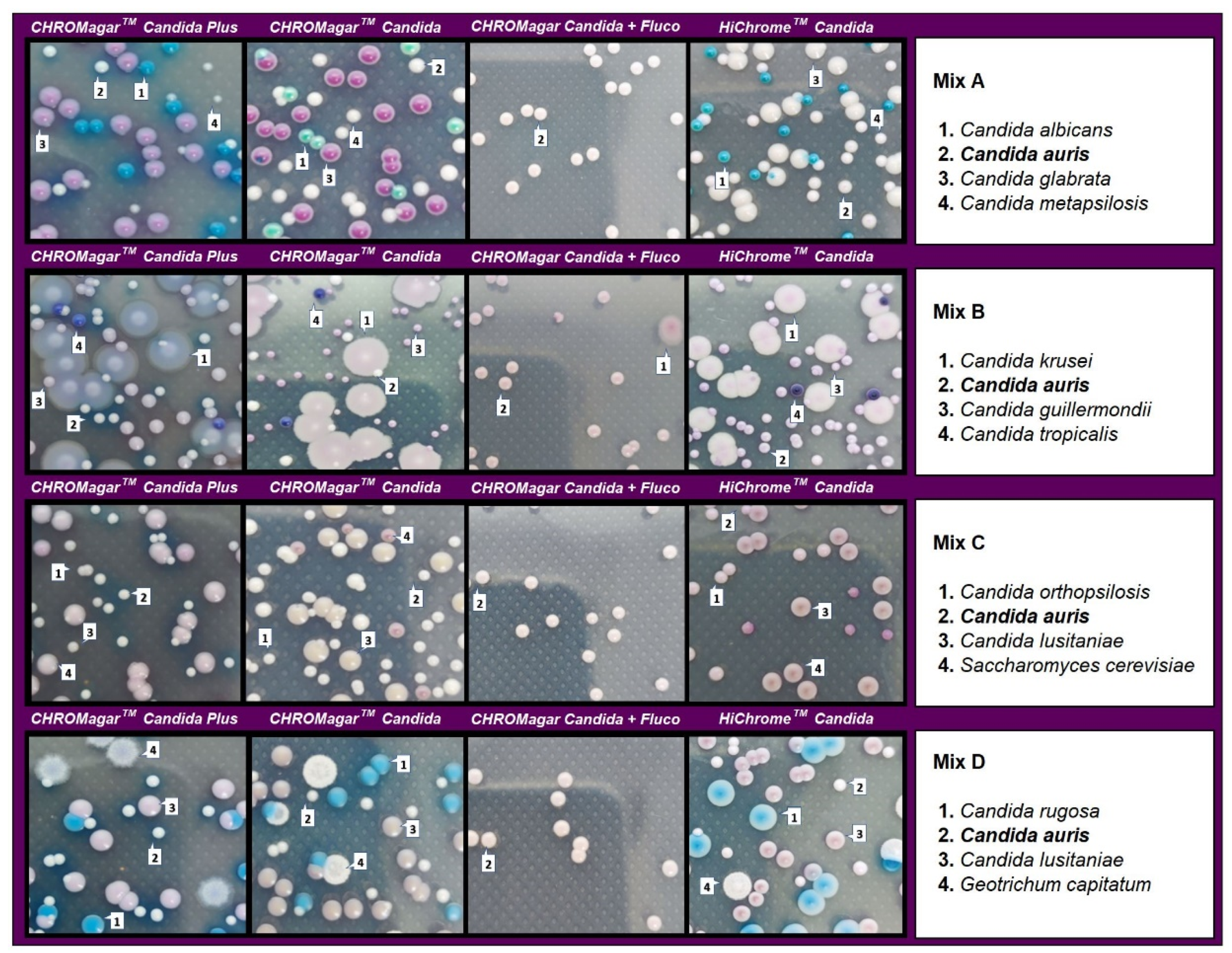
| Mix A | ||||
| C. auris | 142 | 74 | 144 | 112 |
| C. glabrata | 137 | 134 | 137 | NG |
| C. metapsilosis | 32 | 85 | 30 | NG |
| C. albicans | 65 | 65 | 74 | NG |
| Total | 376 | 358 | 385 | 112 |
| C. auris recovered vs. total (%) | 37.77 | 20.67 | 37.40 | 100 |
| Mix B | ||||
| C. auris | 100 | * 325 | 81 | 124 |
| C. guillermondii | 213 | 166 | NG | |
| C. tropicalis | 21 | 37 | 13 | NG |
| C. krusei | 89 | 77 | 67 | 3 |
| Total | 423 | 114 | 327 | 127 |
| C. auris recovered vs. total (%) | 23.64 | ND | 24.77 | 97.64 |
| Mix C | ||||
| C. auris | 105 | 122 | 107 | 103 |
| C. lusitaniae | 108 | 107 | 108 | NG |
| S. cerevisiae | 39 | 35 | 28 | NG |
| C. orthopsilosis | 85 | 20 | 70 | NG |
| Total | 337 | 284 | 313 | 103 |
| C. auris recovered vs. total (%) | 31.16 | 42.96 | 34.19 | 100 |
| Mix D | ||||
| C. auris | 105 | 85 | 72 | 74 |
| C. rugosa | 57 | 57 | 35 | NG |
| C. lusitaniae | 96 | 49 | 130 | NG |
| G. capitatum | 37 | 24 | 19 | NG |
| Total | 295 | 215 | 256 | 74 |
| C. auris recovered vs. total (%) | 35.59 | 39.53 | 28.13 | 100 |
Disclaimer/Publisher’s Note: The statements, opinions and data contained in all publications are solely those of the individual author(s) and contributor(s) and not of MDPI and/or the editor(s). MDPI and/or the editor(s) disclaim responsibility for any injury to people or property resulting from any ideas, methods, instructions or products referred to in the content. |
© 2023 by the authors. Licensee MDPI, Basel, Switzerland. This article is an open access article distributed under the terms and conditions of the Creative Commons Attribution (CC BY) license (https://creativecommons.org/licenses/by/4.0/).
Share and Cite
Ruiz-Gaitán, A.; Sigona-Giangreco, I.; Pérez-Royo, J.M.; Garcia-Bustos, V.; García-Hita, M.; Valentín-Gómez, E.; Almaraz, S.G.; de Groot, P.W.J.; Pemán, J. Usefulness of Chromogenic Media with Fluconazole Supplementation for Presumptive Identification of Candida auris. Diagnostics 2023, 13, 231. https://doi.org/10.3390/diagnostics13020231
Ruiz-Gaitán A, Sigona-Giangreco I, Pérez-Royo JM, Garcia-Bustos V, García-Hita M, Valentín-Gómez E, Almaraz SG, de Groot PWJ, Pemán J. Usefulness of Chromogenic Media with Fluconazole Supplementation for Presumptive Identification of Candida auris. Diagnostics. 2023; 13(2):231. https://doi.org/10.3390/diagnostics13020231
Chicago/Turabian StyleRuiz-Gaitán, Alba, Ignacio Sigona-Giangreco, José Manuel Pérez-Royo, Victor Garcia-Bustos, Marta García-Hita, Eulogio Valentín-Gómez, Salvador Giner Almaraz, Piet W. J. de Groot, and Javier Pemán. 2023. "Usefulness of Chromogenic Media with Fluconazole Supplementation for Presumptive Identification of Candida auris" Diagnostics 13, no. 2: 231. https://doi.org/10.3390/diagnostics13020231
APA StyleRuiz-Gaitán, A., Sigona-Giangreco, I., Pérez-Royo, J. M., Garcia-Bustos, V., García-Hita, M., Valentín-Gómez, E., Almaraz, S. G., de Groot, P. W. J., & Pemán, J. (2023). Usefulness of Chromogenic Media with Fluconazole Supplementation for Presumptive Identification of Candida auris. Diagnostics, 13(2), 231. https://doi.org/10.3390/diagnostics13020231