
Optical Coherence Tomography as a Valuable Tool for the Evaluation of Cutaneous Kaposi Sarcoma Treated with Imiquimod 5% Cream

 ,
, 
 ,
,  and
and 
Abstract
:1. Introduction
2. Case Presentation
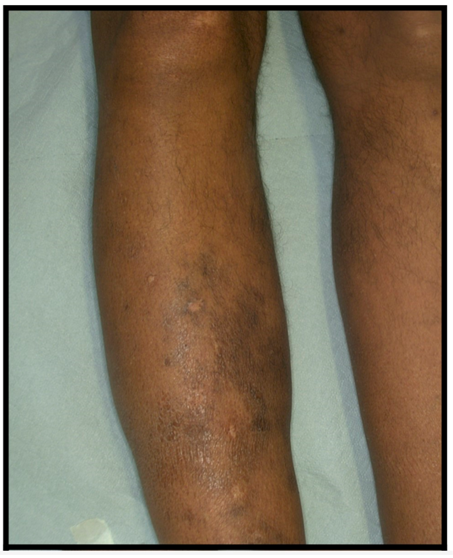
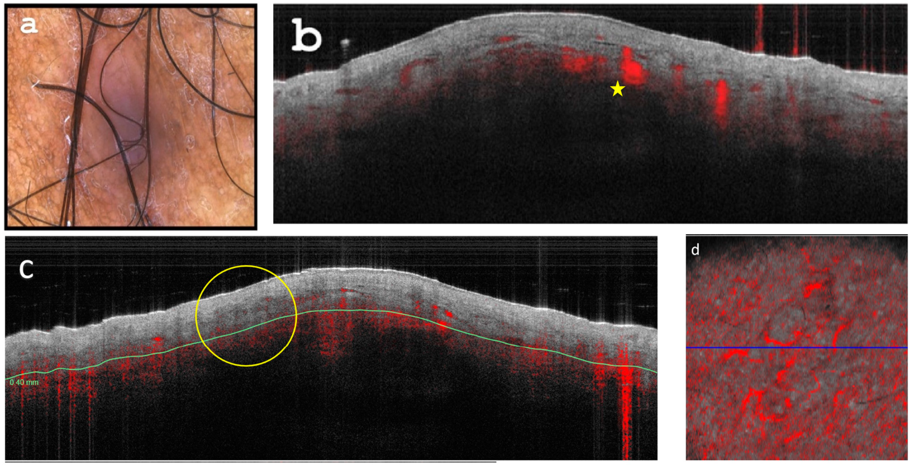
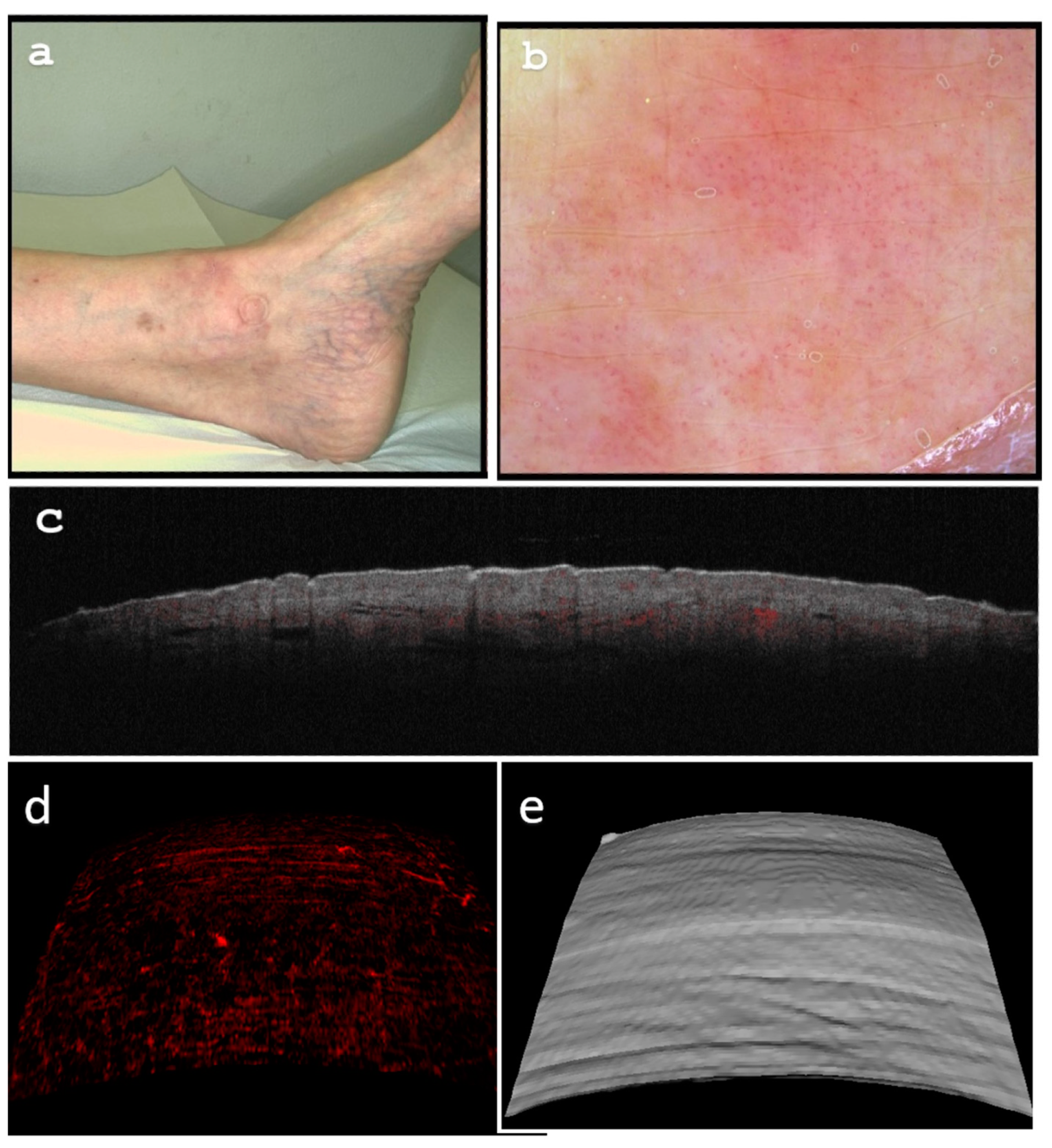
2.1. Case Report 1
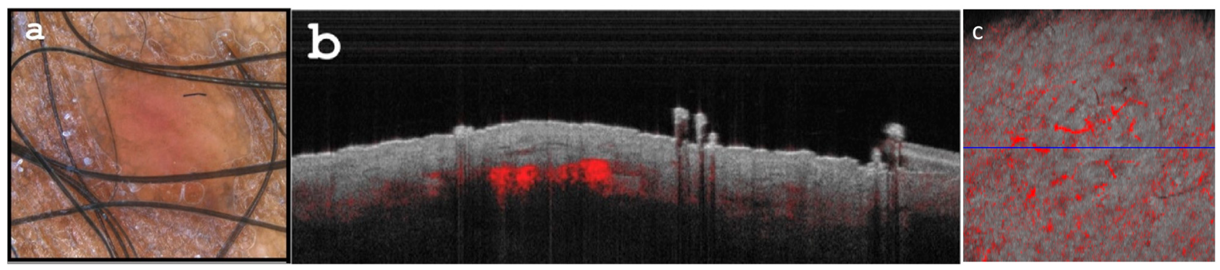
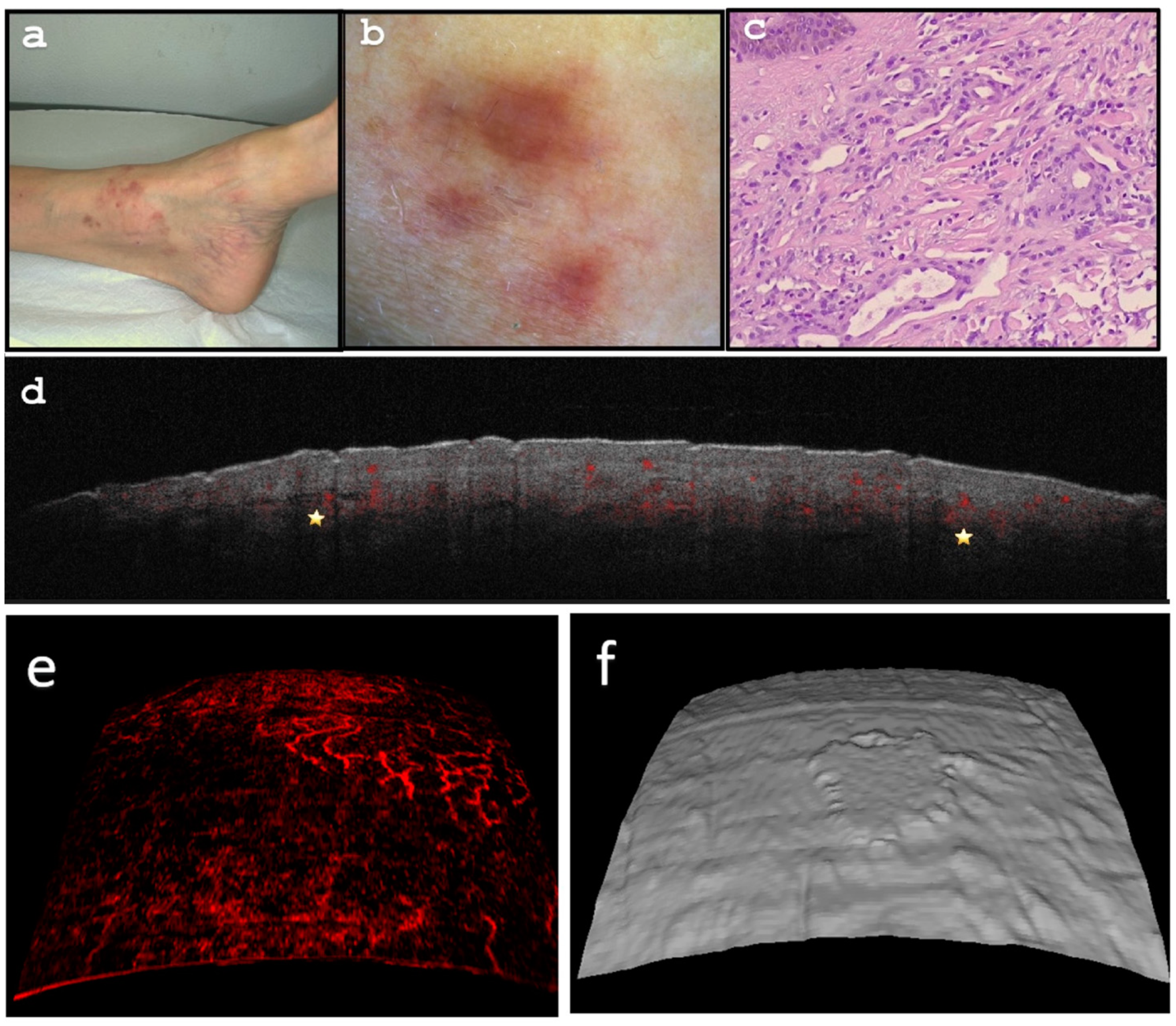
2.2. Case Report 2
3. Discussion
4. Conclusions
Author Contributions
Funding
Institutional Review Board Statement
Informed Consent Statement
Data Availability Statement
Acknowledgments
Conflicts of Interest
References
- Etemad, S.A.; Dewan, A.K. Kaposi sarcoma updates. Dermatol. Clin. 2019, 37, 505–517. [Google Scholar] [CrossRef] [PubMed]
- Buonaguro, F.; Tornesello, M.; Buonaguro, L.; Satriano, R.; Ruocco, E.; Castello, G.; Ruocco, V. Kaposi’s sarcoma: Aetiopathogenesis, histology and clinical features. J. Eur. Acad. Dermatol. Venereol. 2003, 17, 138–154. [Google Scholar] [CrossRef] [PubMed]
- Hu, S.S.; Ke, C.L.; Lee, C.H.; Wu, C.S.; Chen, G.S.; Cheng, S.T. Dermoscopy of Kaposi’s sarcoma: Areas exhibiting the multicoloured ‘rainbow pattern’. J. Eur. Acad. Dermatol. Venereol. 2009, 23, 1128–1132. [Google Scholar] [CrossRef] [PubMed]
- Chang, Y.; Cesarman, E.; Pessin, M.S.; Lee, F.; Culpepper, J.; Knowles, D.M.; Moore, P.S. Identification of herpesvirus-like DNA sequences in AIDS-sssociated kaposi’s sarcoma. Science 1994, 266, 1865–1869. [Google Scholar] [CrossRef] [PubMed]
- Hengge, U.R.; Ruzicka, T.; Tyring, S.K.; Stuschke, M.; Roggendorf, M.; Schwartz, R.A.; Seeber, S. Update on Kaposi’s sarcoma and other HHV8 associated diseases. Part 2: Pathogenesis, Castleman’s disease, and pleural effusion lymphoma. Lancet Infect. Dis. 2002, 2, 344–352. [Google Scholar] [CrossRef] [PubMed]
- Hengge, U.R.; Ruzicka, T.; Tyring, S.K.; Stuschke, M.; Roggendorf, M.; Schwartz, R.A.; Seeber, S. Update on Kaposi’s sarcoma and other HHV8 associated diseases. Part 1: Epidemiology, environmental predispositions, clinical manifestations, and therapy. Lancet Infect. Dis. 2002, 2, 281–292. [Google Scholar] [CrossRef]
- Iscovich, J.; Boffetta, P.; Franceschi, S.; Azizi, E.; Sarid, R. Classic Kaposi sarcoma: Epidemiology and risk factors. Cancer 2000, 88, 500–517. [Google Scholar] [CrossRef]
- Kestens, L.; Melbye, M.; Biggar, R.J.; Stevens, W.J.; Piot, P.; De Muynck, A.; Taelman, H.; De Feyter, M.; Paluku, L.; Gigase, P.L. Endemic African Kaposi’s sarcoma is not associated with immunodeficiency. Int. J. Cancer 1985, 36, 49–54. [Google Scholar] [CrossRef]
- Amerson, E.; Woodruff, C.M.; Forrestel, A.; Wenger, M.; McCalmont, T.; LeBoit, P.; Maurer, T.; Laker-Oketta, M.; Muyindike, W.; Bwana, M. Accuracy of clinical suspicion and pathologic diagnosis of Kaposi sarcoma in East Africa. J. Acquir. Immune Defic. Syndr. 2016, 71, 295. [Google Scholar] [CrossRef]
- Dupin, N.; Fisher, C.; Kellam, P.; Ariad, S.; Tulliez, M.; Franck, N.; Van Marck, E.; Salmon, D.; Gorin, I.; Escande, J.-P. Distribution of human herpesvirus-8 latently infected cells in Kaposi’s sarcoma, multicentric Castleman’s disease, and primary effusion lymphoma. Proc. Natl. Acad. Sci. USA 1999, 96, 4546–4551. [Google Scholar] [CrossRef]
- Schwartz, E.J.; Dorfman, R.F.; Kohler, S. Human herpesvirus-8 latent nuclear antigen-1 expression in endemic Kaposi sarcoma: An immunohistochemical study of 16 cases. Am. J. Surg. Pathol. 2003, 27, 1546–1550. [Google Scholar] [CrossRef] [PubMed]
- Patel, R.M.; Goldblum, J.R.; Hsi, E.D. Immunohistochemical detection of human herpes virus-8 latent nuclear antigen-1 is useful in the diagnosis of Kaposi sarcoma. Mod. Pathol. 2004, 17, 456–460. [Google Scholar] [CrossRef]
- Cheuk, W.; Wong, K.O.; Wong, C.S.; Dinkel, J.; Ben-Dor, D.; Chan, J.K. Immunostaining for human herpesvirus 8 latent nuclear antigen-1 helps distinguish Kaposi sarcoma from its mimickers. Am. J. Clin. Pathol. 2004, 121, 335–342. [Google Scholar] [CrossRef] [PubMed]
- Ramirez, J.A.; Laskin, W.B.; Guitart, J. Lymphangioma-like Kaposi sarcoma. J. Cutan. Pathol. 2005, 32, 286–292. [Google Scholar] [CrossRef]
- Barbato, F.; Carrera, C.; Ferrara, G.; Guilabert, A.; Massi, D.; Moreno-Romero, J.A.; Munoz-Santos, C.; Petrillo, G.; Segura, S.; Soyer, H.P. Dermoscopy improves accuracy of primary care physicians to triage lesions suggestive of skin cancer. J. Clin. Oncol. 2006, 24, 1877–1882. [Google Scholar]
- Rajadhyaksha, M.; Grossman, M.; Esterowitz, D.; Webb, R.H.; Anderson, R.R. In vivo confocal scanning laser microscopy of human skin: Melanin provides strong contrast. J. Investig. Dermatol. 1995, 104, 946–952. [Google Scholar] [CrossRef] [PubMed]
- Olsen, J.; Themstrup, L.; Jemec, G. Optical coherence tomography in dermatology. G. Ital. Dermatol. Venereol. 2015, 150, 603–615. [Google Scholar] [PubMed]
- Argenziano, G.; Albertini, G.; Castagnetti, F.; De Pace, B.; Di Lernia, V.; Longo, C.; Pellacani, G.; Piana, S.; Ricci, C.; Zalaudek, I. Early diagnosis of melanoma: What is the impact of dermoscopy? Dermatol. Ther. 2012, 25, 403–409. [Google Scholar] [CrossRef] [PubMed]
- Banerjee, S.; Singh, S.K.; Chakraborty, A.; Das, A.; Bag, R. Melanoma Diagnosis Using Deep Learning and Fuzzy Logic. Diagnostics 2020, 10, 577. [Google Scholar] [CrossRef] [PubMed]
- Corsetti Grazziotin, T.; Cota, C.; Bortoli Buffon, R.; Araújo Pinto, L.; Latini, A.; Ardigò, M. Preliminary evaluation of in vivo reflectance confocal microscopy features of Kaposi’s sarcoma. Dermatology 2010, 220, 346–354. [Google Scholar] [CrossRef]
- Fercher, A.; Mengedoht, K.; Werner, W. Eye-length measurement by interferometry with partially coherent light. Opt. Lett. 1988, 13, 186–188. [Google Scholar] [CrossRef]
- Huang, D.; Swanson, E.A.; Lin, C.P.; Schuman, J.S.; Stinson, W.G.; Chang, W.; Hee, M.R.; Flotte, T.; Gregory, K.; Puliafito, C.A. Optical coherence tomography. Science 1991, 254, 1178–1181. [Google Scholar] [CrossRef] [PubMed]
- Calin, M.A.; Parasca, S.V.; Savastru, R.; Calin, M.R.; Dontu, S. Optical techniques for the noninvasive diagnosis of skin cancer. J. Cancer Res. Clin. Oncol. 2013, 139, 1083–1104. [Google Scholar] [CrossRef] [PubMed]
- Marghoob, A.A.; Swindle, L.D.; Moricz, C.Z.; Negron, F.A.S.; Slue, B.; Halpern, A.C.; Kopf, A.W. Instruments and new technologies for the in vivo diagnosis of melanoma. J. Am. Acad. Dermatol. 2003, 49, 777–797. [Google Scholar] [CrossRef]
- Lebbe, C.; Garbe, C.; Stratigos, A.J.; Harwood, C.; Peris, K.; Del Marmol, V.; Malvehy, J.; Zalaudek, I.; Hoeller, C.; Dummer, R. Diagnosis and treatment of Kaposi’s sarcoma: European consensus-based interdisciplinary guideline (EDF/EADO/EORTC). Eur. J. Cancer 2019, 114, 117–127. [Google Scholar] [CrossRef] [PubMed]
- Cantisani, C.; Lazic, T.; Richetta, A.G.; Clerico, R.; Mattozzi, C.; Calvieri, S. Imiquimod 5% cream use in dermatology, side effects and recent patents. Recent Pat. Inflamm. Allergy Drug Discov. 2012, 6, 65–69. [Google Scholar] [CrossRef]
- Odyakmaz Demirsoy, E.; Bayramgürler, D.; Çağlayan, Ç.; Bilen, N.; Şikar Aktürk, A.; Kıran, R. Imiquimod 5% cream versus cryotherapy in classic kaposi sarcoma. J. Cutan. Med. Surg. 2019, 23, 488–495. [Google Scholar] [CrossRef]
- Htet, K.Z.; Waul, M.A.; Leslie, K.S. Topical treatments for Kaposi sarcoma: A systematic review. Ski. Health Dis. 2022, 2, e107. [Google Scholar] [CrossRef]
- Schartz, N.E.C.; Chevret, S.; Paz, C.; Kerob, D.; Verola, O.; Morel, P.; Lebbé, C. Imiquimod 5% cream for treatment of HIV-negative Kaposi’s sarcoma skin lesions: A phase I to II, open-label trial in 17 patients. J. Am. Acad. Dermatol. 2008, 58, 585–591. [Google Scholar] [CrossRef]
- Forsea, A.-M.; Carstea, E.M.; Ghervase, L.; Giurcaneanu, C.; Pavelescu, G. Clinical application of optical coherence tomography for the imaging of non–melanocytic cutaneous tumors: A pilot multi–modal study. J. Med. Life 2010, 3, 381. [Google Scholar]
- Markowitz, O.; Schwartz, M.; Feldman, E.; Bienenfeld, A.; Bieber, A.K.; Ellis, J.; Alapati, U.; Lebwohl, M.; Siegel, D.M. Evaluation of optical coherence tomography as a means of identifying earlier stage basal cell carcinomas while reducing the use of diagnostic biopsy. J. Clin. Aesthetic Dermatol. 2015, 8, 14. [Google Scholar]
- Di Stefani, A.; Fionda, B.; Cappilli, S.; Tagliaferri, L.; Peris, K. Extramammary Paget disease imaged by LC-OCT and treated with radiotherapy. Int. J. Dermatol. 2023, 62, e503–e505. [Google Scholar] [CrossRef] [PubMed]
- Verzì, A.E.; Micali, G.; Lacarrubba, F. Line-field confocal optical coherence tomography may enhance monitoring of superficial basal cell carcinoma treated with imiquimod 5% cream: A pilot study. Cancers 2021, 13, 4913. [Google Scholar] [CrossRef] [PubMed]
- Cheng, H.; Guitera, P. Systematic review of optical coherence tomography usage in the diagnosis and management of basal cell carcinoma. Br. J. Dermatol. 2015, 173, 1371–1380. [Google Scholar] [CrossRef]
- Reddy, N.; Nguyen, B. The utility of optical coherence tomography for diagnosis of basal cell carcinoma: A quantitative review. Br. J. Dermatol. 2019, 180, 475–483. [Google Scholar] [CrossRef] [PubMed]
- Cappilli, S.; Suppa, M.; Ricci, C.; Del Marmol, V.; Peris, K.; Di Stefani, A. Line-field confocal optical coherence tomography of cutaneous vascular lesions: Morphological assessment and histopathological correlations. J. Eur. Acad. Dermatol. Venereol. 2023, 37, 1664–1668. [Google Scholar] [CrossRef]
Disclaimer/Publisher’s Note: The statements, opinions and data contained in all publications are solely those of the individual author(s) and contributor(s) and not of MDPI and/or the editor(s). MDPI and/or the editor(s) disclaim responsibility for any injury to people or property resulting from any ideas, methods, instructions or products referred to in the content. |
© 2023 by the authors. Licensee MDPI, Basel, Switzerland. This article is an open access article distributed under the terms and conditions of the Creative Commons Attribution (CC BY) license (https://creativecommons.org/licenses/by/4.0/).
Share and Cite
Cantisani, C.; Baja, A.-V.; Gargano, L.; Rossi, G.; Ardigò, M.; Soda, G.; Boostani, M.; Kiss, N.; Pellacani, G. Optical Coherence Tomography as a Valuable Tool for the Evaluation of Cutaneous Kaposi Sarcoma Treated with Imiquimod 5% Cream. Diagnostics 2023, 13, 2901. https://doi.org/10.3390/diagnostics13182901
Cantisani C, Baja A-V, Gargano L, Rossi G, Ardigò M, Soda G, Boostani M, Kiss N, Pellacani G. Optical Coherence Tomography as a Valuable Tool for the Evaluation of Cutaneous Kaposi Sarcoma Treated with Imiquimod 5% Cream. Diagnostics. 2023; 13(18):2901. https://doi.org/10.3390/diagnostics13182901
Chicago/Turabian StyleCantisani, Carmen, Alexandru-Vasile Baja, Luca Gargano, Giovanni Rossi, Marco Ardigò, Giuseppe Soda, Mehdi Boostani, Norbert Kiss, and Giovanni Pellacani. 2023. "Optical Coherence Tomography as a Valuable Tool for the Evaluation of Cutaneous Kaposi Sarcoma Treated with Imiquimod 5% Cream" Diagnostics 13, no. 18: 2901. https://doi.org/10.3390/diagnostics13182901
APA StyleCantisani, C., Baja, A.-V., Gargano, L., Rossi, G., Ardigò, M., Soda, G., Boostani, M., Kiss, N., & Pellacani, G. (2023). Optical Coherence Tomography as a Valuable Tool for the Evaluation of Cutaneous Kaposi Sarcoma Treated with Imiquimod 5% Cream. Diagnostics, 13(18), 2901. https://doi.org/10.3390/diagnostics13182901