Article

Evaluation of a Sample-to-Result POCKIT Central SARS-CoV-2 PCR System

1
Department of Veterinary Diagnostic and Production Animal Medicine, College of Veterinary Medicine, Iowa State University, Ames, IA 50011, USA
2
GeneReach Biotechnology Corporation, Taichung 407, Taiwan
3
State Hygienic Laboratory, University of Iowa, Coralville, IA 52241, USA
*
Author to whom correspondence should be addressed.
Diagnostics 2023, 13(13), 2219; https://doi.org/10.3390/diagnostics13132219
Submission received: 25 April 2023 / Revised: 15 June 2023 / Accepted: 23 June 2023 / Published: 29 June 2023
(This article belongs to the Section Diagnostic Microbiology and Infectious Disease)

Abstract

:
The emergence of COVID-19 has caused unprecedented impacts on global public health and many other aspects. Meanwhile, many types of methods have been developed to detect the causative agent, SARS-CoV-2; this has greatly advanced the technologies in the diagnostic field. Here, we describe the development and validation of a sample-in-result-out POCKIT Central SARS-CoV-2 PCR system for detecting SARS-CoV-2 in comparison with a commercial reference real-time RT-PCR assay (TaqPath COVID-19 Combo Kit). Both assays were specific and did not cross-react with non-SARS-CoV-2 agents. Both assays were able to detect various SARS-CoV-2 strains including some variants. Based on testing serial dilutions of SARS-CoV-2 USA-WA1/2020 isolate, the limit of detection was 0.8 TCID50/mL (1.87 × 10³ genomic copies/mL) for POCKIT Central SARS-CoV-2 PCR and 0.16 TCID50/mL (3.75 × 10² genomic copies/mL) for the reference PCR. Subsequently, 183 clinical samples were tested by both assays and the diagnostic sensitivity, specificity, and agreement of the POCKIT Central SARS-CoV-2 PCR were 91.7%, 100%, and 94.0%, respectively, when compared to the reference PCR. The compact sample-to-result POCKIT Central SARS-CoV-2 PCR system is a simplified and efficient point-of-care tool for SARS-CoV-2 detection. In addition, this platform can be readily adapted to detect other human and animal viruses.

1. Introduction

The pandemic of novel coronavirus disease (COVID-19), caused by severe acute respiratory syndrome coronavirus 2 (SARS-CoV-2), has resulted in a global public health crisis and severely affected the global economy [1,2,3]. As of 12 April 2023, according to the World Health Organization (WHO) report, there have been >762 million confirmed cases and roughly 6.9 million deaths caused by COVID-19 globally. The causative agent SARS-CoV-2 is a positive-stranded RNA virus belonging to the genus Betacoronavirus of the family Coronaviridae. The SARS-CoV-2 genome in the range of 29.8–29.9 kb in length is composed of multiple open reading frames (ORF). These include ORF1a and ORF1b encoding the polyprotein 1a and polyprotein 1ab which are further processed into 16 non-structural proteins (nsp 1–16), ORFs encoding structural proteins spike (S), envelope (E), membrane (M), and nucleocapsid (N), and some ORFs (e.g., ORFs 3a, 3b, 3c, 3d, 6, 7a, 7b, 8, 9b, 9c, and 10) encoding accessory proteins [4,5].
Coronaviruses are well known for high mutation and recombination rates which drive their genetic diversification [6,7]. In the period of over three years (late 2019 to early 2023), the SARS-CoV-2 virus evolved while rapidly spreading in the human population, resulting in the emergence of various virus variants with different characteristics compared to the ancestral/original strains [8]. Different nomenclature systems have been proposed to describe genetically diverse SARS-CoV-2 strains, and some most notable classification systems include GISAID (www.gisaid.org), Nextstrain (clade.nextstrain.org), and Pango lineages [9,10]. The original SARS-CoV-2 strains in early outbreaks included two Pango lineages A and B [9]. However, with the rapid evolution of SARS-CoV-2, a few thousand Pango lineages have been described (cov-lineages.org). For easy and efficient communications, from May 2021, WHO began to use the Greek alphabet to label the key SARS-CoV-2 variants such as the Alpha (B.1.1.7), Beta (B.1.351), Gamma (P.1, i.e., B.1.1.28.1), Delta (B.1.617.2), Kappa (B.1.617.1), Epsilon (B.1.427 and B.1.429), Eta (B.1.525), Iota (B.1.526), Lambda (C.37), Mu (B.1.621), Omicron (B.1.1.529) variants, and so on (www.who.int; accessed on 15 March 2023).
Rapid, sensitive, and specific methods to detect SARS-CoV-2 and identify the infected individuals are critical to better monitor the infection and limit the spread of COVID-19 [11]. Many advances have been made in laboratory testing for SARS-CoV-2 in the past few years. Molecular diagnostic tools (e.g., real-time PCR, droplet-digital PCR [ddPCR], loop-mediated isothermal amplification [LAMP] assays, and genome sequencing approaches, etc.), rapid antigen tests, antibody tests, and various methods in other formats (e.g., microfluidic and/or biosensor methods) have been developed for detecting or confirming SARS-CoV-2 infection [11,12,13,14,15,16,17,18,19]. Nucleic acid extraction followed by real-time RT-PCR conducted in the central laboratory is considered the gold-standard method for SARS-CoV-2 detection. However, the accuracy of this assay relies on trained personnel and it also requires time-consuming laboratory processes with complex and expensive equipment [11,20]. For these reasons, point-of-care (POC) testing is needed in some resource-limited areas and/or in nursing homes or long-term care facilities [20,21].
Insulated isothermal PCR (iiPCR) is a fluorescent hydrolysis probe-based technology that provides isothermal heating at the bottom of special capillary tubes to induce thermal convection and temperature gradient in an insulated environment to drive PCR reaction within a relatively short amount of time [22,23]. The POCKIT Nucleic Acid Analyzer series, which is a user-friendly iiPCR system (GeneReach Biotech), provide various iiPCR assays for detecting different human pathogens, including malaria, Zika virus, Middle East respiratory syndrome coronavirus (MERS-CoV), and dengue virus [24,25,26,27,28]. The new-generation POCKIT Central system automatically completes both nucleic acid extraction and iiPCR amplification and eventually generates a qualitative result. Simply, it is a sample-in–answer-out equipment that is easy to operate and could be a tool for point-of-care testing.
In 2020, due to the urgent need for SARS-CoV-2 PCR testing, the POCKIT Central SARS-CoV-2 PCR was clinically evaluated on 100 oropharyngeal swab samples [29] in order to apply for emergency use authorization from various agencies. However, in Chang et al. paper [29], the POCKIT Central SARS-CoV-2 PCR was not validated for analytical sensitivity (by testing serial dilutions of a SARS-CoV-2 isolate with known concentration) and specificity (by testing against other non-SARS-CoV-2 pathogens) and was not tested for its capability to detect various SARS-CoV-2 variants. In this study, we thoroughly evaluated the accuracy of the POCKIT Central SARS-CoV-2 orf1ab RT-iiPCR assay in comparison with a commercial TaqPath COVID-19 Combo Kit (Thermo Fisher Scientific).

2. Materials and Methods

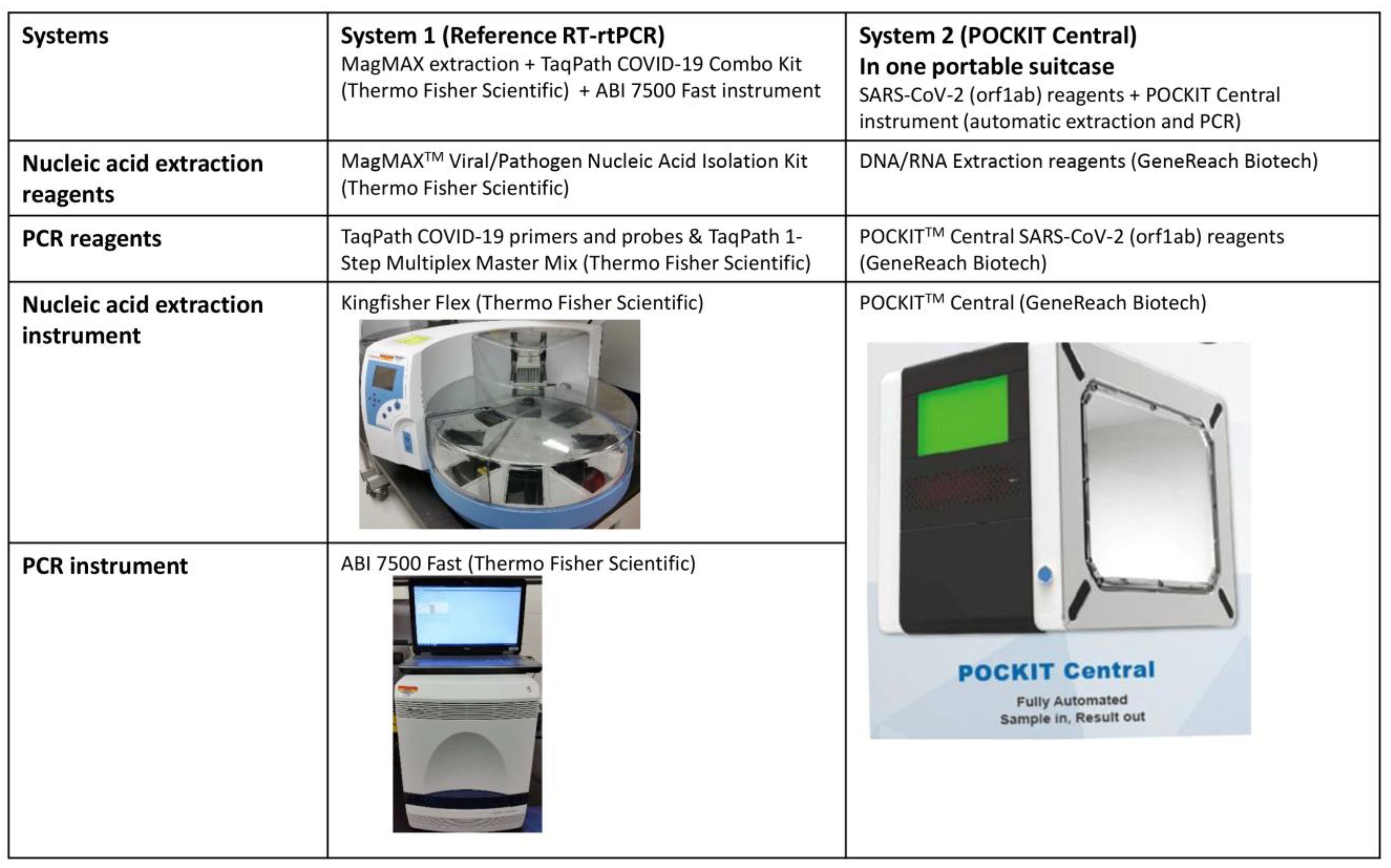
2.1. POCKIT Central SARS-CoV-2 Orf1ab RT-iiPCR System

The POCKIT Central SARS-CoV-2 RT-iiPCR system (GeneReach Biotech, Taichung, Taiwan) used in this study included nucleic acid extraction reagents, SARS-CoV-2 (orf1ab) Premix reagent, and POCKIT Central instrument that allowed to test 8 samples in one run (Figure 1). The PCR assay was designed to target the conserved orf1ab genomic region of SARS-CoV-2 together with a specific Internal Control. The Internal Control template is a plasmid containing a fragment of nucleotides that was artificially designed and synthesized and is not present in any analyzed pathogens or host species. The Internal Control template and the corresponding primers and probe were included in the Premix to monitor the performance of the PCR system. The SARS-CoV-2 primer and probe sequences are: forward primer (ORF1ab-F) 5′-CCCTGTGGGTTTTACACTTAA-3′, reverse primer (ORF1ab-R) 5′-ACGATTGTGCATCAGCTGA-3′, probe (ORF1ab-P) 5′-FAM-CCGTCTGCGGTATGTGGAAAGGTTATGG-BHQ1-3′. The amplicon size is 119 base pairs with the location at nucleotides 13,331–13,449 according to the isolate SARS-CoV-2/human/USA/CA-IGI-0045/2020.
There was one set of Extraction Cartridges, Transfer Cartridges, and SARS-CoV-2 Premix for each sample. Both the nucleic acid extraction and PCR amplification were conducted in one POCKIT Central instrument following the manufacturer’s protocol. Briefly, the Premix vials were snapped into the Transfer Cartridges and placed in the designated slots of the POCKIT Central instrument. Samples (200 µL for each) were loaded into the Extraction Cartridges which were placed in the designated slots of the POCKIT Central instrument. After that, a button was pressed to start the run and it took ~85 min to obtain the results. POCKIT Central SARS-CoV-2 (orf1ab) P(+) Control Reagent (GeneReach Biotech, Taichung, Taiwan) was used as an external positive control and a provided Negative Control Reagent was included.
POCKIT Central system automatically completed nucleic acid extraction, iiPCR amplification, and qualitative result interpretation sequentially. Fluorescent signals at 520 nm and 550 nm were generated, respectively, when the targeted SARS-CoV-2 sequences and the internal control sequences were amplified. For results to be valid, the negative control and the SARS-CoV-2 positive control included in the kit must be correct and all of the samples must be Internal Control positive regardless of the SARS-CoV-2 status in the sample.

2.2. The Reference TaqPath COVID-19 Real-Time RT-PCR System

In the reference TaqPath COVID-19 real-time RT-PCR system, nucleic acid extraction and PCR reaction were conducted in two separate steps (Figure 1).
A MagMAX Viral/Pathogen Nucleic Acid Isolation Kit (Thermo Fisher Scientific, Waltham, MA, USA) and a Kingfisher Flex instrument (Thermo Fisher Scientific, Waltham, MA, USA) were used to extract nucleic acids from viral pathogens and clinical samples. First, 500 μL of Wash Buffer for Wash 1 Plate, 1000 μL of 80% Ethanol for Wash 2 Plate, and 50 μL of Elution Solution for Elution Plate were prepared per well. Then the Binding Bead Mix with 265 μL of Binding Solution and 10 μL of Total Nucleic Acid Magnetic Beads per reaction was prepared. Next, the sample plate was prepared with 5 μL of Proteinase K, 200 μL of sample, and 275 μL of the Binding Bead Mix per well. Subsequently, 5 μL of MS2 Phage Control (Thermo Fisher Scientific) was added to each sample well and the Negative Control well. Eventually, all of the prepared plates were loaded into a Kingfisher Flex instrument and the automated program was run to extract nucleic acids. The extracted nucleic acids were immediately used for TaqPath COVID-19 PCR testing. The remaining nucleic acids were saved at −80 °C freezer.
TaqPath COVID-19 Combo Kit (Thermo Fisher Scientific) was included as the reference PCR assay for evaluation and comparison in this study. For PCR reaction setup, 6.25 µL of TaqPath 1-Step Multiplex Master Mix (No ROX) (4×), 1.25 µL of COVID-19 Real-Time PCR Assay Multiplex, 7.50 µL of nuclease-free water and 10.0 µL of the RNA template were included in a 25 µL reaction on the 96-well reaction plate. The Positive Control of this assay was TaqPath COVID-19 Control (1 × 10⁴ copies/μL) diluted to a working stock of 25 copies/μL provided by the manufacturer. The Negative Control was included for each RT-PCR reaction plate. PCR reactions were performed on the ABI 7500 Fast instrument (Thermo Fisher Scientific) with the following conditions: one cycle of 25 °C for 2 min, one cycle of 53 °C for 10 min, one cycle of 95 °C for 2 min, and 40 cycles of 95 °C for 3 s and 60 °C for 30 s. COVID-19 Interpretive Software 1.5 version was used for analysis. The reference TaqPath COVID-19 assay included primers and probes targeting three SARS-CoV-2 genes (ORF1ab, N, and S) and one internal positive control MS2. For each target, Ct < 37 was considered Positive and Ct ≥ 37 was considered Negative. For the result interpretation, if ≥2 SARS-CoV-2 targets were positive and MS2 was either positive or negative, the sample was considered Positive; if one SARS-CoV-2 target gene was positive and MS2 was either positive or negative, the sample was considered Inconclusive and would be retested; if 3 SARS-CoV-2 targets were all negative and MS2 was positive, the sample was considered Not Detected for SARS-CoV-2; if 3 SARS-CoV-2 targets and MS2 were all negative, the result was Invalid and retest was required.

2.3. Viral and Bacterial Pathogens

To evaluate the cross-reactivity (analytical specificity) of the POCKIT Central SARS-CoV-2 PCR assay and the reference TaqPath COVID-19 assay, a variety of human viral and bacterial pathogens were included for testing (Table 1).
To evaluate the inclusivity of the assays, different SARS-CoV-2 original strains, and variants were tested. These 22 SARS-CoV-2 original strains and variants were obtained from BEI Resources in the format of either genomic RNA or heat-inactivated virus (Table 2). For the heat-inactivated virus strains, 200 µL of the virus was used in the sample-in-result-out POCKIT Central SARS-CoV-2 PCR system; 200 µL of the virus was used for nucleic acid extraction followed by PCR in the TaqPath COVID-19 PCR system. For SARS-CoV genomic RNA obtained from BEI Resources, 10 µL of the genomic RNA was diluted into 190 µL of nuclease-free water and then used in the POCKIT Central SARS-CoV-2 PCR system while 10 µL of the genomic RNA was directly used for TaqPath COVID-19 PCR reaction without going through nucleic acid extraction.
The whole genome sequences of these 22 SARS-CoV-2 strains were obtained from GenBank or GISAID. Then, the 22 whole genome sequences were used as the query sequences at the websites clades.nextstrain.org and gisaid.org to determine the Pango lineage, Nextstrain clade, and GISAID clade information of these strains. In addition, the ORF1ab sequences of 23 SARS-CoV-2 strains (22 strains included in this study and the primary strain Wuhan/WIV04/2019) were aligned and compared using BioEdit software (version 7.2.5) to determine the conservation of POCKIT Central SARS-CoV-2 PCR primer and probe sequences.

2.4. Clinical Samples

To evaluate the diagnostic accuracy of the POCKIT Central SARS-CoV-2 PCR in comparison with the reference TaqPath COVID-19 PCR, a total of 183 human nasopharyngeal swab samples collected and submitted to the State Hygienic Laboratory of University of Iowa or the Iowa State University Public Health Testing Services in 2020 and 2021 were tested. Among the 183 samples, 100 of them were collected in 2020 and the remaining 83 samples were collected in 2021; all of the samples were collected in the USA.

2.5. Limit of Detection of POCKIT Central SARS-CoV-2 PCR Assay and the Reference TaqPath COVID-19 PCR Assay

The heat-inactivated SARS-CoV-2 USA-WA1/2020 isolate (BEI Resources Cat#: NR-52286) with a titer of 1.6 × 10⁵ TCID50/mL before heat-inactivation and 3.75 × 10⁸ genomic copies (GC)/mL determined by digital PCR at BEI Resources were serially diluted and used to determine the limit of detection (LOD) of the POCKIT Central SARS-CoV-2 PCR and the reference PCR with 5 replicates at high concentrations and 20 replicates at low concentrations. For the reference PCR, the serially diluted isolate was first extracted with 200 µL input for each dilution and eluted into 50 µL, and then 10 µL of the extracted RNA was used for PCR setup. For the POCKIT Central SARS-CoV-2 PCR, 200 µL of the sample at each dilution was directly used in the system in which nucleic acid extraction and PCR reaction were combined in one instrument.

3. Results

3.1. Analytical Specificity of POCKIT Central SARS-CoV-2 orf1ab RT-iiPCR Assay and the Reference TaqPath COVID-19 PCR Assay

As shown in Table 1, both POCKIT Central SARS-CoV-2 PCR and the reference PCR had great analytical specificity and did not cross-react with any of the tested non-SARS-CoV-2 viral or bacterial pathogens.

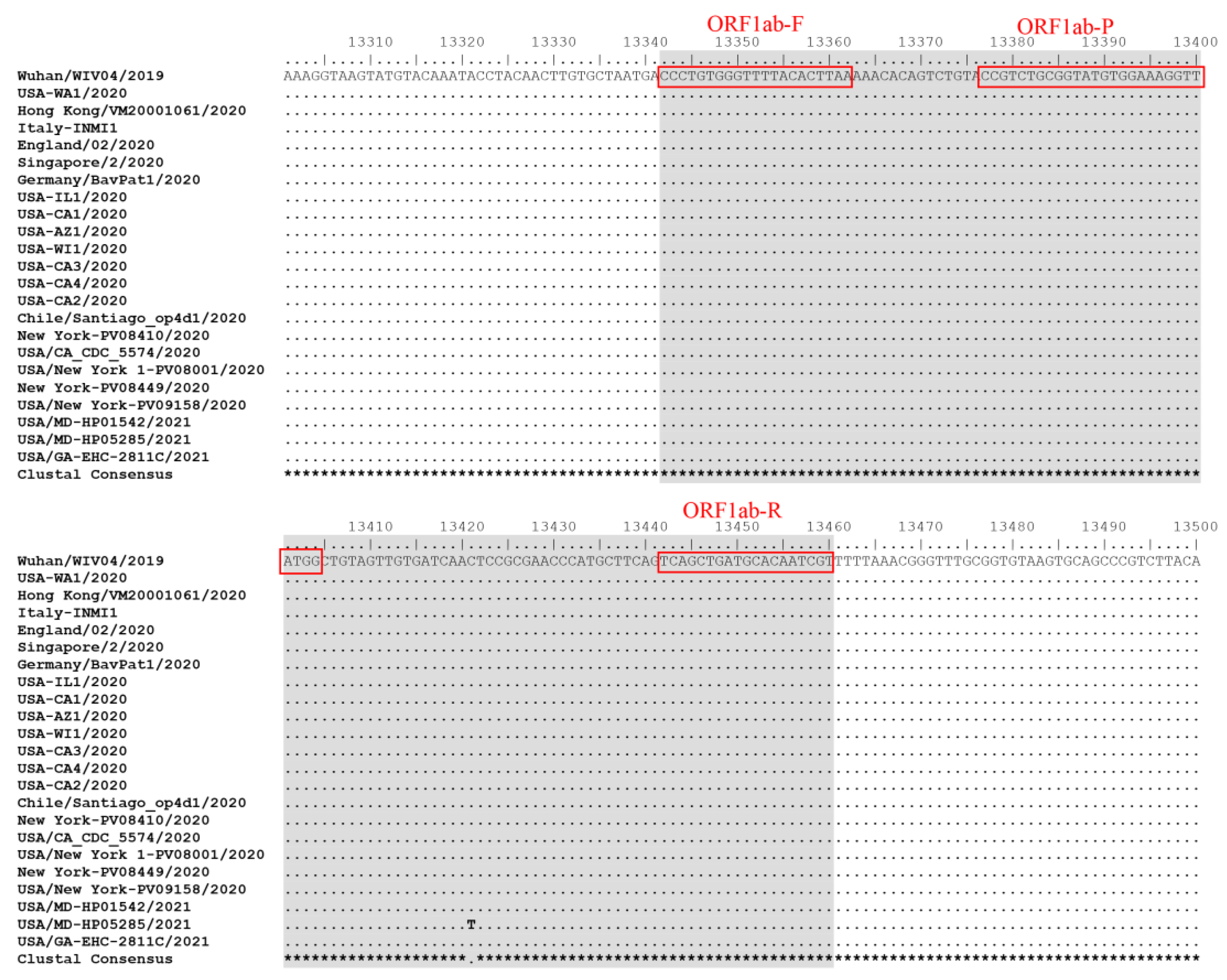
3.2. Inclusivity of POCKIT Central SARS-CoV-2 orf1ab RT-iiPCR Assay and the Reference TaqPath COVID-19 PCR Assay

A total of 22 different SARS-CoV-2 isolates obtained from BEI Resources were tested by both PCR assays. These 22 isolates include the original SARS-CoV-2 strains detected in various countries and regions as well as the SARS-CoV-2 Alpha, Beta, Delta, and Omicron variants. We also determined the Pango lineage, Nextstrain clade, and GISAID clade information of these SARS-CoV-2 isolates with the data summarized in Table 2. All of the 22 isolates tested positive by both PCR assays (Table 2), suggesting that both PCR assays had good coverage to detect genetically diverse SARS-CoV-2 strains. Sequence alignment indicated that the ORF1ab genomic region targeted by POCKIT Central SARS-CoV-2 PCR primers and probe was conserved among all of the evaluated SARS-CoV-2 strains except one SARS-CoV-2 isolate USA/MD-HP05285/2021 which had one nucleotide substitution compared to other SARS-CoV-2 isolates (Figure 2). However, this nucleotide substitution did not affect the detection of USA/MD-HP05285/2021 isolate by POCKIT Central SARS-CoV-2 PCR.

3.3. Limit of Detection of POCKIT Central SARS-CoV-2 orf1ab RT-iiPCR Assay and the Reference TaqPath COVID-19 PCR Assay

Different dilutions of the SARS-CoV-2 isolate USA-WA1/2020 were tested by both the POCKIT Central SARS-CoV-2 PCR and the reference PCR, with 5 replicates at high concentrations and 20 replicates at low concentrations. As shown in Table 3, the limit of detection (at least 95% of reactions were positive) of the POCKIT Central SARS-CoV-2 PCR was 1.87 × 10³ genomic copies/mL (corresponding to 0.8 TCID50/mL) while the limit of detection of the reference PCR was 3.75 × 10² genomic copies/mL (corresponding to 0.16 TCID50/mL) under the conditions of this study.

3.4. Diagnostic Accuracy of POCKIT Central SARS-CoV-2 orf1ab RT-iiPCR Assay

The diagnostic accuracy of the POCKIT Central SARS-CoV-2 PCR was evaluated by testing 183 clinical samples in comparison with the reference TaqPath COVID-19 PCR. The detailed results are shown in Supplemental Table S1. For all 183 samples, the Internal Control of the POCKIT Central SARS-CoV-2 PCR and the internal positive control (MS2 gene) of the reference PCR were all positive; hence, all of the SARS-CoV-2 results of the two PCR assays were valid.
Regarding the reference PCR, 51 samples were negative (Ct ≥ 37) by all of the three SARS-CoV-2 target genes ORF1ab, N, and S, 94 samples were positive (Ct < 37) by all of the three target genes, and 38 samples were positive by the ORF1ab and N target genes but negative by the S target gene (Table S1). According to the interpretation criteria from the kit manufacturer, it was concluded that 51 samples were negative and 132 samples were positive for SARS-CoV-2 by the reference PCR. For the 132 samples positive by SARS-CoV-2 ORF1ab and N target genes of the reference PCR, the Ct values ranged from 9.46–33.86 (ORF1ab target gene) and 8.12–36.97 (N target gene). For the 38 samples that were positive by the ORF1ab and N target genes but negative by the S target gene, their Ct values ranged from 10.04–33.86 (ORF1ab target gene) and 8.81–34.20 (N target gene).
Regarding the POCKIT Central SARS-CoV-2 PCR, 62 samples were negative and 121 samples were positive for SARS-CoV-2. Compared with the reference PCR, the diagnostic sensitivity, specificity, and agreement of the POCKIT Central SARS-CoV-2 PCR were 91.7%, 100%, and 94.0%, respectively, with all of the 183 samples being accounted for (Table 4). There were 11 discrepant results among the 183 clinical samples between the two PCR assays and all of these 11 samples were negative by the POCKIT Central SARS-CoV-2 PCR but positive by the reference PCR. These 11 samples had relatively high Ct values of 29.10–33.86 (ORF1ab target gene) and 29.19–36.97 (N target gene) according to the reference PCR. As shown in Table S1, for 113 clinical samples with TaqPath ORF1ab PCR Ct < 29, POCKIT Central SARS-CoV-2 PCR had 100% positive percent agreement (113/113); for 115 clinical samples with TaqPath ORF1ab PCR Ct < 30, POCKIT Central SARS-CoV-2 PCR had 99.1% positive percent agreement (114/115); for 125 clinical samples with TaqPath ORF1ab PCR Ct ≤ 32.50, POCKIT Central SARS-CoV-2 PCR had 96.8% positive percent agreement (121/125).

4. Discussion

The COVID-19 pandemic has prompted an unprecedented global effort to develop diagnostic methods, vaccines, antiviral drugs, and bioinformatics tools to track virus evolution and spread; all of these have greatly contributed to controlling SARS-CoV-2 infection worldwide. In addition, the knowledge and experience learned from these efforts are invaluable for fighting against other infectious diseases. The speedy development and application of effective mRNA vaccines against COVID-19 is a stellar example of new technology. Similarly, significant advances have been made in the SARS-CoV-2 diagnostic field. According to the 360D× that tracks the commercially available SARS-CoV-2 tests for diagnostic and clinical use (https://www.360dx.com/coronavirus-test-tracker-launched-COVID-19-tests), up to 15 March 2023, there have been 403 different types of PCR tests, 36 isothermal amplification tests, 102 antigen tests, 155 antibody tests, and a few biosensor tests for SARS-CoV-2 worldwide.
Various real-time RT-PCRs targeting ORF1ab, N, S, and E genomic regions have been developed and widely used for detecting SARS-CoV-2, and are considered the gold standard for SARS-CoV-2 testing [11,12,13,19]. However, at the early stage of the COVID-19 outbreak, human medicine diagnostic laboratories did not have sufficient capacity (shortage of supplies, reagents, and instruments for nucleic acid extraction and real-time RT-PCR) to meet the exploding SARS-CoV-2 PCR testing requests. Many veterinary diagnostic laboratories (including our lab at Iowa State University College of Veterinary Medicine) were brought in to test human samples for SARS-CoV-2 by real-time RT-PCR in a high-throughput format. However, the instrument for such high-throughput SARS-CoV-2 real-time RT-PCR is not field deployable and is unsuitable for point-of-care use.
POCKIT Central insulated isothermal PCR system combines nucleic acid extraction and PCR reaction in one instrument and automates the sample-to-answer process within ~85 min; this system can be a useful POC tool. In this study, we evaluated POCKIT Central SARS-CoV-2 PCR system in comparison with a commercial TaqPath COVID-19 real-time RT-PCR which was used as a reference assay. The POCKIT Central SARS-CoV-2 PCR assay showed great analytical specificity and inclusivity. The primers and probe of POCKIT Central SARS-CoV-2 PCR target the conserved ORF1ab genomic region, enabling this assay to successfully detect all of the 22 SARS-CoV-2 original strains and various variants evaluated in this study (Table 2).
The POCKIT Central SARS-CoV-2 PCR (LOD 1.87 × 10³ genomic copies/mL) showed lower analytical sensitivity than the reference TaqPath PCR (LOD 3.75 × 10² genomic copies/mL) based on testing the serial dilutions of heat-inactivated SARS-CoV-2 isolate USA-WA1/2020 (Table 3). For the POCKIT Central SARS-CoV-2 PCR, the equipment automatically processed 200 µL of samples from the beginning to the result interpretation. As for the reference PCR, 200 µL of the samples were first extracted using the MagMAX Viral/Pathogen Nucleic Acid Isolation Kit on a Kingfisher Flex instrument to obtain 50 µL of viral nucleic acids, then 10 µL of the nucleic acid extracts were used to set up the PCR reaction. In order to reasonably compare the LODs of these two PCR assays, we calculated the LODs based on the genomic copies of the SARS-CoV-2 isolate before nucleic acid extraction. We did not calculate the LODs based on the concentration of viral RNA used for the PCR reaction because we could not measure the viral RNA concentration in the POCKIT Central SARS-CoV-2 PCR system due to its automated sample-to-result process. Therefore, it is noteworthy that the LOD of the reference TaqPath COVID-19 PCR determined in this study may be different from the LOD based on the quantitation of viral RNA used for PCR. For example, in this study, if we calculated the LOD in another way, the LOD of the reference TaqPath COVID-19 PCR would be 15 genomic copies per reaction (3.75 × 10² genomic copies/mL × 0.2 mL × 10 µL/50 µL).
In this study, among the 183 clinical samples, 38 samples were positive by the reference TaqPath COVID-19 PCR ORF1ab and N target genes but negative by the reference PCR S target gene (Table S1). But, this is not surprising because the S gene is known to be highly prone to mutation. This also emphasizes that when developing SARS-CoV-2 screening PCR, the S gene should not be the single target for primers and probes. When all of the 183 clinical samples were used to evaluate the diagnostic accuracy, the POCKIT Central SARS-CoV-2 PCR system had 91.7% sensitivity (121/132) and 100% specificity (51/51) compared to the reference TaqPath COVID-19 PCR system (Table 4). The 11 samples that were negative by the POCKIT Central SARS-CoV-2 PCR but positive by the reference PCR had relatively high Ct values of 29.10–33.86 by TaqPath PCR ORF1ab target gene. For clinical samples with lower Ct values by TaqPath ORF1ab PCR, POCKIT Central SARS-CoV-2 PCR had better positive percent agreement (e.g., 100% for Ct < 29, 99.1% for Ct < 30, and 96.8% for Ct ≤ 32.50) (Table S1). In 2020, the POCKIT Central SARS-CoV-2 PCR was clinically evaluated on 100 oropharyngeal swab samples and it had 96.8% diagnostic sensitivity compared to a SARS-CoV-2 real-time RT-PCR assay [29]. Such preliminary data were used to apply for emergency use authorization from various agencies. Specifically, in 2020–2022, POCKIT Central SARS-CoV-2 PCR was successfully registered under Food and Drug Administration for emergency use authorization in Taiwan, under Agência Nacional de Vigilância Sanitária (ANVISA) in Brazil, and under the In Vitro-Diagnostic Medical Devices Directive 98/79/EC (IVDD) in Europe. In our current study, the POCKIT Central SARS-CoV-2 PCR was thoroughly validated and the study can be considered a combination of technical and clinical validations. The data from this study confirms the validity of the authorization to use this test in an emergency setting based on previous incomplete data. This work demonstrates the diagnostic reliability of the test and the data presented in the current study can be used for additional marketing registrations if needed.
According to the literature, various other PCR-based POC devices have also been developed and validated for SARS-CoV-2 testing [19]. For example, when compared with a standard real-time RT-PCR, the Accula SARS-CoV-2 POC PCR had 68% overall positive percent agreement (34/50) but had 100% positive percent agreement (27/27) for samples with Ct < 30 [30]. Similarly, when compared with a standard real-time RT-PCR, the Visby Medical COVID-19 POC PCR had 95% overall positive percent agreement (58/61) but had 100% positive percent agreement (49/49) for samples with Ct < 31 [31]. Many rapid antigen tests have also been developed for SARS-CoV-2 POC testing. In one review study [32], the authors included 93 studies (reported in 88 publications) that evaluated 36 rapid SARS-CoV-2 antigen tests in 104,961 participants and the rapid antigen tests had an overall sensitivity of 75%. In one study, the SARS-CoV-2 antigen test had 78.9% sensitivity among all symptomatic participants but 96.3% sensitivity in the symptomatic participants who had PCR Ct values of <29 [33]. Similarly, other studies showed that SARS-CoV-2 antigen tests had better sensitivity in samples with PCR Ct values of <25 and the sensitivity decreased a lot in samples with Ct values of >30 [17,34]. Hence, if the sensitivities of various assays are simply compared relative to the real-time RT-PCR Ct values, it appears that the POCKIT Central SARS-CoV-2 PCR had at least comparable sensitivity when compared to other point-of-care PCR and antigen tests.

5. Conclusions

In this study, we described a novel, automated sample-to-result POCKIT Central SARS-CoV-2 orf1ab RT-iiPCR system that combined nucleic acid extraction and PCR reaction in one instrument but still had comparable diagnostic accuracy to the reference real-time RT-PCR, especially for samples with Ct < 30. The compact POCKIT Central SARS-CoV-2 PCR system can be easily set up and implemented in local clinics, health centers, nursing homes, naval ships, cruises, and remote areas when high-throughput testing of a large number of samples is not needed. This system will allow timely (~85 min from loading samples to obtaining the results) on-site detection without transporting samples to a central diagnostic laboratory. In addition, this platform can be readily adapted to detect other human and animal viruses.

Supplementary Materials

The following supporting information can be downloaded at: https://www.mdpi.com/article/10.3390/diagnostics13132219/s1, Table S1: Clinical samples tested by POCIT Central SARS-CoV-2 orf1ab PCR and the reference TaqPath COVID-19 PCR.

Author Contributions

Conceptualization, S.C., Y.-L.T. and J.Z.; methodology and investigation, J.-H.Z., P.-H.C., C.-H.T., C.-F.P., L.B. and J.Z.; data analysis, J.-H.Z. and J.Z.; writing—original draft preparation, J.-H.Z.; writing—review and editing, J.Z., C.-H.T., C.-F.P., P.-H.C., Y.-L.T., S.C., L.B., M.P. and P.G. All authors have read and agreed to the published version of the manuscript.

Funding

This research received no external funding.

Institutional Review Board Statement

The study was conducted according to the guidelines of the Declaration of Helsinki and was approved by the Iowa State University Institutional Biosafety Committee (IBC-20-079).

Informed Consent Statement

Not applicable. The samples used for evaluation in this study were already submitted to the State Hygienic Laboratory of University of Iowa or Iowa State University Public Health Testing Services for testing.

Data Availability Statement

The data set(s) supporting the results of this article are included within the article.

Acknowledgments

We sincerely thank faculty and staff at the State Hygienic Laboratory of University of Iowa and at the Public Health Testing Services within Iowa State University College of Veterinary Medicine for providing clinical samples, some reagents, and technical help.

Conflicts of Interest

C.-H.T., C.-F.P., P.-H.C., Y.-L.T. and S.C. were employee of GeneReach at the time this study was performed. However, this does not alter our adherence to all the policies of Diagnostics on sharing data and materials. The other authors declare that they have no conflicts of interests.

References

  1. Pak, A.; Adegboye, O.A.; Adekunle, A.I.; Rahman, K.M.; McBryde, E.S.; Eisen, D.P. Economic Consequences of the COVID-19 Outbreak: The Need for Epidemic Preparedness. Front. Public. Health 2020, 8, 241. [Google Scholar] [CrossRef]
  2. Mackenzie, J.S.; Smith, D.W. COVID-19-A Novel Zoonotic Disease: A Review of the Disease, the Virus, and Public Health Measures. Asia Pac. J. Public. Health 2020, 32, 145–153. [Google Scholar] [CrossRef] [PubMed]
  3. Onyeaka, H.; Anumudu, C.K.; Al-Sharify, Z.T.; Egele-Godswill, E.; Mbaegbu, P. COVID-19 pandemic: A review of the global lockdown and its far-reaching effects. Sci. Prog. 2021, 104, 368504211019854. [Google Scholar] [CrossRef]
  4. Ellis, P.; Somogyvari, F.; Virok, D.P.; Noseda, M.; McLean, G.R. Decoding Covid-19 with the SARS-CoV-2 Genome. Curr. Genet. Med. Rep. 2021, 9, 1–12. [Google Scholar] [CrossRef]
  5. Redondo, N.; Zaldivar-Lopez, S.; Garrido, J.J.; Montoya, M. SARS-CoV-2 Accessory Proteins in Viral Pathogenesis: Knowns and Unknowns. Front. Immunol. 2021, 12, 708264. [Google Scholar] [CrossRef] [PubMed]
  6. Baric, R.S.; Fu, K.; Chen, W.; Yount, B. High recombination and mutation rates in mouse hepatitis virus suggest that coronaviruses may be potentially important emerging viruses. Adv. Exp. Med. Biol. 1995, 380, 571–576. [Google Scholar] [PubMed] [Green Version]
  7. Goldstein, S.A.; Brown, J.; Pedersen, B.S.; Quinlan, A.R.; Elde, N.C. Extensive Recombination-driven Coronavirus Diversification Expands the Pool of Potential Pandemic Pathogens. Genome Biol. Evol. 2022, 14. [Google Scholar] [CrossRef]
  8. Hillary, V.E.; Ceasar, S.A. An update on COVID-19: SARS-CoV-2 variants, antiviral drugs, and vaccines. Heliyon 2023, 9, e13952. [Google Scholar] [CrossRef]
  9. Rambaut, A.; Holmes, E.C.; O’Toole, A.; Hill, V.; McCrone, J.T.; Ruis, C.; du Plessis, L.; Pybus, O.G. A dynamic nomenclature proposal for SARS-CoV-2 lineages to assist genomic epidemiology. Nat. Microbiol. 2020, 5, 1403–1407. [Google Scholar] [CrossRef]
  10. O’Toole, A.; Scher, E.; Underwood, A.; Jackson, B.; Hill, V.; McCrone, J.T.; Colquhoun, R.; Ruis, C.; Abu-Dahab, K.; Taylor, B.; et al. Assignment of epidemiological lineages in an emerging pandemic using the pangolin tool. Virus Evol. 2021, 7, veab064. [Google Scholar] [CrossRef]
  11. Ciotti, M.; Benedetti, F.; Zella, D.; Angeletti, S.; Ciccozzi, M.; Bernardini, S. SARS-CoV-2 Infection and the COVID-19 Pandemic Emergency: The Importance of Diagnostic Methods. Chemotherapy 2021, 66, 17–23. [Google Scholar] [CrossRef]
  12. Rotondo, J.C.; Martini, F.; Maritati, M.; Caselli, E.; Gallenga, C.E.; Guarino, M.; De Giorgio, R.; Mazziotta, C.; Tramarin, M.L.; Badiale, G.; et al. Advanced Molecular and Immunological Diagnostic Methods to Detect SARS-CoV-2 Infection. Microorganisms 2022, 10, 1193. [Google Scholar] [CrossRef]
  13. La Marca, A.; Capuzzo, M.; Paglia, T.; Roli, L.; Trenti, T.; Nelson, S.M. Testing for SARS-CoV-2 (COVID-19): A systematic review and clinical guide to molecular and serological in-vitro diagnostic assays. Reprod. Biomed. Online 2020, 41, 483–499. [Google Scholar] [CrossRef] [PubMed]
  14. Li, M.; Ge, H.; Sun, Z.; Fu, J.; Cao, L.; Feng, X.; Meng, G.; Peng, Y.; Liu, Y.; Zhao, C. A loop-mediated isothermal amplification-enabled analytical assay for the detection of SARS-CoV-2: A review. Front. Cell. Infect. Microbiol. 2022, 12, 1068015. [Google Scholar] [CrossRef] [PubMed]
  15. Drain, P.K. Rapid Diagnostic Testing for SARS-CoV-2. N. Engl. J. Med. 2022, 386, 264–272. [Google Scholar] [CrossRef] [PubMed]
  16. A, R.; Wang, H.; Wang, W.; Tan, W. Summary of the Detection Kits for SARS-CoV-2 Approved by the National Medical Products Administration of China and Their Application for Diagnosis of COVID-19. Virol. Sin. 2020, 35, 699–712. [Google Scholar] [CrossRef]
  17. Suzuki, H.; Akashi, Y.; Kato, D.; Takeuchi, Y.; Kiyasu, Y.; Terada, N.; Kurihara, Y.; Kuwahara, M.; Muramatsu, S.; Ueda, A.; et al. Analytical performance of the rapid qualitative antigen kit for the detection of SARS-CoV-2 during widespread circulation of the Omicron variant. J. Infect. Chemother. 2023, 29, 257–262. [Google Scholar] [CrossRef]
  18. Tessaro, L.; Aquino, A.; Panzenhagen, P.; Joshi, N.; Conte-Junior, C.A. A systematic review of the advancement on colorimetric nanobiosensors for SARS-CoV-2 detection. J. Pharm. Biomed. Anal. 2023, 222, 115087. [Google Scholar] [CrossRef]
  19. Dhar, B.C. Diagnostic assay and technology advancement for detecting SARS-CoV-2 infections causing the COVID-19 pandemic. Anal. Bioanal. Chem. 2022, 414, 2903–2934. [Google Scholar] [CrossRef]
  20. Zeng, R.J.; Qiu, M.H.; Wan, Q.; Huang, Z.S.; Liu, X.L.; Tang, D.P.; Knopp, D. Smartphone-Based Electrochemical Immunoassay for Point-of-Care Detection of SARS-CoV-2 Nucleocapsid Protein. Anal. Chem. 2022, 94, 15155–15161. [Google Scholar] [CrossRef]
  21. Kruger, L.J.; Klein, J.A.F.; Tobian, F.; Gaeddert, M.; Lainati, F.; Klemm, S.; Schnitzler, P.; Bartenschlager, R.; Cerikan, B.; Neufeldt, C.J.; et al. Evaluation of accuracy, exclusivity, limit-of-detection and ease-of-use of LumiraDx (TM): An antigen-detecting point-of-care device for SARS-CoV-2. Infection 2022, 50, 395–406. [Google Scholar] [CrossRef]
  22. Li, H.; Te, S.H.; Tavakoli, Y.; Zhang, J.J.; Gin, K.Y.H.; He, Y.L. Rapid detection methods and modelling simulations provide new insights into cyanobacteria detection and bloom management in a tropical reservoir. J. Environ. Manag. 2023, 326, 116730. [Google Scholar] [CrossRef] [PubMed]
  23. Tsai, Y.L.; Wang, H.T.; Chang, H.F.; Tsai, C.F.; Lin, C.K.; Teng, P.H.; Su, C.; Jeng, C.C.; Lee, P.Y. Development of TaqMan probe-based insulated isothermal PCR (iiPCR) for sensitive and specific on-site pathogen detection. PLoS ONE 2012, 7, e45278. [Google Scholar] [CrossRef] [PubMed] [Green Version]
  24. Chua, K.H.; Lee, P.C.; Chai, H.C. Development of insulated isothermal PCR for rapid on-site malaria detection. Malar. J. 2016, 15, 134. [Google Scholar] [CrossRef] [PubMed] [Green Version]
  25. Go, Y.Y.; Kim, Y.S.; Cheon, S.; Nam, S.; Ku, K.B.; Kim, M.; Cho, N.H.; Park, H.; Alison Lee, P.Y.; Lin, Y.C.; et al. Evaluation and Clinical Validation of Two Field-Deployable Reverse Transcription-Insulated Isothermal PCR Assays for the Detection of the Middle East Respiratory Syndrome-Coronavirus. J. Mol. Diagn. 2017, 19, 817–827. [Google Scholar] [CrossRef] [PubMed] [Green Version]
  26. Go, Y.Y.; Rajapakse, R.; Kularatne, S.A.M.; Lee, P.A.; Ku, K.B.; Nam, S.; Chou, P.H.; Tsai, Y.L.; Liu, Y.L.; Chang, H.G.; et al. A Pan-Dengue Virus Reverse Transcription-Insulated Isothermal PCR Assay Intended for Point-of-Need Diagnosis of Dengue Virus Infection by Use of the POCKIT Nucleic Acid Analyzer. J. Clin. Microbiol. 2016, 54, 1528–1535. [Google Scholar] [CrossRef] [PubMed] [Green Version]
  27. Carossino, M.; Li, Y.; Lee, P.A.; Tsai, C.F.; Chou, P.H.; Williams, D.; Skillman, A.; Frank Cook, R.; Brown, G.; Chang, H.G.; et al. Evaluation of a field-deployable reverse transcription-insulated isothermal PCR for rapid and sensitive on-site detection of Zika virus. BMC Infect. Dis. 2017, 17, 778. [Google Scholar] [CrossRef] [Green Version]
  28. Tsai, J.J.; Liu, W.L.; Lin, P.C.; Huang, B.Y.; Tsai, C.Y.; Chou, P.H.; Lee, F.C.; Ping, C.F.; Lee, P.A.; Liu, L.T.; et al. An RT-PCR panel for rapid serotyping of dengue virus serotypes 1 to 4 in human serum and mosquito on a field-deployable PCR system. PLoS ONE 2019, 14, e0214328. [Google Scholar] [CrossRef] [Green Version]
  29. Chang, P.L.; Lin, C.Y.; Chen, C.P.; Lin, Y.C.; Hu, H.C.; Cheng, S.H.; Cheng, C.Y. Clinical validation of an automated reverse transcription-insulated isothermal PCR assay for the detection of severe acute respiratory syndrome coronavirus 2. J. Microbiol. Immunol. Infect. 2021, 54, 522–526. [Google Scholar] [CrossRef]
  30. Hogan, C.A.; Garamani, N.; Lee, A.S.; Tung, J.K.; Sahoo, M.K.; Huang, C.; Stevens, B.; Zehnder, J.; Pinsky, B.A. Comparison of the Accula SARS-CoV-2 Test with a Laboratory-Developed Assay for Detection of SARS-CoV-2 RNA in Clinical Nasopharyngeal Specimens. J. Clin. Microbiol. 2020, 58, e01072-20. [Google Scholar] [CrossRef]
  31. Renzoni, A.; Perez, F.; Ngo Nsoga, M.T.; Yerly, S.; Boehm, E.; Gayet-Ageron, A.; Kaiser, L.; Schibler, M. Analytical Evaluation of Visby Medical RT-PCR Portable Device for Rapid Detection of SARS-CoV-2. Diagnostics 2021, 11, 813. [Google Scholar] [CrossRef] [PubMed]
  32. Veroniki, A.A.; Tricco, A.C.; Watt, J.; Tsokani, S.; Khan, P.A.; Soobiah, C.; Negm, A.; Doherty-Kirby, A.; Taylor, P.; Lunny, C.; et al. Rapid antigen-based and rapid molecular tests for the detection of SARS-CoV-2: A rapid review with network meta-analysis of diagnostic test accuracy studies. BMC Med. 2023, 21, 110. [Google Scholar] [CrossRef] [PubMed]
  33. Ford, L.; Lee, C.; Pray, I.W.; Cole, D.; Bigouette, J.P.; Abedi, G.R.; Bushman, D.; Delahoy, M.J.; Currie, D.W.; Cherney, B.; et al. Epidemiologic Characteristics Associated with Severe Acute Respiratory Syndrome Coronavirus 2 (SARS-CoV-2) Antigen-Based Test Results, Real-Time Reverse Transcription Polymerase Chain Reaction (rRT-PCR) Cycle Threshold Values, Subgenomic RNA, and Viral Culture Results From University Testing. Clin. Infect. Dis. 2021, 73, e1348–e1355. [Google Scholar] [PubMed]
  34. Barrera-Avalos, C.; Mena, J.; Luraschi, R.; Rojas, P.; Mateluna-Flores, C.; Vallejos-Vidal, E.; Imarai, M.; Sandino, A.M.; Valdes, D.; Vera, R.; et al. Sensitivity analysis of rapid antigen tests for the Omicron SARS-CoV-2 variant detection from nasopharyngeal swab samples collected in Santiago of Chile. Front. Public. Health 2022, 10, 976875. [Google Scholar] [CrossRef]
Figure 1. Flow chart of two SARS-CoV-2 PCR systems evaluated in this study.
Diagnostics 13 02219 g001
Figure 2. Nucleotide sequence alignment of 23 SARS-CoV-2 ORF1ab genomic regions covering the primers and probe of POCKIT Central SARS-CoV-2 orf1ab RT-iiPCR. The fragment amplified by POCKIT Central SARS-CoV-2 orf1ab RT-iiPCR is highlighted in gray color and the sequences of primers and probes are shown in red boxes.
Diagnostics 13 02219 g002
Table 1. Analytical specificity of POCKIT Central SARS-CoV-2 RT-iiPCR assay and the reference TaqPath COVID-19 PCR assay.
PathogenATCC or BEI ResourcesPOCKIT Central SARS-CoV-2 (orf 1ab) PCR ResultTaqPath COVID-19 RT-PCR Result
Pseudomonas aeruginosaATCC 27853NegativeNegative
Staphylococcus aureus subsp. Aureus RosenbachATCC 8095NegativeNegative
Influenza A Virus (H1N1)ATCC VR-1469NegativeNegative
Influenza B VirusATCC VR-1931NegativeNegative
SARS coronavirus Urbani strain RNABEI NR-52346NegativeNegative
MERS coronavirus EMC/2012, heat-inactivatedBEI NR-50171NegativeNegative
Human coronavirus NL63ATCC VR-3263SDNegativeNegative
Human coronavirus 229EATCC VR-740NegativeNegative
Human coronavirus OC43ATCC VR-1558DNegativeNegative
Human adenovirus 5ATCC VR-5NegativeNegative
Respiratory syncytial virusATCC VR-1540NegativeNegative
Human rhinovirus 1AATCC VR-1559NegativeNegative
Human parainfluenza 1ATCC VR-94NegativeNegative
Human parainfluenza 2ATCC VR-92NegativeNegative
Human parainfluenza 3ATCC VR-93NegativeNegative
Table 2. Inclusivity of POCKIT Central SARS-CoV-2 RT-iiPCR assay and the reference TaqPath COVID-19 PCR assay for detecting different SARS-CoV-2 strains.
SARS-CoV-2 StrainType of MaterialBEI ResourcesGenBank Accession Number or GISAID EPI_SET IDWHO LabelPango LineageNext strain CladeGISAID cladePOCKIT Central SARS-CoV-2 PCRTaqPath COVID-19 RT-PCR
USA-WA1/2020 virusHeat-inactivated NR-52286MT576653 A19BSPositivePositive
Hong Kong/VM20001061/2020Genomic RNANR-52388MT644268 A19BSPositivePositive
Italy-INMI1Genomic RNANR-52498MT077125 B19AVPositivePositive
England/02/2020Genomic RNANR-52499EPI_ISL_407073 A19BSPositivePositive
Singapore/2/2020Genomic RNANR-52501EPI_ISL_407987 B19ALPositivePositive
Germany/BavPat1/2020Genomic RNANR-52502MT270101 B.120AGPositivePositive
USA-IL1/2020Genomic RNANR-52503MN988713 B19AOtherPositivePositive
USA-CA1/2020Genomic RNANR-52504MN994467 A19BSPositivePositive
USA-AZ1/2020Genomic RNANR-52505MN997409 A19BSPositivePositive
USA-WI1/2020Genomic RNANR-52506MT039887 B19ALPositivePositive
USA-CA3/2020Genomic RNANR-52507MT027062 B19ALPositivePositive
USA-CA4/2020Genomic RNANR-52508MT027063 B19ALPositivePositive
USA-CA2/2020Genomic RNANR-52509MN994468 B19AOtherPositivePositive
Chile/Santiago_op4d1/2020Genomic RNANR-52510EPI_ISL_415661 A.219BSPositivePositive
New Yor-PV08410/2020Genomic RNANR-53518MT370900 B.120CGHPositivePositive
USA/New York 1-PV08001/2020Genomic RNANR-52389MT370904 B.419AOtherPositivePositive
New York-PV08449/2020Genomic RNANR-53519MT370902 B.1.31920CGHPositivePositive
USA/New York-PV09158/2020Genomic RNANR-53520MT371034 B.120CGHPositivePositive
USA/CA_CDC_5574/2020 virusHeat-inactivated NR-55245EPI_ISL_751801Alpha variantB.1.1.720IGRYPositivePositive
USA/MD-HP01542/2021 virusHeat-inactivated NR-55350EPI_ISL_890360Beta variantB.1.35120HGHPositivePositive
USA/MD-HP05285/2021 virusHeat-inactivated NR-56128EPI_ISL_2103264Delta variantB.1.617.221IGKPositivePositive
USA/GA-EHC-2811C/2021 virusHeat-inactivated NR-56495OL744074Omicron variantB.1.1.529 / BA.121KGRAPositivePositive
Notes: Pango lineage and Nextstrain clade were determined by entering the query sequences at https:clades.nextstrain.org on 27 March 2023. GISAID clade was determined by entering the query sequences at gisaid.org on 27 March 2023.
Table 3. Limit of Detection of POCKIT Central SARS-CoV-2 RT-iiPCR assay and the reference TaqPath COVID-19 PCR assay. The detection endpoint of each assay was shown in bold.
Isolate Concentration (TCID50/mL)Isolate Concentration (Genomic Copies/mL)POCKIT Central SARS-CoV-2
(orf 1ab) PCR
TaqPath COVID-19 RT-PCR
% (No. of Pos for Target)% (No. of Pos for Internal Control)% (No. of Pos for Target)% (No. of Pos for Internal Control)
163.75 × 104100% (5/5)100% (5/5)100% (5/5)100% (5/5)
122.81 × 104100% (5/5)100% (5/5)100% (5/5)100% (5/5)
81.87 × 104100% (5/5)100% (5/5)100% (5/5)100% (5/5)
49.37 × 103100% (5/5)100% (5/5)100% (5/5)100% (5/5)
1.63.75 × 103100% (5/5)100% (5/5)100% (5/5)100% (5/5)
0.81.87 × 103100% (20/20)100% (20/20)100% (20/20)100% (20/20)
0.49.37 × 10290% (18/20)100% (20/20)100% (20/20)100% (20/20)
0.163.75 × 10260% (12/20)100% (20/20)100% (20/20)100% (20/20)
0.081.87 × 10215% (3/20)100% (20/20)90% (18/20)100% (20/20)
0.049.37 × 101 65% (13/20)100% (20/20)
0.012.34 × 101 10% (2/20)100% (20/20)
Table 4. Diagnostic accuracy of POCKIT Central SARS-CoV-2 RT-iiPCR assay in comparison with the reference TaqPath COVID-19 PCR assay.
TaqPath COVID-19 RT-PCRTotal
PositiveNegative
POCKIT Central SARS-CoV-2 (orf 1ab) PCRPositive1210121
Negative115162
Total13251183
Sensitivity 91.7%; specificity 100%; agreement 94.0%
Disclaimer/Publisher’s Note: The statements, opinions and data contained in all publications are solely those of the individual author(s) and contributor(s) and not of MDPI and/or the editor(s). MDPI and/or the editor(s) disclaim responsibility for any injury to people or property resulting from any ideas, methods, instructions or products referred to in the content.

