Article

Community-Acquired Methicillin-Resistant Staphylococcus aureus in Hospitals: Age-Specificity and Potential Zoonotic–Zooanthroponotic Transmission Dynamics

by
Ahmed Alsolami
1,
Naif Saad ALGhasab
2,
Mohammed S. M. Alharbi
1,
Abdelhafiz I. Bashir
3,
Mohd Saleem
4,
Azharuddin Sajid Syed Khaja
4,
Dakheel F. Aldakheel
5,
Ehab Rakha
6,7,
Jabar Aziz Alshammari
4,
Taha E. Taha
8,
Ziyad Melibari
4,
Yaseer H. Alharbi
4,
Ali A. Almutlag
4 and
Kamaleldin B. Said
4,9,*,† on behalf of the Ha’il COM Research Unit Group
1
Department of Internal Medicine, College of Medicine, University of Ha’il, Ha’il 55476, Saudi Arabia
2
Department of Cardiology, College of Medicine, University of Ha’il, Ha’il 55476, Saudi Arabia
3
Department of Physiology, College of Medicine, University of Ha’il, Ha’il 55476, Saudi Arabia
4
Department of Pathology, College of Medicine, University of Ha’il, Ha’il 55476, Saudi Arabia
5
Medical Coordination Unit, Ha’il General Hospital, Ha’il 55428, Saudi Arabia
6
Departments of Microbiology, King Khalid Hospital, Ha’il 55421, Saudi Arabia
7
Clinical Pathology Department, Faculty of Medicine, Mansoura University, Mansoura 35516, Egypt
8
Department of Epidemiology, John Hopkins Bloomberg School of Public Health, Baltimore, MD 21205, USA
9
Genomics, Bioinformatics and Systems Biology, Carleton University, 1125 Colonel-By Drive, Ottawa, ON K1S 5B6, Canada
*
Author to whom correspondence should be addressed.
Ha’il College of Medicine (COM) Research Unit is a research-intensive hub that, in addition to housing advanced research initiatives, co-ordinates medical and clinical research in the college, and organizes, follows up, and facilitates Deanship Funded Research Programs such as annual “Research Groups” series, Badi, and other institutional local, national and regional program.
Diagnostics 2023, 13(12), 2089; https://doi.org/10.3390/diagnostics13122089
Submission received: 8 May 2023 / Revised: 29 May 2023 / Accepted: 8 June 2023 / Published: 16 June 2023
(This article belongs to the Section Diagnostic Microbiology and Infectious Disease)

Abstract

:
Methicillin-resistant Staphylococcus aureus (MRSA) lineages are a devastating clinical and public health issue. Data on local lineage profiles are limited. We report on the frequency of community-acquired and hospital-acquired cases (CA-MRSA, HA-MRSA). We studied 147 isolates from King Khalid tertiary care hospitals (KKH), each from a case in a patient and including 33 patients at the Maternity and Children’s Hospital (MCH). Of the 147 isolates, 87 males (59%) and 60 females (41%) were in KKH. The overwhelming majority (80%; n = 119/147) were CA-MRSA in KKH. Intriguingly, despite significant differences between males (70%) and females (53%), lineage-acquisition remained age-specific around 58–60 years in both genders. However, while CA-MRSA dominated early in life (0–20, 70% MCH), it increased with age in KKH adults; 21–50 (28%), >50 (59%) until the overall 80% (n = 144/180). Major specimens included skin-wounds, surgeries (70.3%), blood (13.5%), sputum (8.8%), very rarely urine (4.1%), and nasal (3.4%), albeit most patients showed severe enteritis and necrotizing pneumonia. Antibiograms showed high beta lactam resistances, including amoxicillin–clavulanate (83%), oxacillin (84%), cefoxitin FOX (100%), penicillin and ampicillin (~100%), as well as high resistance (82%) to carbapenem. Fortunately, high susceptibility was seen to non-beta lactams and, to a lesser extent, gentamicin, erythromycin, and fusidic acid; 33%, 34%, and 38%, respectively, in KKH. A similar pattern was seen in MCH except for a low resistance pattern to gentamicin CN, clindamycin CD, erythromycin E, and tobramycin TOB; 34%, 31%, 39%, and 41%, respectively, except for fusidic acid. These findings have significant clinical implications for MRSA patient management strategies. Clinical- and lineage-profiles imply host-selection and zoonotic–zooanthroponotic transmission dynamics. Future molecular typing, sequencing, and characterization of dominant clone(s) is imperative.

1. Introduction

The devastating emergence and re-emergence of Staphylococcus aureus (S. aureus) lineages in hospitals and communities have been significant public health concerns globally. The World Health Organization (WHO) listed MRSA lineages as a priority pathogen and labeled them as one of the indicators for antimicrobial resistance in the Sustainable Development Goals connected to the health target 3.d [1]. Since the global MRSA pandemics a decade ago, outbreaks with high morbidities and mortalities are still being reported globally not in spite of, but because of advances in medicine [2,3]. Despite enormous efforts, it is not yet clear how a species with a clonal genome continuously evolves. It has become critical to evaluate the adaptive distribution of S. aureus in the contexts of host factors and genome plasticity. The previously established patterns of evolutionary history in S. aureus across decades implies that only a handful of subtle mechanisms are responsible for the rapid emergence of host-specific lineages, despite a stable genome. We and others have previously provided solid evidence, both from core genome and highly polymorphic loci, that S. aureus from humans and animals has a common genomic background and gene content [4,5,6,7]. Yet, lineages continue to emerge into sub-clonal populations specifically associated with humans and animals, as we have shown earlier [8,9]. One of the major reasons why that has not been fully explored with regard to continued emergence is the human host intracellular factor, which differentiates selective expression of virulence in community-associated lineages [10]. This stems from several striking observations that expression patterns of intrinsic host factors remained the same in methicillin-sensitive and resistant S. aureus (MSSA and MRSA) lineages of the same genotype, supporting the idea that gene transfer and resistance alone, although they may aid in transmission, do not account for the entire virulence per se [11,12,13]. Therefore, it has become imperative to study S. aureus lineages in the context of both human demographics and antibiogram patterns.
Staphylococcus aureus is among the first described pathogens that still continues to cause serious infections and is carried in the nasal mucosa and skin surfaces. Since first isolated by Ogston A (1881) [14] from surgical wound infection, S. aureus continues to re-emerge with enhanced virulence. This includes skin and soft tissue infections (SSTIs) as bullous impetigo, furuncles, and cellulitis, central-line-associated bloodstream infections (CLABSI), systemic infections such as bloodstream infection (BSI) leading to sepsis, bacteremic pneumonia, and endocarditis, in addition to toxin-associated diseases (such as toxic shock syndrome and food poisoning) [15,16,17]. The source of these infections is mostly endogenous, since 20% and 30% of individuals have been well-known to carry S. aureus either permanently or occasionally. [18,19]. Several reasons explain why S. aureus is a deadly pathogen in human medicine. Lineages possess highly similar genomes that allow gene transfer and rapid host-specific adaptations by plastic accessory elements that code for a repertoire of mosaic virulence factors and antimicrobial resistance, inducing the emergence of virulent host- and organ-specific clones [4,5,6,7,8]. The evolution of virulence in these clones is initiated by genetic and cellular regulation of fitness, selection, and virulence factors in different types of host-specific microenvironments, leading to the coordinative production of several factors, including toxin production, biofilm formation, immune evasions, tissue invasion, internalization, intracellular persistence, and/or emergence of diseases [20]. This is particularly clear in the evolution of mastitis-associated S. aureus strains [9]. Therefore, the major reason for the dissemination of resistance has been that common ancestors differentiated into different host species, allowing the transfer of strains. In fact, only two years after the introduction of penicillin, S. aureus developed antimicrobial resistance [21]. Consequently, the overuse of the semisynthetic methicillin produced in late 1950s led to the emergence of methicillin-resistant S. aureus (MRSA) in early 1960 [22]. Then this led to waves of outbreaks by strains resistant to antimicrobials [23]. Evolutionary mechanisms continued in the same pattern throughout history, producing different types of S. aureus strains with unique antibiotic resistances [24,25]. For instance, the initially multidrug-resistant S. aureus lineage appeared to resist penicillin G, chloramphenicol, tetracycline, and erythromycin by producing penicillinase (penicillin-hydrolyzing enzyme in the mid-1940s). Since then, the widespread and heavy usage of antimicrobial agents has led to the emergence of several drug-resistant S. aureus strains, most notably, the MRSA lineage [26], which survives in hospitals and has adapted to so many drugs that it has been called (HA)-MRSA. It has been inferred from high-resolution evolutionary models of parsimony that MRSA has evolved a minimum of 20 times from its methicillin-sensitive S. aureus (MSSA) ancestor since its acquisition of the methicillin resistance determinant, which is carried on a mobile genetic element, the staphylococcal cassette chromosome mec (SCCmec) [27,28]. Most of antibiotic resistance in S. aureus is attributed to horizontal gene transfers of resistant cassettes as well as intrinsic chromosomal mutations [29]. In addition, a recent study revealed that the use of beta-lactams enhances MRSA virulence mechanisms controlled by the SarA gene family [30].
The excessive use of antimicrobials in hospitals, farms, veterinary work, and agriculture has led to the emergence of multidrug resistant strains in hospitals and communities. For example, the emergence of colistin-resistant Enterobacteriaceae occurred after the introduction of selective digestive tract decontamination in an intensive care unit [31]. Similarly, an increasing trend in the outbreaks of resistant hypervirulent Acinetobacter strains in public hospitals in southwest Iran [32], and campylobacter in broiler chickens in north Lebanon [33], are a few examples of zoonotic transmission. Perhaps one of the most devastating transmissions was that of the zoonotic and reverse-zoonotic evolutions of community-associated S. aureus (CA-MRSA) lineages with enhanced virulence in young and otherwise healthy individuals. As a result, waves of global pandemics with significant economic and public health losses occurred [30,34,35,36,37,38,39,40,41,42]. Many of the reasons for the emergence of virulence were due to the low-level usage of antimicrobial substances [43,44]. However, the mechanisms of CA-MRSA entailed a highly complex, orchestrated evolutionary convergence of enhanced virulence, high-resistance, and low-susceptibility to non-beta lactams [44,45,46]. In contrast, high levels of multi-drug resistance in HA-MRSA lineages have been associated with mortalities due to hospital-acquired bacteremia [47]. At present, the exchange of these lineages between communities, hospitals, and livestock animals [48] has posed challenges for planning and clinical management strategies. The most striking difference in these lineages is their clinical and antibiogram patterns in response to treatment options, which is of concern in this study. It has become so blurred that it is not clear which of the two patterns, HA-MRSA or CA-MRSA, dominates the region.
Methicillin resistance in S. aureus is defined as an oxacillin minimum inhibitory concentration (MIC) of greater than or equal to 4 micrograms/mL, based on antibiotic susceptibility. A standard definition of HA-MRSA refers to those isolated from in-hospital patients who tested negative to nasal screening on admission or MRSA isolated from inpatients for 48–72 h or more after their stay or after discharge [17,49,50,51]. According to epidemiology, CA-MRSA is defined as MRSA isolated from community or hospital outpatients without a recent hospitalization history and without any other known risk factors for MRSA infection. The acquisition of mecA or mecC elements classified into 13 types and carried on the staphylococcal cassette chromosome mec (SCCmec) confers resistance to most beta-lactams [52,53]. CA-MRSA strains generally harbor SCCmec type IV or V and are susceptible to non-beta-lactam antimicrobials. The HA-MRSA strains commonly harbor SCCmec types I, II, or III, which contain genes that confer broad resistance to antimicrobials [54]. These distinct differences in the antibiotic susceptibility and resistance patterns between the two lineages have become one of the most important distinguishing features. These two lineages have, as their name suggests, originated in hospitals and communities, albeit they are well-known to coexist in both settings, making it difficult to successfully diagnose and initiate an empiric therapy. This is separate from several livestock sub-lineages.
Reports on the exact frequency of HA-MRSA and CA-MRSA distributions in communities and hospitals in Saudi Arabia and Gulf countries are limited. In fact, there is a severe paucity in high quality data on rates, frequencies, and clinical pictures of infections by each lineage. A comprehensive review of articles on MRSA in the Gulf countries during 2011–2021 reported on all countries, including Kuwait (44%), Saudi Arabia (28%), and the United Arab Emirates (10%). A clear emergence in antibiotic resistance, especially against fusidic acid, ciprofloxacin, and clindamycin, was demonstrated. The regional prevalence of MRSA is reported as 25–35%, with clear dominance of community-acquired (CA)-MRSA. Panton–Valentine leucocidin (PVL)-producing strains accounted for 35–45%, with a clear association with CA-MRSA emergence, but there were some sporadic reports of the incorporation of PVL in healthcare-associated (HA)-MRSA. They reported dominant strains included EUST80, USA1100, and WA-MRSA-51. Novel strains are more likely to produce PVL and show fusidic acid resistance [55]. However, the state of these lineages in each country is not well-defined at different geographic region(s). Thus, it has become imperative to understand the patterns, distributions, demographics, clinical pictures, and potential outbreak situations in each region for local strain profiling, infection control planning, and patient management strategies. Some, although not many, widely reported cases have laid solid foundations for profiles of lineages in the region as a result of increased mortality, morbidity, and hospital stays [56]. For instance, comprehensive reports on strategic planning of the Gulf Co-operation Council Center for Infection Control (GCC-IC) have placed the emergence of antimicrobial resistance (AMR) on the top of its agenda since 2014 [57]. Consequently, a second meeting reported on the “One Health” concept from the Gulf Cooperation Council Countries (“Part Two: A Focus on Human Health”) [58]. Nevertheless, despite enormous efforts, and as is the case globally, calls for regional MRSA surveillance programs were made especially with the emergence of strains that require no underlying risk factors to cause illness, as well as the propagation of chimeric resistance elements in both HA-MRSA and CA-MRSA [55]. Since then, there have been several reports on the rise of S. aureus-resistant strains in the region that lacked objectivity and accuracy. However, many were useful and indicated that the rise in the isolates were predominantly from skin, soft tissue, wounds, and nasal swabs [59,60,61,62,63,64]. The antibiotics most commonly used for MRSA infections (skin and soft tissue infection) included fusidic acid, mupirocin, vancomycin, and clindamycin. Most resistances were found in the eastern region of Saudi Arabia, compared to Riyadh and other cities [59,65], while resistance has increased by three times compared to the usual rate in the UAE and Gulf countries, which have suffered higher rates of resistance [65,66,67,68,69]. For instance, MRSA genotypes with unique virulence, including a high prevalence of PVL and fusidic acid resistance in Kuwait hospitals, were reported [70,71,72].

2. Materials and Methods

2.1. Microbiology of Specimens and Antibiograms of Staphylococcus aureus Lineages from Patients

Hospital clinical and laboratory records were collected for S. aureus isolates, types of lineages, and antimicrobial susceptibility and patient demographics data. The major selection was based on positive specimens for S. aureus isolates where infections were clinically characterized in patients and specimens processed in the laboratory. All types of specimens from all patient age and gender groups were included. All records of clinical specimens from different hospital departments were processed using standard protocols from different hospital departments in Ha’il from the last quarter of 2021 to beyond the middle of 2022. Briefly, routine bacteriology and antibiograms were followed by molecular detection, characterization, and profiling of S. aureus lineages. Specimens were used immediately upon receipt or stored until processed. Most of the samples, such as swabs or aspirations, were subjected to processing immediately by identification on primary media and standard conditions of incubation at 37 °C for 18 to 24 h. Portions of isolate inoculums were stored immediately in liquid media at −30 °C for future downstream analysis. Automated testing and ID susceptibility on automated systems were used simultaneously. Most of this phase was performed on the BD Phoenix system (BD Biosciences, Franklin Lakes, NJ, USA) and MicroScan plus (Beckman Coulter, Brea, CA, USA). When required, sensitivities were confirmed by in vitro cultures in agar diffusions interpreted by zone interpretive standards for this region. The susceptibility testing and breakpoint interpretive standards were carried out in accordance with the recommendations of the Clinical and Laboratory Standard Institute (CLSI document M100S-26) [73].

2.2. Standard Definitions for Classification of Resistance in S. aureus as Multi-Drug Resistant Bacteria (MDR)

According to the recommendations of the “Center for Disease Control (CDC) and European Centre for Disease Control (ECDC)”, the following definitions are usually accepted as standard. According to Magiorakos et al. [74], the standard definition classifications considers MRSA isolates as multi drug-resistant (MDR) for their methicillin resistance and resistance to beta-lactams, except for the community-acquired lineages (CA-MRSA), since the latter is susceptible to beta-lactams. These criteria define resistances as follows: MDR = non-susceptibility to at least one agent in three or more antimicrobial categories; XDR = non-susceptibility to at least one agent in all but two or fewer antimicrobial categories (i.e., bacterial isolates remain susceptible to only one or two categories); PDR = non-susceptibility to all agents in all antimicrobial categories as reported. Known intrinsic resistances to particular drugs were not included. Thus, the MRSA criteria for defining MDR S. aureus classifications must include one or more of the following to apply: (1) hospital-acquired MRSA is always considered MDR by virtue of being an MRSA; (2) non- susceptibility to ≥1 agent in >3 antimicrobial categories.

2.3. Molecular Profiling of S. aureus Lineages by Multi-Gene Systems (GeneXpert)

The latest version of the Cepheid GeneXpert® Dx system with specific all-in-one cartilages of the SA Complete and MRSA assay kits was used for the characterization and profiling of S. aureus isolates following the manufacturers’ recommendations and using the codes included in each kit. Depending on the kit used, assay definition files were imported into the software, such as the Blood Culture assay definition file. Then, when sample patient IDs were scanned, the kit barcodes re-populated the boxes with Assay, Reagent Lot ID, Cartridge SN, and Expiration Date. Certain types of specimens, such as sputum, require a few simple steps, such as homogenizations and mixing, before loading into cartridges. However, the general protocol for all kits is concise, automated, user-friendly, and highly robust. For instance, Xpert MRSA/SA SSTI (skin and soft-tissue infections, as well as wounds and surgical infections) swabs from deep tissue, surgical sites, and wound infections were inserted into the sample reagent vial (break-in). Then, for the vortex well, particularly for samples with mucus contents or debris, dispense the sample into Port-S, insert the cartridge, and start the test. Multi-gene primers, probes, and reagents in kits allow for robust automated direct confirmation of S. aureus at the species level with subsequent differentiation into S. aureus lineages directly from specimens. This is accomplished by the built-in primers for nuc spa, mecA, and the mec (SCCmec) gene and their direct detection from specimens utilizing automated real-time polymerase chain reaction (PCR) in a single-use, disposable, self-contained cartridge with PCR reagents inserted and inoculated directly with swabs/samples. Therefore, the influence of laboratory media that is known to trigger sensing genes on S. aureus is minimized, leading to adaptive genome expressions and in turn the emergence of different types. Of importance is that it reduces cross-contamination between specimens as well as cross-sequence contaminations in molecular tests. Furthermore, a sample processing control (SPC) and a probe check control (PCC) are also included. The SPC is present to control for adequate processing of the target bacteria and to monitor the presence of inhibitor(s) in the PCR reaction. The PCC verifies reagent rehydration, PCR tube filling in the cartridge, probe integrity, and dye stability. Real-time PCR assays for mecA, nuc, and lukPV were consistent as previously performed [4] using primers luk pvl-1 [gcatcaastgtattggatagcaaaagc and luk pvl-2 atcattaggtaaaatgtctggacatggtcc with TET-probe atttgtaaacagaaattacacagtta].

2.4. Statistical Analysis

The Statistical Package for Social Sciences software (IBM SPSS; Version 24 SPSS version 23.0 for Windows (SPSS, Inc., Chicago, IL, USA) was used. The study was descriptive and stratified analyses were conducted. In this study, we present absolute numbers, proportions, and graphical distributions. We conducted exact statistical tests for proportions and show p-values (based on Chi square test values) where appropriate (a p-value < 0.05 was considered statistically significant).

3. Results

In the present study, we have analyzed the antibiotic-resistant pattern of MRSA obtained from different clinical specimens of 147 patients from King Khalid tertiary care hospitals (KKH) and 33 patients from the Maternity and Children’s Hospital (MCH) in the Hail region in the last quarter of 2021 and the middle of 2022. The patients’ characteristics, types of specimens, hospital wards, and antibiogram increments are all presented in Table 1. Most patients showed enteritis and necrotizing pneumonia profiles. In KKH, out of 147 patients there were 87 males (59.2%) and 60 females (41%) in our study, with 58 years being the median age of the study participants. The MCH specimens were all received and recorded under MCH samples as they all had similar descriptions that are common after birth and treated with pediatric protocol. The major types of clinical specimens targeted were those that were likely to have a skin or nasopharyngeal origin, such as skin wounds and surgeries (70.3%), blood (13.5%), sputum (8.8%), very rarely urine (4.1%), and nasal samples (3.4%). Figure 1 shows the antibiogram patterns of all non-beta lactam and beta lactam antimicrobials of isolates from King Khalid Hospital (KKH). A significantly higher number of isolates were resistant to all beta lactam antibiotics, including amoxicillin–clavulanate (83%), oxacillin (84%), cefoxitin FOX (100%), and penicillin and ampicillin (almost 100%). More important is that a higher number of isolates developed high resistance (82%) to the carbapenem antibiotic imipenem IMI. However, isolates were more susceptible to all of the non-beta lactam antibiotics with relatively lesser resistance patterns to gentamicin, erythromycin, and fusidic acid; 33%, 34%, and 38%, respectively. Furthermore, the overwhelming majority (80%; n = 119/148) of MRSA identified were CA-MRSA in KKH, while only 20% were HA-MRSA.
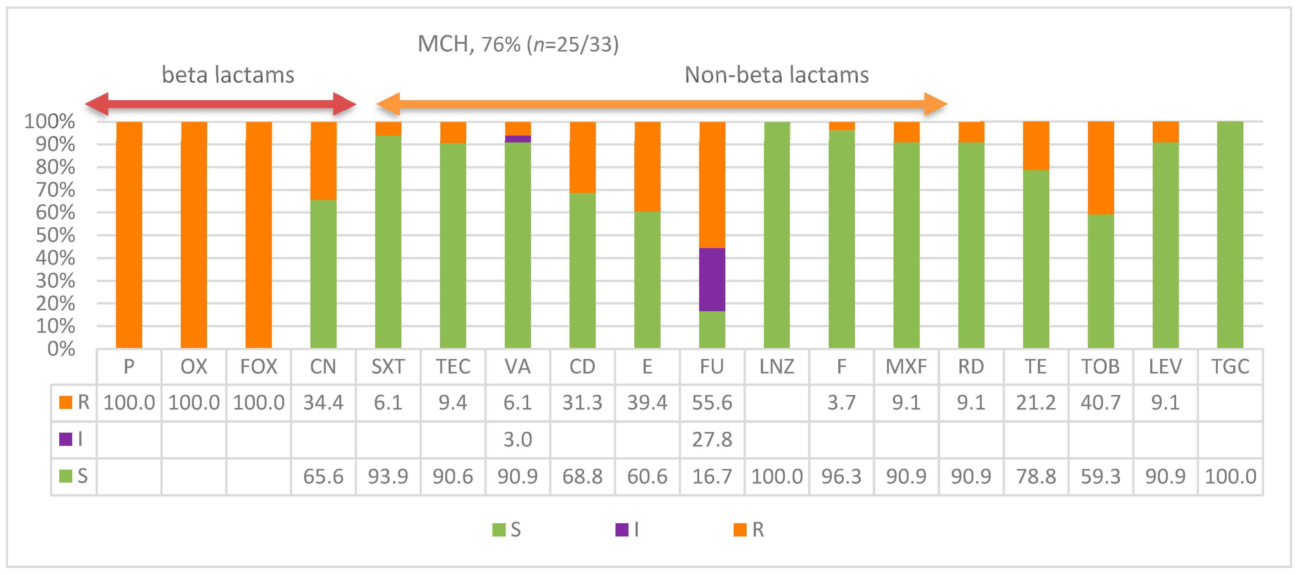
Antibiogram susceptibility patterns of S. aureus isolates from the MCH are presented in Figure 2. Although the prescription protocol for pediatrics differs, this figure presented a much more unique pattern from that of KKH while showing similar responses of isolates to beta lactam antibiotics against which isolates were highly resistant. Isolates were fully and completely resistant to beta lactam antimicrobials used in this protocol with 100% resistance to all three: penicillin P, oxacillin OX, and cefoxitin FOX. However, similar to adult hospitals, high susceptibility of isolates was also observed for non-beta lactam antibiotics, except for low resistance patterns to gentamicin CN, clindamycin CD, erythromycin E, fusidic acid FU, and tobramycin TOB; 34%, 31%, 39%, 56%, and 41% respectively. Isolates for the rest of all non-beta lactam antimicrobials used showed a very a high pattern of susceptibility. A unique exception was the higher fusidic acid resistance compared to the MCH isolates, with higher intermediate resistance to the drug compared to all other antibiotics used in this study. In pediatrics, MCH 76% (n = 25/33) were CA-MRSA and 24% were HA-MRSA. Overall, 80% (n = 144/180) of MRSA that were CA-MRSA had the patterns.
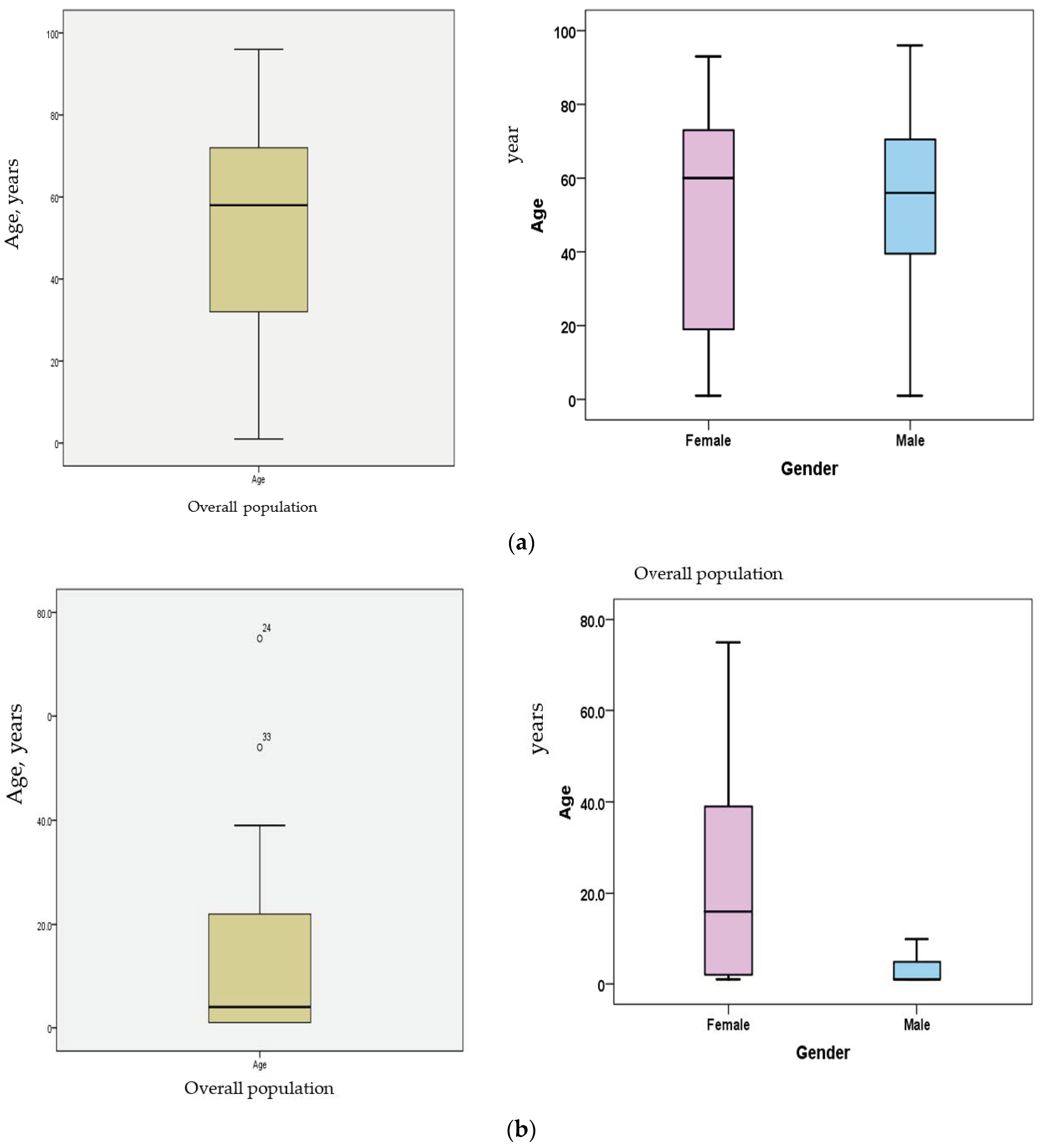
The age- and gender-specific distributions reveal unique characteristics of potential patient groups with respect to the acquisition of specific S. aureus lineage infections. Males’ overall frequency (53%) was always 10% higher than that of females (43%) in contracting MRSA lineages. Most patients were in the mid age groups; the overall mean for ages was 58. The median age for male patients was 58 years while it was 60 for female patients (Figure 3a,b). The distributions of infections within these age groups in Table 1 were as follows: age groups (0 to 20) and (21 to 50) had 23% and 27% of S. aureus infections, respectively. While the former group included an almost similar number of isolates from MCH (n = 23) and KKH (n = 19), the latter group had the majority of isolates (n = 41) from KKH and only seven from MCH. The sum of isolates in these two groups constituted 50% of isolates from age 0 to 50. However, the age group of over 50 years reported 49% of isolates, which was almost equal to the sum of the isolates reported in the two younger groups. In KKH isolates, 80% (n = 119) that were susceptible to non-beta lactam antimicrobials confirm the CA-MRSA pattern. Of the isolates of this lineage, 49.5% (n = 59/119) were mostly isolated from younger age groups, from 0 to around 40 years of age, while almost all of the HA-MRSA isolates resistant to all drugs belonged to much more senior age groups of patients. Similarly, isolates of MCH were from pediatric patients and therefore, except for eight HA-MRSA, all were from children with high susceptibility to non-beta lactams. In KKH, the highest number of isolates were made on specimens received from these departments, respectively: the ICU (27%), ER (14%), men’s medical wards (11%), female surgical wards (10%), and other units with lower isolation rates.

4. Discussion

There have been drastic epidemiological changes in S. aureus lineage evolutionary patterns, resulting in new strains with enhanced virulence in health care systems globally as well as in this country. New distinct lineages constantly emerge and transmit between both human and animals, crossing all host-species barriers. In the Kingdom of Saudi Arabia, MRSA has been reported as one of the common superbugs associated with severe public health infections [55,75,76,77]. We attempted to address the enhanced virulence and epidemicity in the context of host-selection, resistance, and transmission dynamics. However, there is a severe paucity of high-quality data about the profiles of circulating lineages and demographic patterns. Our data depicted a change in diversity and frequencies of HA-MRSA and MRSA (CA-MRSA) in the northern-central regions of Saudi Arabia. There are several other studies in the region and neighboring countries reporting on the sporadic evolution of community lineages with no clear epidemic profiles [77,78,79,80,81,82]. This potentially relates to the socio-economic migration across Saudi Arabia as a major global economic hub and as well as a holy shrine for pilgrimage. Thus, it has become imperative to understand the reasons behind enhanced virulence associated with specific age groups and the frequency of MRSA profiles across different age- and gender-specific groups in this study to substantiate selective susceptibility among the human host population structure.
In the current study, we show the replacement of HA-MRSA by the CA-MRSA lineages particularly in younger age groups in the Ha’il region of Saudi Arabia in comparison to regional variations. The MRSA isolated from KKH showed an extreme resistance against beta lactams, including amoxicillin–clavulanate and oxacillin at a rate of 83% and nearly absolute resistance against cefoxitin (100%), penicillin (98.6%), and ampicillin (98.6%). More important is that a significantly higher number of isolates developed high resistance (82%) to the commonly used imipenem antibiotic. At the same time, there was a high susceptibility to non-beta lactams, often reaching full susceptibility, such as linezolid (99.3%), teicoplanin (98.6%), rifampicin (98.6%), daptomycin (97.9%), vancomycin (97.3%), nitrofuran (96.6%), and mupirocin (96.6%). These findings indicate the dominance of CA-MRSA lineages in the hospitals consistent with our isolate characterizations. The antibiotic susceptibility profile observed in this study is in line with other studies [82,83]. All CA-MRSA isolates reported in this study were highly susceptible to linezolid, teicoplanin, and vancomycin, which is considered a last resort in MRSA treatment; this stands in agreement with other studies. Hence, this drug should not be used as empirical therapy in the treatment of Gram-positive bacterial infections except in extreme cases. Fortunately, in this study high susceptibility was reported towards TGC, TEI, and SXT. In addition, while there is a prevalence of beta-lactam-resistant MRSA, there is a higher frequency of non-beta lactam susceptibility to gentamicin, erythromycin, and fusidic acid; 33%, 34%, and 38%, respectively, was found. These findings implied that the overwhelming majority (80%; n = 119/148) of MRSA identified were CA-MRSA, while only 20% were HA-MRSA. Nevertheless, this contrasted with the scenario in the western regions a decade ago where CA-MRSA infections represented 31.5% of community S. aureus infections, while HA-MRSA accounted for 52.6% of hospital S. aureus, which was mainly associated with seniors (p = 0.0029) [83]. However, an eleven year (2009–2019) surveillance reported an increasing HA-MRSA trend (5% to 14%) among 1338 isolates in the eastern regions, consisting mainly of skin and soft tissue, with a significant reduction in susceptibility to clindamycin (p = 0.003), trimethoprim-sulfamethoxazole (p = 0.001), and rifampin (p < 0.0001) [84]. Unfortunately, data on age and gender specificity of CA-MRSA lineages in different regions for comparison are limited.
An important finding in this study was the age-specific gradual evolutionary replacement of HA-MRSA by the CA-MRSA lineages irrespective of gender differences. This is supported by several findings; for instance, in Table 1, while at the young age group 0 to 20 both adults and children had similar low MRSA counts (MCH n = 23; KKH n = 19), a significant shift in the MRSA increasing load showed at the age group 21 to 50, with (n = 41) from KKH and only seven from MCH. This increasing shift continued, reaching its maximum at the age group >50 with 49%, which is the sum of isolates in the two previous young groups. Intriguingly, despite the significant difference between male (59.2%; n = 87) and female (41%; n = 60) counts in KKH, as well as the overall 10% difference between genders (male 53%, female 43%), the highest HA-MRSA acquisition point remained age-specific around 58–60 years irrespective of gender differences, as shown in Figure 3a. On the other hand, CA-MRSA initiates early in life and continues to replace the former lineage. These findings have established a solid foundation in support of the established understanding that only a handful of subtle evolutionary mechanisms are involved in the emergence of CA-MRSA epidemics at a young age and HA-MRSA later, apart from resistance. These include host microenvironmental changes at different age-groups that differentiate and select emergence of best fit lineages. While acquired resistance increases epidemicity, it does not significantly alter the inherent adaptive evolution of virulence per se. We and others have established previously that host-specificity, despite genome clonality, is the key in the acquisition of S. aureus lineages [8,85]. This strongly supports the notion that fitness in a specific host is usually reached through processes of cellular differentiation and/or the acquisition of lineage-specific trait(s) [10,86]. Genome sequencing revealed variations in a single cohesive population, showing changes in only 30 SNP and four indels accumulating at a steady rate over a 13-mo period, except for a specific cluster of eight mutations preceding the transition to disease that made the new bloodstream variant [87]. Although host-specificity related to age is evident in this study, it is still imperative to understand the original sources of these isolates since S. aureus from animals and humans has similar population genetic structures, permitting cross-host transmissions.
The nature of isolates, patient demographics, infection types and sources, and antibiograms all imply novel evolutionary mechanisms in the emergence of CA-MRSA lineages, consistent with previous research. Unfortunately, the bias towards sequencing human-MRSA has created several gaps that hinder understanding of human- and livestock-associated lineages. The CA-MRSA lineage virulence and transmission dynamics is a multifaced mechanism involving the increased use of beta lactams and other antimicrobials in hospitals, agriculture, livestock, and domestic animals. This allows for zoonotic and anthroponoses transmission of animal lineages that are by nature methicillin-sensitive [4,8,9] and has led to the evolution of community-associated S. aureus (CA-MRSA) lineages with enhanced virulence in young and otherwise healthy individuals. As a result, waves of global pandemics with significant economic and public health losses have occurred [30,31,32,33,34,35,36,37,38,39]. Many of the reasons for the emergence of virulence were due to the low-level usage of antimicrobial substances [43,44]. For instance, the increased past usage of beta-lactam antibiotics is potentially behind the enhanced virulence which has recently been found to be controlled by the sarA gene family [30]. We have conducted a genome-wide expression of dairy and human isolates (unpublished data) under mammary-mimicked hypoxic conditions and revealed specific host factors such as nutritional and glucose impulse, consistent with others [88], and host-tissue specific adherence factors [89,90] that precisely selected bovine-mammary lineages. These factors were controlled by agr induction through the environment-sensor, the DNA binding protein of the SarA family, consistent with earlier descriptions [91]. This shows that the intracellular microenvironment of the host cell conditions the emergence of toxigenic strains while repressing protein A (spa) [89], a property that distinguishes human and animal lineages, making it imperative to test these isolates in the future for zoonotic transmission potentials.

5. Conclusions

We reported on the frequency of CA-MRSA and HA-MRSA infection rates in hospitals with respect to disease patterns, patient demographics, transmission dynamics, and antibiogram patterns. We further identified a potentially host-specific selective replacement of HA-MRSA lineages by the CA-MRSA. Furthermore, irrespective of gender, the acquisition of HA-MRSA infection is largely age-dependent, occurring predominantly around 58–60 years, while CA-MRSA starts early in life and continues to dominate patients, with the most striking disease profiles of this lineage in most patients being the enteritis and necrotizing pneumonia profiles. The prevalence and mechanisms of the emergences of CA-MRSA and MRSA lineages entail a highly complex, orchestrated evolutionary convergence of enhanced virulence, high-resistance, and low-susceptibility to non-beta lactams, in agreement with others [41,42,43]. In contrast, high levels of multi-drug resistance in HA-MRSA lineages confined to hospitals have been associated with mortalities due to hospital-acquired bacteremia, in agreement with others [44]. At present, the exchange of these lineages between the community, hospitals, and livestock animals [45] has made it difficult for planning and clinical management strategies. The most striking difference in these lineages is their patterns in response to treatment options and their acquisition of human-specific markers such as spa, clfB, and clfA—a property that accurately distinguishes human and animal lineages, making it imperative to test these isolates in the future for zoonotic transmission potentials. This study is limited by the confined area of the two hospitals; the inclusion of other hospitals in the neighboring cities as well as nationwide would have allowed for more insights into S. aureus infection patterns to be gained. In addition, a comparative analysis with reference to veterinary strains would have been useful; however, the specific local strain profiling that is necessary is available only in remote regions and therefore it carries the risk of regional differences.

Author Contributions

Data curation, A.A., N.S.A., M.S.M.A., A.I.B., M.S., A.S.S.K., D.F.A., E.R., J.A.A., T.E.T., Z.M., Y.H.A., A.A.A. and K.B.S.; formal analysis, A.A., N.S.A., M.S.M.A., A.I.B., M.S., A.S.S.K., D.F.A., E.R., J.A.A., T.E.T., Z.M., Y.H.A., A.A.A. and K.B.S.; funding acquisition, A.A. and K.B.S.; investigation, A.A. and K.B.S.; methodology, A.A., N.S.A., M.S.M.A., A.I.B., M.S., A.S.S.K., D.F.A., E.R., J.A.A., T.E.T., Z.M., Y.H.A., A.A.A. and K.B.S.; project administration, A.A. and K.B.S.; resources, A.A., N.S.A., M.S.M.A., A.I.B., M.S., A.S.S.K., D.F.A., E.R., J.A.A., T.E.T., Z.M., Y.H.A., A.A.A. and K.B.S.; software, A.A., N.S.A., M.S.M.A., A.I.B., M.S., A.S.S.K., D.F.A., E.R., J.A.A., T.E.T., Z.M., Y.H.A., A.A.A. and K.B.S.; supervision, A.A. and K.B.S.; validation, A.A., N.S.A., M.S.M.A., A.I.B., M.S., A.S.S.K., D.F.A., E.R., J.A.A., T.E.T., Z.M., Y.H.A., A.A.A. and K.B.S.; visualization, A.A., N.S.A., M.S.M.A., A.I.B., M.S., A.S.S.K., D.F.A., E.R., J.A.A., T.E.T., Z.M., Y.H.A., A.A.A. and K.B.S.; writing—original draft, A.A. and K.B.S.; writing—review & editing, A.A., N.S.A., M.S.M.A., A.I.B., M.S., A.S.S.K., D.F.A., E.R., J.A.A., T.E.T., Z.M., Y.H.A., A.A.A. and K.B.S. All authors have read and agreed to the published version of the manuscript.

Funding

This research has been funded by the Scientific Research Deanship at the University of Ha’il, Saudi Arabia, through the project number RG20184.

Institutional Review Board Statement

This project (number RG20184) has been approved by the Research Ethical Committee (H-2020–248, 361–7849) (REC at the University of Ha’il, dated 17 December 2020) and endorsed by the university president’s letter number 23561/5/5/42, dated 05/05/1442.

Informed Consent Statement

Informed consent was obtained from all subjects involved in the study.

Data Availability Statement

There is no additional data deposited on any other site other than those in this manuscript.

Acknowledgments

We acknowledge the University of Ha’il for the support and encouragement through the Deanship of Scientific Research.

Conflicts of Interest

The authors declare no conflict of interest.

References

  1. WHO. Global Antimicrobial Resistance and Use Surveillance System (GLASS) Report. 2015. Available online: https://www.who.int/initiatives/glass (accessed on 8 May 2023).
  2. Fowler, V.G.; Miro, J.M.; Hoen, B.; Cabell, C.H.; Abrutyn, E.; Rubinstein, E.; Corey, G.R.; Spelman, D.; Bradley, S.F.; Barsic, B.; et al. Staphylococcus aureus Endocarditis: A Consequence of Medical Progress. JAMA 2005, 293, 3012–3021. [Google Scholar] [CrossRef] [PubMed] [Green Version]
  3. Hassoun, A.; Linden, P.K.; Friedman, B. Incidence, prevalence, and management of MRSA bacteremia across patient populations—A review of recent developments in MRSA management and treatment. Crit. Care 2017, 21, 211. [Google Scholar] [CrossRef] [PubMed] [Green Version]
  4. Said, K.B.; Ismail, J.; Campbell, J.; Mulvey, M.R.; Bourgault, A.-M.; Messier, S.; Zhao, X. Regional Profiling for Determination of Genotype Diversity of Mastitis-Specific Staphylococcus aureus Lineage in Canada by Use of Clumping Factor A, Pulsed-Field Gel Electrophoresis, and spa Typing. J. Clin. Microbiol. 2010, 48, 375–386. [Google Scholar] [CrossRef] [PubMed] [Green Version]
  5. Said, K.B.; Ramotar, K.; Zhu, G.; Zhao, X. Repeat-based subtyping and grouping of Staphylococcus aureus from human infections and bovine mastitis using the R-domain of the clumping factor A gene. Diagn. Microbiol. Infect. Dis. 2009, 63, 24–37. [Google Scholar] [CrossRef] [PubMed]
  6. Kuhn, G.; Francioli, P.; Blanc, D.S. Evidence for Clonal Evolution among Highly Polymorphic Genes in Methicillin-Resistant Staphylococcus aureus. J. Bacteriol. 2006, 188, 169–178. [Google Scholar] [CrossRef] [Green Version]
  7. Feil, E.J.; Cooper, J.E.; Grundmann, H.; Robinson, D.A.; Enright, M.C.; Berendt, T.; Peacock, S.J.; Smith, J.M.; Murphy, M.; Spratt, B.G.; et al. How Clonal Is Staphylococcus aureus? J. Bacteriol. 2003, 185, 3307–3316. [Google Scholar] [CrossRef] [Green Version]
  8. Said, K.B.; Zhu, G.; Zhao, X. Organ- and Host-Specific Clonal Groups of Staphylococcus aureus from Human Infections and Bovine Mastitis Revealed by the Clumping Factor A Gene. Foodborne Pathog. Dis. 2010, 7, 111–119. [Google Scholar] [CrossRef]
  9. Van Leeuwen, W.B.; Melles, D.C.; Alaidan, A.; Al-Ahdal, M.; Boelens, H.A.M.; Snijders, S.V.; Wertheim, H.; van Duijkeren, E.; Peeters, J.K.; van der Spek, P.J.; et al. Host- and Tissue-Specific Pathogenic Traits of Staphylococcus aureus. J. Bacteriol. 2005, 187, 4584–4591. [Google Scholar] [CrossRef] [Green Version]
  10. Josefsson, E.; Kubica, M.; Mydel, P.; Potempa, J.; Tarkowski, A. In vivo sortase A and clumping factor A mRNA expression during Staphylococcus aureus infection. Microb. Pathog. 2008, 44, 103–110. [Google Scholar] [CrossRef]
  11. Rossney, A.S.; Shore, A.C.; Morgan, P.M.; Fitzgibbon, M.M.; O’Connell, B.; Coleman, D.C. The Emergence and Importation of Diverse Genotypes of Methicillin-Resistant Staphylococcus aureus (MRSA) Harboring the Panton-Valentine Leukocidin Gene (pvl) Reveal that pvl Is a Poor Marker for Community-Acquired MRSA Strains in Ireland. J. Clin. Microbiol. 2007, 45, 2554–2563. [Google Scholar] [CrossRef] [Green Version]
  12. Li, M.; Diep, B.A.; Villaruz, A.E.; Braughton, K.R.; Jiang, X.; DeLeo, F.R.; Chambers, H.F.; Lu, Y.; Otto, M. Evolution of virulence in epidemic community-associated methicillin-resistant Staphylococcus aureus. Proc. Natl. Acad. Sci. USA 2009, 106, 5883–5888. [Google Scholar] [CrossRef] [Green Version]
  13. Wardenburg, J.B.; Palazzolo-Ballance, A.M.; Otto, M.; Schneewind, O.; DeLeo, F.R. Panton-Valentine Leukocidin Is Not a Virulence Determinant in Murine Models of Community-Associated Methicillin-Resistant Staphylococcus aureus Disease. J. Infect. Dis. 2008, 198, 1166–1170. [Google Scholar] [CrossRef] [Green Version]
  14. Ogston, A. Report upon Micro-Organisms in Surgical Diseases. BMJ 1881, 1, 369–377. [Google Scholar] [CrossRef] [Green Version]
  15. Tong, S.Y.C.; Davis, J.S.; Eichenberger, E.; Holland, T.L.; Fowler, V.G., Jr. Staphylococcus aureus Infections: Epidemiology, Pathophysiology, Clinical Manifestations, and Management. Clin. Microbiol. Rev. 2015, 28, 603–661. [Google Scholar] [CrossRef] [Green Version]
  16. Tenover, F.C.; Gorwitz, R.J. The Epidemiology of Staphylococcus Infections. Gram-Posit. Pathog. 2014, 526–534. [Google Scholar] [CrossRef]
  17. Lowy, F.D. Staphylococcus aureus Infections. N. Engl. J. Med. 1998, 339, 520–532. [Google Scholar] [CrossRef] [PubMed]
  18. Williams, R.E.O.; Jevons, M.P.; Shooter, R.A.; Hunter, C.J.W.; Girling, J.A.; Griffiths, J.D.; Taylor, G.W. Nasal Staphylococci and Sepsis in Hospital Patients. BMJ 1959, 2, 658–662. [Google Scholar] [CrossRef] [PubMed] [Green Version]
  19. Wertheim, H.F.; Melles, D.C.; Vos, M.C.; van Leeuwen, W.; van Belkum, A.; Verbrugh, H.A.; Nouwen, J.L. The role of nasal carriage in Staphylococcus aureus infections. Lancet Infect. Dis. 2005, 5, 751–762. [Google Scholar] [CrossRef] [PubMed]
  20. Cheung, G.Y.C.; Bae, J.S.; Otto, M. Pathogenicity and virulence of Staphylococcus aureus. Virulence 2021, 12, 547–569. [Google Scholar] [CrossRef]
  21. Kirby, W.M.M. Extraction of a Highly Potent Penicillin Inactivator from Penicillin Resistant Staphylococci. Science 1944, 99, 452–453. [Google Scholar] [CrossRef]
  22. Jevons, M.P. “Celbenin”—Resistant Staphylococci. BMJ 1961, 1, 124. [Google Scholar] [CrossRef]
  23. Chambers, H.F.; DeLeo, F.R. Waves of resistance: Staphylococcus aureus in the antibiotic era. Nat. Rev. Microbiol. 2009, 7, 629–641. [Google Scholar] [CrossRef] [PubMed]
  24. Projan, S.J.; Ruzin, A. Antibiotic Resistance in the Staphylococci. Gram-Posit. Pathog. 2014, 587–597. [Google Scholar] [CrossRef]
  25. Bryskier, A. Penicillins. Antimicrob. Agents 2014, 113–162. [Google Scholar] [CrossRef]
  26. Ito, T.; Hiramatsu, K.; Oliveira, D.C.; de Lencastre, H.; Zhang, K.; Westh, H.; O’Brien, F.; Giffard, P.M.; Coleman, D.; Tenover, F.C.; et al. Classification of Staphylococcal Cassette Chromosome mec (SCC mec): Guidelines for Reporting Novel SCC mec Elements. Antimicrob. Agents Chemother. 2009, 53, 4961–4967. [Google Scholar] [CrossRef] [Green Version]
  27. Enright, M.C.; Robinson, D.A.; Randle, G.; Feil, E.J.; Grundmann, H.; Spratt, B.G. The evolutionary history of methicillin-resistant Staphylococcus aureus (MRSA). Proc. Natl. Acad. Sci. USA 2002, 99, 7687–7692. [Google Scholar] [CrossRef] [Green Version]
  28. Robinson, D.A.; Enright, M.C. Evolutionary Models of the Emergence ofMethicillin-Resistant Staphylococcusaureus. Antimicrob. Agents Chemother. 2003, 47, 3926–3934. [Google Scholar] [CrossRef] [Green Version]
  29. Ito, T.; Okuma, K.; Ma, X.X.; Yuzawa, H.; Hiramatsu, K. Insights on antibiotic resistance of Staphylococcus aureus from its whole genome: Genomic island SCC. Drug Resist. Updat. 2003, 6, 41–52. [Google Scholar] [CrossRef]
  30. Shang, W.; Rao, Y.; Zheng, Y.; Yang, Y.; Hu, Q.; Hu, Z.; Yuan, J.; Peng, H.; Xiong, K.; Tan, L.; et al. β-Lactam Antibiotics Enhance the Pathogenicity of Methicillin-Resistant Staphylococcus aureus via SarA-Controlled Lipoprotein-Like Cluster Expression. mBio 2019, 10, e00880-19. [Google Scholar] [CrossRef] [Green Version]
  31. Halaby, T.; al Naiemi, N.; Kluytmans, J.; van der Palen, J.; Vandenbroucke-Grauls, C.M. Emergence of Colistin Resistance in Enterobacteriaceae after the Introduction of Selective Digestive Tract Decontamination in an Intensive Care Unit. Antimicrob. Agents Chemother. 2013, 57, 3224–3229. [Google Scholar] [CrossRef] [Green Version]
  32. Namdari, S.; Farhadi, A.; Khademalhoseini, A.; Behzad-Behbahani, A.; Moaddeb, A. Emergence of Highly Multidrug-Resistant Bacteria Isolated from Patients with Infections Admitted to Public Hospitals in Southwest Iran. Interdiscip. Perspect. Infect. Dis. 2021, 2021, 5265379. [Google Scholar] [CrossRef] [PubMed]
  33. Ghssein, G.; Awada, R.; Salami, A.; Bahmad, H.F.; Awad, A.; Joumaa, W.H.; El Roz, A. Prevalence, Laboratory Findings and Clinical Characteristics of Campylobacteriosis Agents among Hospitalized Children with Acute Gastroenteritis in Lebanon. Pediatr. Gastroenterol. Hepatol. Nutr. 2021, 24, 346–356. [Google Scholar] [CrossRef] [PubMed]
  34. Grundmann, H.; Aires-De-Sousa, M.; Boyce, J.; Tiemersma, E. Emergence and resurgence of meticillin-resistant Staphylococcus aureus as a public-health threat. Lancet 2006, 368, 874–885. [Google Scholar] [CrossRef] [PubMed] [Green Version]
  35. Boucher, H.W.; Corey, G.R. Epidemiology of Methicillin-Resistant Staphylococcus aureus. Clin. Infect. Dis. 2008, 46 (Suppl. S5), S344–S349. [Google Scholar] [CrossRef] [PubMed] [Green Version]
  36. Kuehnert, M.J.; Hill, H.A.; Kupronis, B.A.; Tokars, J.I.; Solomon, S.L.; Jernigan, D.B. Methicillin-resistant–Staphylococcus aureus Hospitalizations, United States. Emerg. Infect. Dis. 2005, 11, 868–872. [Google Scholar] [CrossRef]
  37. Klevens, R.M.; Morrison, M.A.; Nadle, J.; Petit, S.; Gershman, K.; Ray, S.; Harrison, L.H.; Lynfield, R.; Dumyati, G.; Townes, J.M.; et al. Invasive Methicillin-Resistant Staphylococcus aureus Infections in the United States. JAMA 2007, 298, 1763–1771. [Google Scholar] [CrossRef] [Green Version]
  38. Louise Gerberding, J.; Director, M.; Cohen, M.; Ronald Valdiserri, P.O.; Acting Director, M.; Janssen, R.S. Centers for Disease Control and Prevention. Available online: https://www.cdc.gov/hiv/pdf/library/reports/surveillance/cdc-hiv-surveillance-report-2004-vol-16.pdf (accessed on 7 June 2023).
  39. FastStats—Viral Hepatitis. Available online: https://www.cdc.gov/nchs/fastats/hepatitis.htm (accessed on 19 March 2022).
  40. Ito, T.; Iijima, M.; Fukushima, T.; Nonoyama, M.; Ishii, M.; Baranovich, T.; Otsuka, T.; Takano, T.; Yamamoto, T. Pediatric Pneumonia Death Caused by Community-acquired Methicillin-Resistant Staphylococcus aureus, Japan. Emerg. Infect. Dis. 2008, 14, 1312–1314. [Google Scholar] [CrossRef]
  41. Zetola, N.; Francis, J.S.; Nuermberger, E.L.; Bishai, W.R. Community-acquired meticillin-resistant Staphylococcus aureus: An emerging threat. Lancet Infect. Dis. 2005, 5, 275–286. [Google Scholar] [CrossRef]
  42. Deurenberg, R.H.; Stobberingh, E.E. The evolution of Staphylococcus aureus. Infect. Genet. Evol. 2008, 8, 747–763. [Google Scholar] [CrossRef]
  43. Yamamoto, T.; Hung, W.-C.; Takano, T.; Nishiyama, A. Genetic nature and virulence of community-associated methicillin-resistant Staphylococcus aureus. Biomed. Pharmacother. 2013, 3, 2–18. [Google Scholar] [CrossRef]
  44. Uhlemann, A.-C.; Otto, M.; Lowy, F.D.; DeLeo, F.R. Evolution of community- and healthcare-associated methicillin-resistant Staphylococcus aureus. Infect. Genet. Evol. 2014, 21, 563–574. [Google Scholar] [CrossRef] [Green Version]
  45. Diep, B.A.; Stone, G.G.; Basuino, L.; Graber, C.J.; Miller, A.; Etages, S.D.; Jones, A.; Palazzolo-Ballance, A.M.; Perdreau-Remington, F.; Sensabaugh, G.F.; et al. The Arginine Catabolic Mobile Element and Staphylococcal Chromosomal Cassette mec Linkage: Convergence of Virulence and Resistance in the USA300 Clone of Methicillin-Resistant Staphylococcus aureus. J. Infect. Dis. 2008, 197, 1523–1530. [Google Scholar] [CrossRef] [Green Version]
  46. Levin, B.R. Minimizing Potential Resistance: A Population Dynamics View. Clin. Infect. Dis. 2001, 33 (Suppl. S3), S161–S169. [Google Scholar] [CrossRef] [PubMed] [Green Version]
  47. Aratani, T.; Tsukamoto, H.; Higashi, T.; Kodawara, T.; Yano, R.; Hida, Y.; Iwasaki, H.; Goto, N. Association of methicillin resistance with mortality of hospital-acquired Staphylococcus aureus bacteremia. J. Int. Med. Res. 2021, 49, 03000605211058872. [Google Scholar] [CrossRef]
  48. Haag, A.F.; Fitzgerald, J.R.; Penadés, J.R. Staphylococcus aureus in Animals. Microbiol. Spectr. 2019, 7, GPP3-0060-2019. [Google Scholar] [CrossRef] [PubMed]
  49. Vandenesch, F.; Naimi, T.; Enright, M.C.; Lina, G.; Nimmo, G.R.; Heffernan, H.; Liassine, N.; Bes, M.; Greenland, T.; Reverdy, M.-E.; et al. Community-Acquired Methicillin-Resistant Staphylococcus aureus Carrying Panton-Valentine Leukocidin Genes: Worldwide Emergence. Emerg. Infect. Dis. 2003, 9, 978–984. [Google Scholar] [CrossRef] [PubMed]
  50. Brumfitt, W.; Hamilton-Miller, J. Methicillin-Resistant Staphylococcus aureus. N. Engl. J. Med. 1989, 320, 1188–1196. [Google Scholar] [CrossRef] [PubMed] [Green Version]
  51. Naimi, T.S.; LeDell, K.H.; Como-Sabetti, K.; Borchardt, S.M.; Boxrud, D.J.; Etienne, J.; Johnson, S.K.; Vandenesch, F.; Fridkin, S.; O’Boyle, C.; et al. Comparison of Community- and Health Care–Associated Methicillin-Resistant Staphylococcus aureus Infection. JAMA 2003, 290, 2976–2984. [Google Scholar] [CrossRef] [Green Version]
  52. Da Costa, T.M.; De Oliveira, C.R.; Chambers, H.F.; Chatterjee, S.S. PBP4: A New Perspective on Staphylococcus aureus β-Lactam Resistance. Microorganisms 2018, 6, 57. [Google Scholar] [CrossRef] [Green Version]
  53. Geisinger, E.; Isberg, R.R. Interplay Between Antibiotic Resistance and Virulence During Disease Promoted by Multidrug-Resistant Bacteria. J. Infect. Dis. 2017, 215 (Suppl. S1), S9–S17. [Google Scholar] [CrossRef] [Green Version]
  54. Kateete, D.P.; Bwanga, F.; Seni, J.; Mayanja, R.; Kigozi, E.; Mujuni, B.; Ashaba, F.K.; Baluku, H.; Najjuka, C.F.; Källander, K.; et al. CA-MRSA and HA-MRSA coexist in community and hospital settings in Uganda. Antimicrob. Resist. Infect. Control 2019, 8, 1–9. [Google Scholar] [CrossRef]
  55. Al-Saleh, A.; Shahid, M.; Farid, E.; Bindayna, K. Trends in methicillin-resistant Staphylococcus aureus in the Gulf Cooperation Council countries: Antibiotic resistance, virulence factors and emerging strains. East. Mediterr. Health J. 2022, 28, 434–443. [Google Scholar] [CrossRef]
  56. Turner, N.A.; Sharma-Kuinkel, B.K.; Maskarinec, S.A.; Eichenberger, E.M.; Shah, P.P.; Carugati, M.; Holland, T.L.; Fowler, V.G., Jr. Methicillin-resistant Staphylococcus aureus: An overview of basic and clinical research. Nat. Rev. Microbiol. 2019, 17, 203–218. [Google Scholar] [CrossRef] [PubMed]
  57. Balkhy, H.H.; Assiri, A.; Al Mousa, H.; Al-Abri, S.S.; Al-Katheeri, H.; Alansari, H.; Abdulrazzaq, N.M.; Aidara-Kane, A.; Pittet, D.; Erlacher-Vindel, E.; et al. The strategic plan for combating antimicrobial resistance in Gulf Cooperation Council States. J. Infect. Public Health 2016, 9, 375–385. [Google Scholar] [CrossRef]
  58. Balkhy, H.H.; Zowawi, H.M.; Alshamrani, M.M.; Allegranzi, B.; Srinivasan, A.; Al-Abdely, H.M.; Somily, A.M.; Al-Quwaizani, M.A.; Al-Maani, A.S.; Al-Katheeri, H.A.; et al. Antimicrobial resistance: A round table discussion on the “One Health” concept from the Gulf Cooperation Council Countries. Part Two: A focus on Human Health. J. Infect. Public Health 2018, 11, 778–783. [Google Scholar] [CrossRef] [PubMed]
  59. Udo, E.E. Community-Acquired Methicillin-Resistant Staphylococcus aureus: The New Face of an Old Foe? Med. Princ. Pract. 2013, 22 (Suppl. S1), 20–29. [Google Scholar] [CrossRef]
  60. Vali, L.; Dashti, A.A.; Mathew, F.; Udo, E.E. Characterization of Heterogeneous MRSA and MSSA with Reduced Susceptibility to Chlorhexidine in Kuwaiti Hospitals. Front. Microbiol. 2017, 8, 1359. [Google Scholar] [CrossRef] [PubMed] [Green Version]
  61. Alfouzan, W.; Udo, E.E.; Modhaffer, A.; Alosaimi, A. Molecular Characterization of Methicillin- Resistant Staphylococcus aureus in a Tertiary Care hospital in Kuwait. Sci. Rep. 2019, 9, 18527. [Google Scholar] [CrossRef] [Green Version]
  62. Senok, A.; Nassar, R.; Celiloglu, H.; Nabi, A.; Alfaresi, M.; Weber, S.; Rizvi, I.; Müller, E.; Reissig, A.; Gawlik, D.; et al. Genotyping of methicillin resistant Staphylococcus aureus from the United Arab Emirates. Sci. Rep. 2020, 10, 18551. [Google Scholar] [CrossRef]
  63. Al Jalaf, M.; Fadali, H.; Alanee, R.; Najjar, F.; Al Deesi, Z.; Seliem, R.M.; Nilles, E.J. Methicillin resistant Staphylococcus Aureus in emergency department patients in the United Arab Emirates. BMC Emerg. Med. 2018, 18, 12. [Google Scholar] [CrossRef]
  64. Alrahmany, D.; Albeloushi, A.; Alreesi, I.; Alzaabi, A.; Alreesi, M.; Pontiggia, L.; Ghazi, I.M. Exploring bacterial resistance in Northern Oman, a foundation for implementing evidence-based antimicrobial stewardship program. Int. J. Infect. Dis. 2019, 83, 77–82. [Google Scholar] [CrossRef] [Green Version]
  65. Eed, E.M.; Ghonaim, M.M.; Hussein, Y.M.; Saber, T.M.; Khalifa, A.S. Phenotypic and molecular characterization of HA-MRSA in Taif hospitals, Saudi Arabia. J. Infect. Dev. Ctries. 2015, 9, 298–303. [Google Scholar] [CrossRef] [Green Version]
  66. Monecke, S.; Skakni, L.; Hasan, R.; Ruppelt, A.; Ghazal, S.S.; Hakawi, A.; Slickers, P.; Ehricht, R. Characterisation of MRSA strains isolated from patients in a hospital in Riyadh, Kingdom of Saudi Arabia. BMC Microbiol. 2012, 12, 146. [Google Scholar] [CrossRef] [Green Version]
  67. Alkharsah, K.R.; Rehman, S.; Alkhamis, F.; AlNimr, A.; Diab, A.; Al-Ali, A.K. Comparative and molecular analysis of MRSA isolates from infection sites and carrier colonization sites. Ann. Clin. Microbiol. Antimicrob. 2018, 17, 7. [Google Scholar] [CrossRef] [Green Version]
  68. Sonnevend, Á.; Blair, I.; Alkaabi, M.; Jumaa, P.; al Haj, M.; Ghazawi, A.; Akawi, N.; Jouhar, F.S.; Hamadeh, M.B.; Pál, T. Change in meticillin-resistant Staphylococcus aureus clones at a tertiary care hospital in the United Arab Emirates over a 5-year period. J. Clin. Pathol. 2012, 65, 178–182. [Google Scholar] [CrossRef]
  69. Senok, A.; Somily, A.M.; Nassar, R.; Garaween, G.; Sing, G.K.; Müller, E.; Reissig, A.; Gawlik, D.; Ehricht, R.; Monecke, S. Emergence of novel methicillin-resistant Staphylococcus aureus strains in a tertiary care facility in Riyadh, Saudi Arabia. Infect. Drug Resist. 2019, 12, 2739–2746. [Google Scholar] [CrossRef] [Green Version]
  70. Boswihi, S.S.; Udo, E.E.; Monecke, S.; Mathew, B.; Noronha, B.; Verghese, T.; Tappa, S.B. Emerging variants of methicillin-resistant Staphylococcus aureus genotypes in Kuwait hospitals. PLoS ONE 2018, 13, e0195933. [Google Scholar] [CrossRef] [Green Version]
  71. Pollitt, E.J.G.; Szkuta, P.T.; Burns, N.; Foster, S.J. Staphylococcus aureus infection dynamics. PLoS Pathog. 2018, 14, e1007112. [Google Scholar] [CrossRef] [PubMed] [Green Version]
  72. De Jong, N.W.M.; van Kessel, K.P.M.; van Strijp, J.A.G. Immune Evasion by Staphylococcus aureus. Microbiol. Spectr. 2019, 7, GPP3-0061-2019. [Google Scholar] [CrossRef] [PubMed]
  73. Performance Standards for Antimicrobial Susceptibility Testing An Informational Supplement for Global Application Devel-Oped through the Clinical and Laboratory Standards Institute Consensus Process. M100; 26th ANSI Web Store. Available online: https://webstore.ansi.org/standards/clsi/clsim100s26 (accessed on 7 June 2023).
  74. Magiorakos, A.-P.; Srinivasan, A.; Carey, R.B.; Carmeli, Y.; Falagas, M.E.; Giske, C.G.; Harbarth, S.; Hindler, J.F.; Kahlmeter, G.; Olsson-Liljequist, B.; et al. Multidrug-resistant, extensively drug-resistant and pandrug-resistant bacteria: An international expert proposal for interim standard definitions for acquired resistance. Clin. Microbiol. Infect. 2012, 18, 268–281. [Google Scholar] [CrossRef] [PubMed] [Green Version]
  75. Patil, S.S.; Suresh, K.P.; Shinduja, R.; Amachawadi, R.G.; Chandrashekar, S.; Pradeep, S.; Kollur, S.P.; Syed, A.; Sood, R.; Roy, P.; et al. Prevalence of Methicillin-resistant Staphylococcus Aureus in India: A Systematic Review and Meta-analysis. Oman Med. J. 2022, 37, e440. [Google Scholar] [CrossRef] [PubMed]
  76. Algammal, A.M.; Hetta, H.F.; Elkelish, A.; Alkhalifah, D.H.H.; Hozzein, W.N.; Batiha, G.E.-S.; El Nahhas, N.; Mabrok, M.A. Methicillin-Resistant Staphylococcus aureus (MRSA): One Health Perspective Approach to the Bacterium Epidemiology, Virulence Factors, Antibiotic-Resistance, and Zoonotic Impact. Infect. Drug Resist. 2020, 13, 3255–3265. [Google Scholar] [CrossRef]
  77. Taha, A.E.; Al-Ruwaili, N.M.; El-Masry, E.A.; Saad, A.E.; Taher, I.A. MRSA as an indicator of infection control measures in Turaif General Hospital, Northern Area-Saudi Arabia. J. Infect. Dev. Ctries. 2022, 16, 1037–1044. [Google Scholar] [CrossRef] [PubMed]
  78. Mohamad Farook, N.A.; Argimón, S.; Abdul Samat, M.N.; Salleh, S.A.; Sulaiman, S.; Tan, T.L.; Periyasamy, P.; Lau, C.L.; Ismail, Z.; Muhammad Azami, N.A.; et al. Diversity and Dissemination of Methicillin-Resistant Staphylococcus aureus (MRSA) Genotypes in Southeast Asia. Trop. Med. Infect. Dis. 2022, 7, 438. [Google Scholar] [CrossRef] [PubMed]
  79. Chen, H.; Yin, Y.; van Dorp, L.; Shaw, L.P.; Gao, H.; Acman, M.; Yuan, J.; Chen, F.; Sun, S.; Wang, X.; et al. Drivers of methicillin-resistant Staphylococcus aureus (MRSA) lineage replacement in China. Genome Med. 2021, 13, 171. [Google Scholar] [CrossRef]
  80. Abroo, S.; Hosseini Jazani, N.; Sharifi, Y. Methicillin-resistant Staphylococcus aureus nasal carriage between healthy students of medical and nonmedical universities. Am. J. Infect. Control 2017, 45, 709–712. [Google Scholar] [CrossRef]
  81. Ahmadi, E.; Khojasteh, M.; Mortazavi, S.M.; Khan-Mohammadi, F.; Kazemnia, A.; Beheshtipour, J.; Raeeszadeh, M. Prevalence of and risk factors for methicillin-resistant Staphylococcus aureus nasal carriage in the West of Iran: A population-based cross-sectional study. BMC Infect. Dis. 2019, 19, 899. [Google Scholar] [CrossRef]
  82. Qodrati, M.; SeyedAlinaghi, S.A.; Dehghan Manshadi, S.A.; Abdollahi, A.; Dadras, O. Antimicrobial susceptibility testing of Staphylococcus aureus isolates from patients at a tertiary hospital in Tehran, Iran, 2018–2019. Eur. J. Med. Res. 2022, 27, 152. [Google Scholar] [CrossRef]
  83. El Amin, N.M.; Faidah, H.S. Methicillin-resistant Staphylococcus aureus in the western region of Saudi Arabia: Prevalence and antibiotic susceptibility pattern. Ann. Saudi Med. 2012, 32, 513–516. [Google Scholar] [CrossRef]
  84. Al Musawi, S.; Alkhaleefa, Q.; Alnassri, S.; Alamri, A.M.; Alnimr, A. Eleven-Year Surveillance of Methicillin-Resistant Staphylococcus aureus Infections at an Academic Health Centre. J. Prev. Med. Hyg. 2022, 63, E132–E138. [Google Scholar] [CrossRef]
  85. Verstrepen, K.J.; Jansen, A.; Lewitter, F.; Fink, G.R. Intragenic tandem repeats generate functional variability. Nat. Genet. 2005, 37, 986–990. [Google Scholar] [CrossRef] [PubMed] [Green Version]
  86. Sakwinska, O.; Giddey, M.; Moreillon, M.; Morisset, D.; Waldvogel, A.; Moreillon, P. Staphylococcus aureus Host Range and Human-Bovine Host Shift. Appl. Environ. Microbiol. 2011, 77, 5908–5915. [Google Scholar] [CrossRef] [PubMed] [Green Version]
  87. Young, B.C.; Golubchik, T.; Batty, E.M.; Fung, R.; Larner-Svensson, H.; Votintseva, A.A.; Miller, R.R.; Godwin, H.; Knox, K.; Everitt, R.G.; et al. Evolutionary dynamics of Staphylococcus aureus during progression from carriage to disease. Proc. Natl. Acad. Sci. USA 2012, 109, 4550–4555. [Google Scholar] [CrossRef] [PubMed] [Green Version]
  88. Seidl, K.; Müller, S.; François, P.; Kriebitzsch, C.; Schrenzel, J.; Engelmann, S.; Bischoff, M.; Berger-Bächi, B. Effect of a glucose impulse on the CcpA regulon in Staphylococcus aureus. BMC Microbiol. 2009, 9, 95. [Google Scholar] [CrossRef] [PubMed] [Green Version]
  89. Atkins, K.L.; Burman, J.D.; Chamberlain, E.S.; Cooper, J.E.; Poutrel, B.; Bagby, S.; Jenkins, A.T.A.; Feil, E.J.; van den Elsen, J.M.H.S. aureus IgG-binding proteins SpA and Sbi: Host specificity and mechanisms of immune complex formation. Mol. Immunol. 2008, 45, 1600–1611. [Google Scholar] [CrossRef] [PubMed]
  90. Wertheim, H.F.L.; Walsh, E.; Choudhurry, R.; Melles, D.C.; Boelens, H.A.M.; Miajlović, H.; Verbrugh, H.A.; Foster, T.; Van Belkum, A. Key Role for Clumping Factor B in Staphylococcus aureus Nasal Colonization of Humans. PLoS Med. 2008, 5, 0104–0112. [Google Scholar] [CrossRef] [Green Version]
  91. Yarwood, J.M.; Paquette, K.M.; Tikh, I.B.; Volper, E.M.; Greenberg, E.P. Generation of Virulence Factor Variants in Staphylococcus aureus Biofilms. J. Bacteriol. 2007, 189, 7961–7967. [Google Scholar] [CrossRef] [Green Version]
Figure 1. Antimicrobial susceptibility patterns of S. aureus isolates recovered from King Khalid Hospital across 22 antimicrobials showing (arrow heads) antibiogram patterns for beta lactam and non-beta lactam antibiotics. S = susceptible; I = intermediate resistance; R = resistance.
Diagnostics 13 02089 g001
Figure 2. Antimicrobial susceptibility patterns of S. aureus isolates recovered from the Maternity and Children’s Hospital across 22 antimicrobials showing (arrow heads) antibiogram patterns for beta lactam and non-beta lactam antibiotics. S = susceptible; I = intermediate resistance; R = resistance.
Diagnostics 13 02089 g002
Figure 3. (a) Age- and gender-specific distributions of Stapylococcus aureus isolates recovered from clinical infections in King Khalid Hospital, Ha’il, Saudi Arabia. (b) Age- and gender-specific distributions of Stapylococcus aureus isolates recovered from clinical infections in Maternity and Children’s Hospital, Ha’il, Saudi Arabia.
Diagnostics 13 02089 g003
Table 1. Patients’ demographics, specimen types, and hospital wards of Staphylococcus isolates recovered from Hospitals in Ha’il, Saudi Arabia.
Patient AgeMCHKKHTotals Frequency
0 to 2023 (70%)19 (13%)4223%
21 to 50 741 (28%)4827%
>50287 (59%)8949%
Not mentioned 112
Total33
(76% CA-MRSA,
n = 25)
148
(80% CA-MRSA,
n = 119)
18180%
overall CA-MRSA, n = 144
Gender Total
Male158710270%
Female18607853%
Not mentioned 011
Total33148181
Ward KKH (n = 148) Total
ICU 4040/14827%
Men surgical ward 1616/14811%
Female surgical ward 1515/14810%
Female medical ward 66/1484%
Medical neuropathy 11
Men medical ward 1616/14811%
Alkabtonuria 77/1485%
ER 2121/14814%
PSW 22
PIW 11
Pediatric specimens 33 33
Total33148181
Disclaimer/Publisher’s Note: The statements, opinions and data contained in all publications are solely those of the individual author(s) and contributor(s) and not of MDPI and/or the editor(s). MDPI and/or the editor(s) disclaim responsibility for any injury to people or property resulting from any ideas, methods, instructions or products referred to in the content.