Diagnostic Accuracy of Dual-Energy CT Material Decomposition Technique for Assessing Bone Status Compared with Quantitative Computed Tomography
Abstract
1. Introduction
2. Materials and Methods
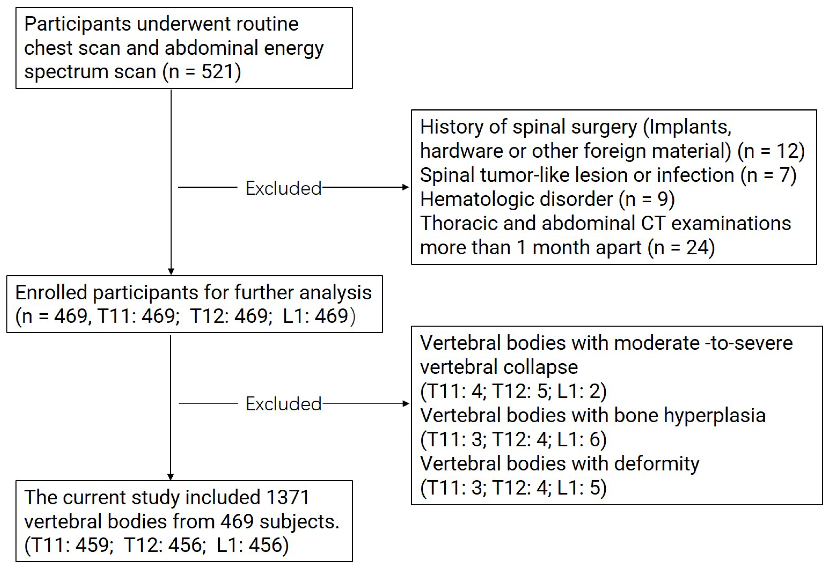
2.1. Population and Study Design
2.2. CT Imaging Protocol
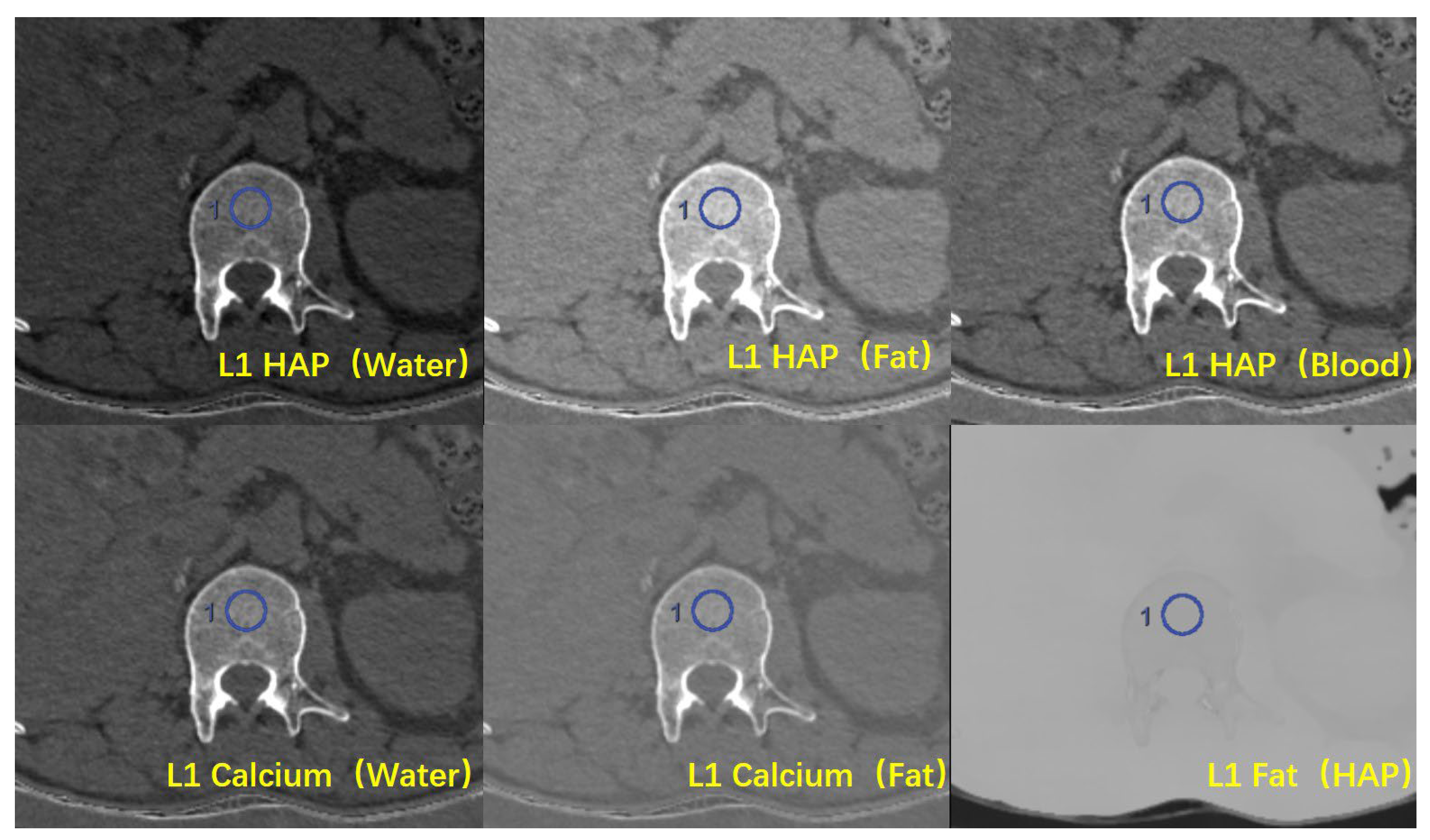
2.3. Image Reconstruction and Quantitative Data Measurement
2.4. Statistical Analysis
3. Results
3.1. Populations
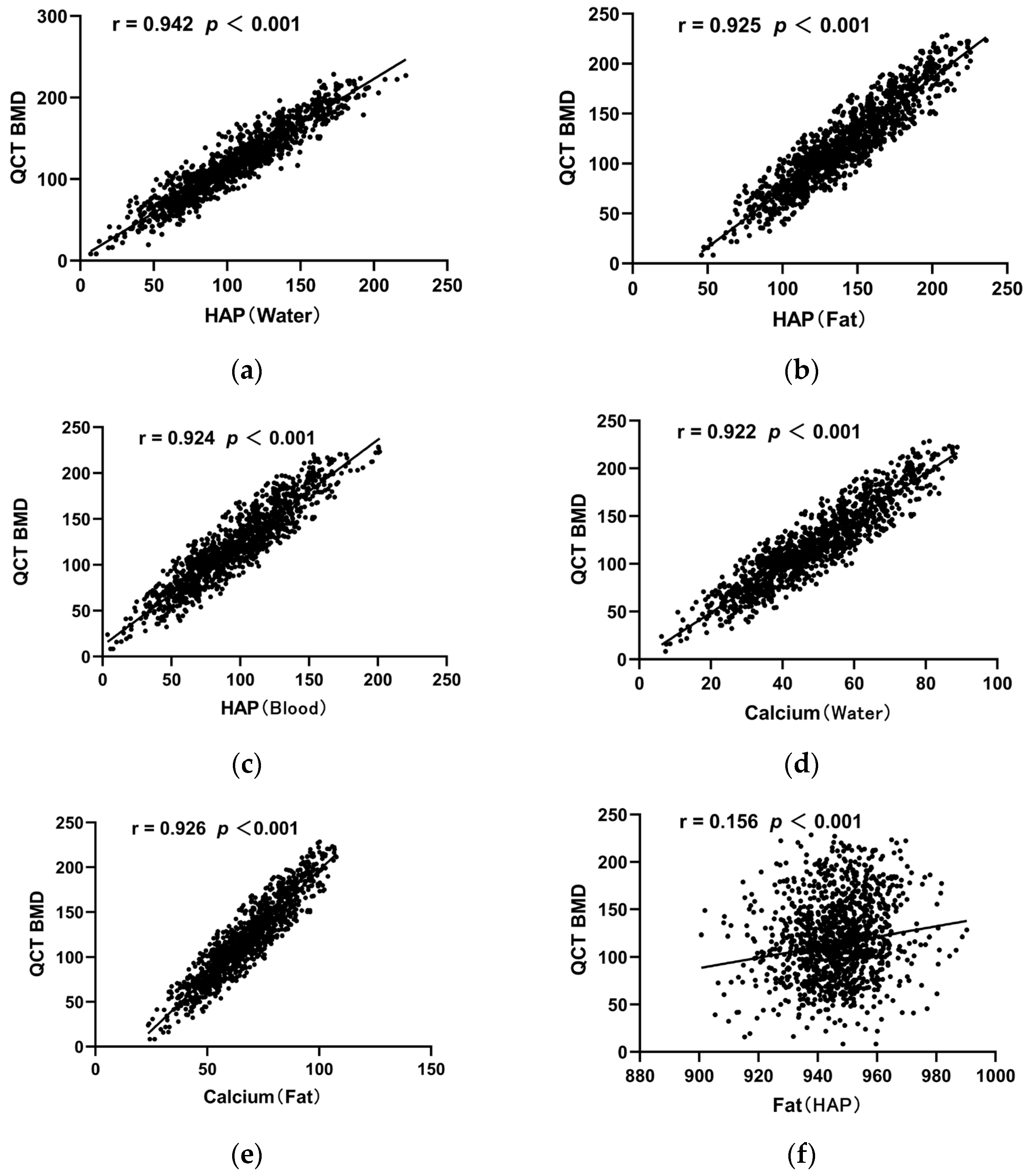
3.2. The Difference among Base Material Pairs and Correlation between DECT- and QCT-Derived BMD
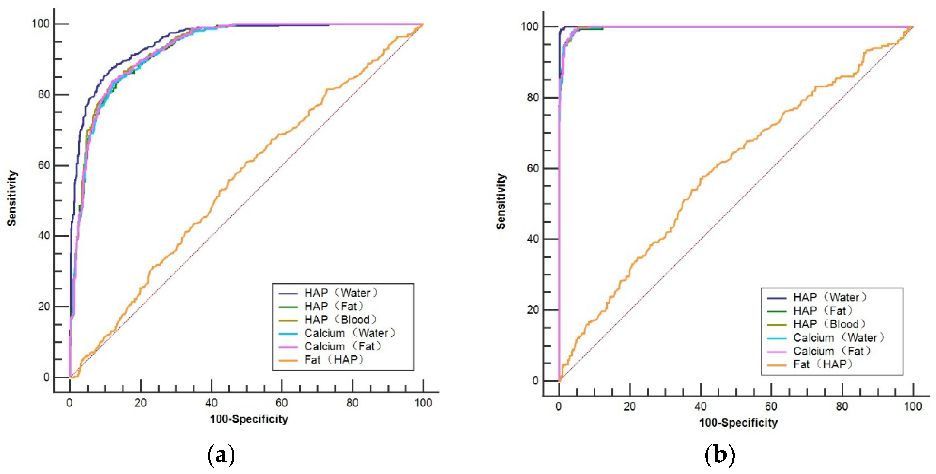
3.3. Diagnostic Effectiveness Evaluation Using Base Material Pairs Derived from DECT
4. Discussion
Author Contributions
Funding
Institutional Review Board Statement
Informed Consent Statement
Data Availability Statement
Conflicts of Interest
References
- Holmberg, T.; Möller, S.; Rothmann, M.J.; Gram, J.; Herman, A.P.; Brixen, K.; Tolstrup, J.S.; Høiberg, M.; Bech, M.; Rubin, K.H. Socioeconomic status and risk of osteoporotic fractures and the use of DXA scans: Data from the Danish population-based ROSE study. Osteoporos. Int. 2019, 30, 343–353. [Google Scholar] [CrossRef] [PubMed]
- Zeng, Q.; Li, N.; Wang, Q.; Feng, J.; Sun, D.; Zhang, Q.; Huang, J.; Wen, Q.; Hu, R.; Wang, L.; et al. The Prevalence of Osteoporosis in China, a Nationwide, Multicenter DXA Survey. J. Bone Miner. Res. 2019, 34, 1789–1797. [Google Scholar] [CrossRef] [PubMed]
- Stuckey, B.; Magraith, K.; Opie, N.; Zhu, K. Fracture risk prediction and the decision to treat low bone density. Aust. J. Gen. Pract. 2021, 50, 165–170. [Google Scholar] [CrossRef] [PubMed]
- Brown, C. Osteoporosis: Staying strong. Nature 2017, 550, S15–S17. [Google Scholar] [CrossRef]
- Wait, J.M.; Cody, D.; Jones, A.K.; Rong, J.; Baladandayuthapani, V.; Kappadath, S.C. Performance Evaluation of Material Decomposition with Rapid-Kilovoltage-Switching Dual-Energy CT and Implications for Assessing Bone Mineral Density. Am. J. Roentgenol. 2015, 204, 1234–1241. [Google Scholar] [CrossRef]
- Berry, M.E. Using DXA to Identify and Treat Osteoporosis in Pediatric Patients. Radiol. Technol. 2018, 89, 312–317. [Google Scholar] [PubMed]
- Bolotin, H.H. DXA in vivo BMD methodology: An erroneous and misleading research and clinical gauge of bone mineral status, bone fragility, and bone remodelling. Bone 2007, 41, 138–154. [Google Scholar] [CrossRef]
- Engelke, K.; Lang, T.; Khosla, S.; Qin, L.; Zysset, P.; Leslie, W.D.; Shepherd, J.A.; Schousboe, J.T. Clinical Use of Quantitative Computed Tomography (QCT) of the Hip in the Management of Osteoporosis in Adults: The 2015 ISCD Official Positions-Part I. J. Clin. Densitom. 2015, 18, 338–358. [Google Scholar] [CrossRef]
- Löffler, M.T.; Jacob, A.; Valentinitsch, A.; Rienmüller, A.; Zimmer, C.; Ryang, Y.M.; Baum, T.; Kirschke, J.S. Improved prediction of incident vertebral fractures using opportunistic QCT compared to DXA. Eur. Radiol. 2019, 29, 4980–4989. [Google Scholar] [CrossRef]
- Cicero, G.; Ascenti, G.; Albrecht, M.H.; Blandino, A.; Cavallaro, M.; D’Angelo, T.; Carerj, M.L.; Vogl, T.J.; Mazziotti, S. Extra-abdominal dual-energy CT applications: A comprehensive overview. Radiol. Med. 2020, 125, 384–397. [Google Scholar] [CrossRef]
- Rajiah, P.; Sundaram, M.; Subhas, N. Dual-Energy CT in Musculoskeletal Imaging: What Is the Role Beyond Gout? Am. J. Roentgenol. 2019, 213, 493–505. [Google Scholar] [CrossRef] [PubMed]
- Patino, M.; Prochowski, A.; Agrawal, M.D.; Simeone, F.J.; Gupta, R.; Hahn, P.F.; Sahani, D.V. Material Separation Using Dual-Energy CT: Current and Emerging Applications. Radiographics 2016, 36, 1087–1105. [Google Scholar] [CrossRef] [PubMed]
- Li, X.; Li, X.; Li, J.; Jiao, X.; Jia, X.; Zhang, X.; Fan, G.; Yang, J.; Guo, J. The accuracy of bone mineral density measurement using dual-energy spectral CT and quantitative CT: A comparative phantom study. Clin. Radiol. 2020, 75, 320.e9–320.e15. [Google Scholar] [CrossRef]
- Yue, D.; Li Fei, S.; Jing, C.; Ru Xin, W.; Rui Tong, D.; Ai Lian, L.; Luo, Y.H. The relationship between calcium (water) density and age distribution in adult women with spectral CT: Initial result compared to bone mineral density by dual-energy X-ray absorptiometry. Acta Radiol. 2019, 60, 762–768. [Google Scholar] [CrossRef]
- Wang, P.; She, W.; Mao, Z.; Zhou, X.; Li, Y.; Niu, J.; Jiang, M.; Huang, G. Use of routine computed tomography scans for detecting osteoporosis in thoracolumbar vertebral bodies. Skelet. Radiol. 2021, 50, 371–379. [Google Scholar] [CrossRef] [PubMed]
- Cheng, X.; Yuan, H.; Cheng, J.; Weng, X.; Xu, H.; Gao, J.; Huang, M.; Wáng, Y.X.J.; Wu, Y.; Xu, W.; et al. Chinese expert consensus on the diagnosis of osteoporosis by imaging and bone mineral density. Quant. Imaging Med. Surg. 2020, 10, 2066–2077. [Google Scholar] [CrossRef]
- Buenger, F.; Eckardt, N.; Sakr, Y.; Senft, C.; Schwarz, F. Correlation of Bone Density Values of Quantitative Computed Tomography and Hounsfield Units Measured in Native Computed Tomography in 902 Vertebral Bodies. World Neurosurg. 2021, 151, e599–e606. [Google Scholar] [CrossRef]
- Budoff, M.J.; Khairallah, W.; Li, D.; Gao, Y.L.; Ismaeel, H.; Flores, F.; Child, J.; Carson, S.; Mao, S.S. Trabecular bone mineral density measurement using thoracic and lumbar quantitative computed tomography. Acad. Radiol. 2012, 19, 179–183. [Google Scholar] [CrossRef]
- Szulc, P. Vertebral Fracture: Diagnostic Difficulties of a Major Medical Problem. J. Bone Miner. Res. 2018, 33, 553–559. [Google Scholar] [CrossRef]
- Anderson, P.A.; Polly, D.W.; Binkley, N.C.; Pickhardt, P.J. Clinical Use of Opportunistic Computed Tomography Screening for Osteoporosis. The Journal of bone and joint surgery. Am. Vol. 2018, 100, 2073–2081. [Google Scholar]
- Laval-Jeantet, A.M.; Roger, B.; Bouysee, S.; Bergot, C.; Mazess, R.B. Influence of vertebral fat content on quantitative CT density. Radiology 1986, 159, 463–466. [Google Scholar] [CrossRef] [PubMed]
- Gruenewald, L.D.; Koch, V.; Martin, S.S.; Yel, I.; Eichler, K.; Gruber-Rouh, T.; Lenga, L.; Wichmann, J.L.; Alizadeh, L.S.; Albrecht, M.H.; et al. Diagnostic accuracy of quantitative dual-energy CT-based volumetric bone mineral density assessment for the prediction of osteoporosis-associated fractures. Eur. Radiol. 2022, 32, 3076–3084. [Google Scholar] [CrossRef] [PubMed]
- Zhou, S.; Zhu, L.; You, T.; Li, P.; Shen, H.; He, Y.; Gao, H.; Yan, L.; He, Z.; Guo, Y.; et al. In vivo quantification of bone mineral density of lumbar vertebrae using fast kVp switching dual-energy CT: Correlation with quantitative computed tomography. Quant. Imaging Med. Surg. 2021, 11, 341–350. [Google Scholar] [CrossRef] [PubMed]
- Wichmann, J.L.; Booz, C.; Wesarg, S.; Kafchitsas, K.; Bauer, R.W.; Kerl, J.M.; Lehnert, T.; Vogl, T.J.; Khan, M.F. Dual-energy CT-based phantomless in vivo three-dimensional bone mineral density assessment of the lumbar spine. Radiology 2014, 271, 778–784. [Google Scholar] [CrossRef]
- Burian, E.; Grundl, L.; Greve, T.; Junker, D.; Sollmann, N.; Löffler, M.; Makowski, M.R.; Zimmer, C.; Kirschke, J.S.; Baum, T. Local Bone Mineral Density, Subcutaneous and Visceral Adipose Tissue Measurements in Routine Multi Detector Computed Tomography-Which Parameter Predicts Incident Vertebral Fractures Best? Diagnostics 2021, 11, 240. [Google Scholar] [CrossRef] [PubMed]
- Yamamoto, M.; Yamauchi, M.; Sugimoto, T. Prevalent vertebral fracture is dominantly associated with spinal microstructural deterioration rather than bone mineral density in patients with type 2 diabetes mellitus. PLoS ONE 2019, 14, e0222571. [Google Scholar] [CrossRef]
- Zhao, Y.; Huang, M.; Ding, J.; Zhang, X.; Spuhler, K.; Hu, S.; Li, M.; Fan, W.; Chen, L.; Zhang, X.; et al. Prediction of Abnormal Bone Density and Osteoporosis from Lumbar Spine MR Using Modified Dixon Quant in 257 Subjects with Quantitative Computed Tomography as Reference. J. Magn. Reson. Imaging 2019, 49, 390–399. [Google Scholar] [CrossRef]
- Cheng, X.; Blake, G.M.; Guo, Z.; Keenan Brown, J.; Wang, L.; Li, K.; Xu, L. Correction of QCT vBMD using MRI measurements of marrow adipose tissue. Bone 2019, 120, 504–511. [Google Scholar] [CrossRef]


| Participants (n = 469) | Sex | Male (n = 256) |
|---|---|---|
| Female (n = 213) | ||
| Average Age (y) | 63.24 (22~90) | |
| Vertebral bodies (n = 1371) | Osteoporosis (n = 393) | T11:103; T12:134; L1:156 |
| Osteopenia (n = 442) | T11:137; T12:145; L1:160 | |
| Normal (n = 536) | T11:219; T12:177; L1:140 |
| ICC | ICC 95% ICC | F | p | |
|---|---|---|---|---|
| QCT BMD (mg/cm3) | 0.990 | (0.984, 0.992) | 798.488 | 0.001 |
| HAP (water) (mg/cm3) | 0.977 | (0.952, 0.988) | 154.252 | 0.001 |
| HAP (fat) (mg/cm3) | 0.981 | (0.947, 0.991) | 209.703 | 0.001 |
| HAP (blood) (mg/cm3) | 0.978 | (0.936, 0.989) | 189.071 | 0.001 |
| Ca (water) (mg/cm3) | 0.976 | (0.954, 0.991) | 206.772 | 0.001 |
| Ca (fat) (mg/cm3) | 0.985 | (0.957, 0.990) | 172.767 | 0.001 |
| Fat (HAP) (mg/cm3) | 0.980 | (0.973, 0.985) | 60.168 | 0.001 |
| Bone States | HAP (Water) | HAP (Fat) | HAP (Blood) | Ca (Water) | Ca (Fat) | Fat (HAP) |
|---|---|---|---|---|---|---|
| (mg/cm3) | (mg/cm3) | (mg/cm3) | (mg/cm3) | (mg/cm3) | (mg/cm3) | |
| Normal (n = 536) | 134.78 ± 24.41 | 168.49 ± 23.31 | 123.19 ± 24.25 | 61.59 ± 10.74 | 80.52 ± 10.98 | 948.06 ± 12.42 |
| Osteopenia (n = 442) | 91.12 ± 14.89 | 127.92 ± 15.38 | 82.01 ± 14.87 | 42.85 ± 7.09 | 61.34 ± 7.07 | 946.13 ± 11.57 |
| Osteoporosis (n = 393) | 59.47 ± 15.65 | 97.72 ± 16.35 | 52.03 ± 15.99 | 28.63 ± 7.61 | 46.63 ± 7.83 | 943.91 ± 13.38 |
| Statistical value | 956.396 | 899.58 | 910.072 | 901.6 | 908.129 | 25.979 |
| p | 0.001 | 0.001 | 0.001 | 0.001 | 0.001 | 0.001 |
| HAP (Water) | HAP (Fat) | HAP (Blood) | Ca (Water) | Ca (Fat) | Fat (HAP) | |
|---|---|---|---|---|---|---|
| (mg/cm3) | (mg/cm3) | (mg/cm3) | (mg/cm3) | (mg/cm3) | (mg/cm3) | |
| AUC | 0.953 | 0.930 | 0.934 | 0.930 | 0.932 | 0.556 |
| 95% CI | 0.938–0.965 | 0.913–0.945 | 0.917–0.949 | 0.913–0.945 | 0.915–0.947 | 0.525–0.587 |
| p value | <0.0001 | <0.0001 | <0.0001 | <0.0001 | <0.0001 | <0.0001 |
| Youden index J | 0.7579 | 0.7045 | 0.7148 | 0.7086 | 0.7182 | 0.1103 |
| Criterion | ≤107.4 | ≤144.3 | ≤97.35 | ≤50.04 | ≤68.35 | ≤949 |
| Sensitivity | 86.88 | 84.62 | 83.94 | 83.48 | 83.94 | 60.86 |
| Specificity | 88.91 | 85.84 | 87.54 | 87.37 | 87.88 | 50.17 |
| HAP (Water) | HAP (Fat) | HAP (Blood) | Ca (Water) | Ca (Fat) | Fat (HAP) | |
|---|---|---|---|---|---|---|
| (mg/cm3) | (mg/cm3) | (mg/cm3) | (mg/cm3) | (mg/cm3) | (mg/cm3) | |
| AUC | 0.999 | 0.996 | 0.997 | 0.997 | 0.997 | 0.594 |
| 95% CI | 0.995–1.000 | 0.990–0.999 | 0.991–0.999 | 0.991–0.999 | 0.991–0.999 | 0.563–0.625 |
| p value | <0.0001 | <0.0001 | <0.0001 | <0.0001 | <0.0001 | <0.0001 |
| Youden index J | 0.9876 | 0.9001 | 0.8758 | 0.9514 | 0.9498 | 0.1732 |
| Criterion | ≤89.62 | ≤122.3 | ≤86.91 | ≤44.85 | ≤63.16 | ≤945.9 |
| Sensitivity | 99.24 | 95.67 | 99.24 | 99.24 | 99.24 | 57.25 |
| Specificity | 99.53 | 94.33 | 88.35 | 95.91 | 95.75 | 60.07 |
Disclaimer/Publisher’s Note: The statements, opinions and data contained in all publications are solely those of the individual author(s) and contributor(s) and not of MDPI and/or the editor(s). MDPI and/or the editor(s) disclaim responsibility for any injury to people or property resulting from any ideas, methods, instructions or products referred to in the content. |
© 2023 by the authors. Licensee MDPI, Basel, Switzerland. This article is an open access article distributed under the terms and conditions of the Creative Commons Attribution (CC BY) license (https://creativecommons.org/licenses/by/4.0/).
Share and Cite
Wang, X.; Li, B.; Tong, X.; Fan, Y.; Wang, S.; Liu, Y.; Fang, X.; Liu, L. Diagnostic Accuracy of Dual-Energy CT Material Decomposition Technique for Assessing Bone Status Compared with Quantitative Computed Tomography. Diagnostics 2023, 13, 1751. https://doi.org/10.3390/diagnostics13101751
Wang X, Li B, Tong X, Fan Y, Wang S, Liu Y, Fang X, Liu L. Diagnostic Accuracy of Dual-Energy CT Material Decomposition Technique for Assessing Bone Status Compared with Quantitative Computed Tomography. Diagnostics. 2023; 13(10):1751. https://doi.org/10.3390/diagnostics13101751
Chicago/Turabian StyleWang, Xu, Beibei Li, Xiaoyu Tong, Yong Fan, Shigeng Wang, Yijun Liu, Xin Fang, and Lei Liu. 2023. "Diagnostic Accuracy of Dual-Energy CT Material Decomposition Technique for Assessing Bone Status Compared with Quantitative Computed Tomography" Diagnostics 13, no. 10: 1751. https://doi.org/10.3390/diagnostics13101751
APA StyleWang, X., Li, B., Tong, X., Fan, Y., Wang, S., Liu, Y., Fang, X., & Liu, L. (2023). Diagnostic Accuracy of Dual-Energy CT Material Decomposition Technique for Assessing Bone Status Compared with Quantitative Computed Tomography. Diagnostics, 13(10), 1751. https://doi.org/10.3390/diagnostics13101751


