Article

SNP rs2920280 in PSCA Is Associated with Susceptibility to Gastric Mucosal Atrophy and Is a Promising Biomarker in Japanese Individuals with Helicobacter pylori Infection

1
Department of Gastroenterology and Nephrology, Faculty of Medicine, Tottori University, 36-1 Nishi-cho, Yonago 683-8504, Japan
2
Department of Gastroenterology and Hepatology, Nagasaki University Graduate School of Biological Sciences, 1-7-1 Sakamoto, Nagasaki 852-8501, Japan
3
Department of Pharmacotherapeutics, Nagasaki University Graduate School of Biomedical Sciences, 1-7-1 Sakamoto, Nagasaki 852-8501, Japan
4
Department of Surgical Oncology, Nagasaki University Graduate School of Biological Sciences, 1-7-1 Sakamoto, Nagasaki 852-8501, Japan
5
Fukuda Yutaka Clinic, 3-5 Hamaguchi-machi, Nagasaki 852-8107, Japan
6
Shunkaikai Inoue Hospital, 6-12 Takara-machi, Nagasaki 850-0045, Japan
7
Department of Gastroenterology, Nagasaki Medical Harbor Center, 6-39 Shinchi-machi, Nagasaki 850-8555, Japan
*
Authors to whom correspondence should be addressed.
These authors contributed equally to this work.
Diagnostics 2022, 12(8), 1988; https://doi.org/10.3390/diagnostics12081988
Submission received: 2 July 2022 / Revised: 31 July 2022 / Accepted: 1 August 2022 / Published: 16 August 2022
(This article belongs to the Section Pathology and Molecular Diagnostics)

Abstract

:
Helicobacter pylori infection results in gastric cancer (GC) with gastric mucosal atrophy (GMA). Some single-nucleotide polymorphisms (SNPs) in the prostate stem cell antigen gene (PSCA) are associated with GC and duodenal ulcers. However, the relationship of other identified SNPs in PSCA with these diseases remains unclear. Herein, the association between PSCA SNPs and GMA among 195 Japanese individuals with H. pylori infection was evaluated. The definition of GMA or non-GMA was based on serum pepsinogen levels or endoscopic findings. Five tag PSCA SNPs were analyzed using PCR high-resolution melting curve analysis with nonlabelled probes. The frequencies of alleles and the genotypes of each tag SNP were compared between the GMA and non-GMA groups. Subsequently, a genetic test was performed using associated SNPs as biomarkers to detect patients developing GMA. Two tag PSCA SNPs (rs2920280 and rs2294008) were related to GMA susceptibility. Individuals with the rs2920280 G/G genotype or the rs2294008 T/T genotype in PSCA had 3.5- and 2.1-fold susceptibility to GMA, respectively. In conclusion, SNP rs2920280 is a possible biomarker for detecting individuals developing GMA. PSCA polymorphisms may be useful biomarkers for predicting GMA linked to GC risk and a screening endoscopy strategy to detect GC related to early stage H. pylori associated GMA.

1. Introduction

Helicobacter pylori colonizes the gastric mucosa and infects no less than half of the entire human population [1]. Persistent H. pylori infection causes chronic stomach inflammation, eventually leading to gastric mucosal atrophy (GMA) [2]. Gastric cancer develops through a multistep process known as the persistent gastritis–GMA carcinogenic pathway, which is triggered by H. pylori infection [3]. Some individuals infected with this bacterium develop gastric cancer with advanced GMA or a precursor toward gastric cancer in a particular intestinal type [3,4]. This may be due to virulence differences among the H. pylori strains; environmental factors, such as salt intake and smoking; and the host’s genetic predisposition, including various single-nucleotide morphisms (SNPs) [5].
Prostate stem cell antigen (PSCA) is a membrane protein binding to cell surfaces through a glycosylphosphatidylinositol anchor [6]. PSCA expression is upregulated in prostate, bladder, and pancreatic cancer, but is downregulated in esophageal and gastric cancer [7,8,9,10,11,12,13,14]. PSCA knockdown in bladder cancer suppresses cancer cell proliferation and induces the expression of anti-inflammatory cytokines, such as interferon α/β (oncogenic effect) [15]. However, PSCA has growth inhibition activity on gastric cancer cells. This suggests that the physiological functions of PSCA include both oncogenic and tumor-suppressor effects in tissue- and cell-specific manners [6,7,8,9,10,11,12,13,14,15,16].
Genome-wide studies have revealed the association of SNP rs2294008 in PSCA with representative H. pylori related diseases, including gastric cancer and duodenal ulcer [7,8]. Although most studies on the relationship between PSCA genetic predisposition and gastroduodenal diseases and gastric cancers have focused on rs2294008 in PSCA, there is information on the association between the risk of gastric cancer and other PSCA genetic variations in the literature [17]. In the present work, an association study was conducted between several SNPs in PSCA and GMA susceptibility in Japanese individuals with H. pylori infection to identify new host genetic factors. The associated SNPs in PSCA can be used as new genetic biomarkers for predicting GMA development, which is a precancer lesion. In this study, we aimed to detect the useful stratification marker to assess gastric cancer risk related to H. pylori associated GMA.

2. Materials and Methods

2.1. Study Participants

We enrolled 195 individuals diagnosed as H. pylori positive with a serum H. pylori antibody titer ≥ 10 U/mL (E-plate Eiken H. pylori antibody II; Eiken Chemical, Tokyo, Japan) among 502 individuals who underwent upper gastrointestinal endoscopy for a healthy checkup between August and December 2013. The study requirements stated that participants must be below 80 years old without a history of stomach surgery or H. pylori eradication therapy. The study included 109 women and 86 men, with a mean age of 56.9 (29–77) years.

2.2. GMA Classification

GMA-positive individuals were determined using two methods: the serological pepsinogen (PG) method and the Kimura–Takemoto endoscopic classification system [18,19]. Participants with serum PG I levels ≤ 70 μg/L and a serum PG I/II ratio ≤ 3.0 were classified into the GMA group, while those who did not meet any of these requirements were classified into the non-GMA group [18].
Participants who were diagnosed through the endoscopic findings of C3 or O1 to O3 according to the Kimura–Takemoto endoscopic classification were classified into the GMA group, and those who were diagnosed through those of C1 or C2 were classified into the non-GMA group [19].

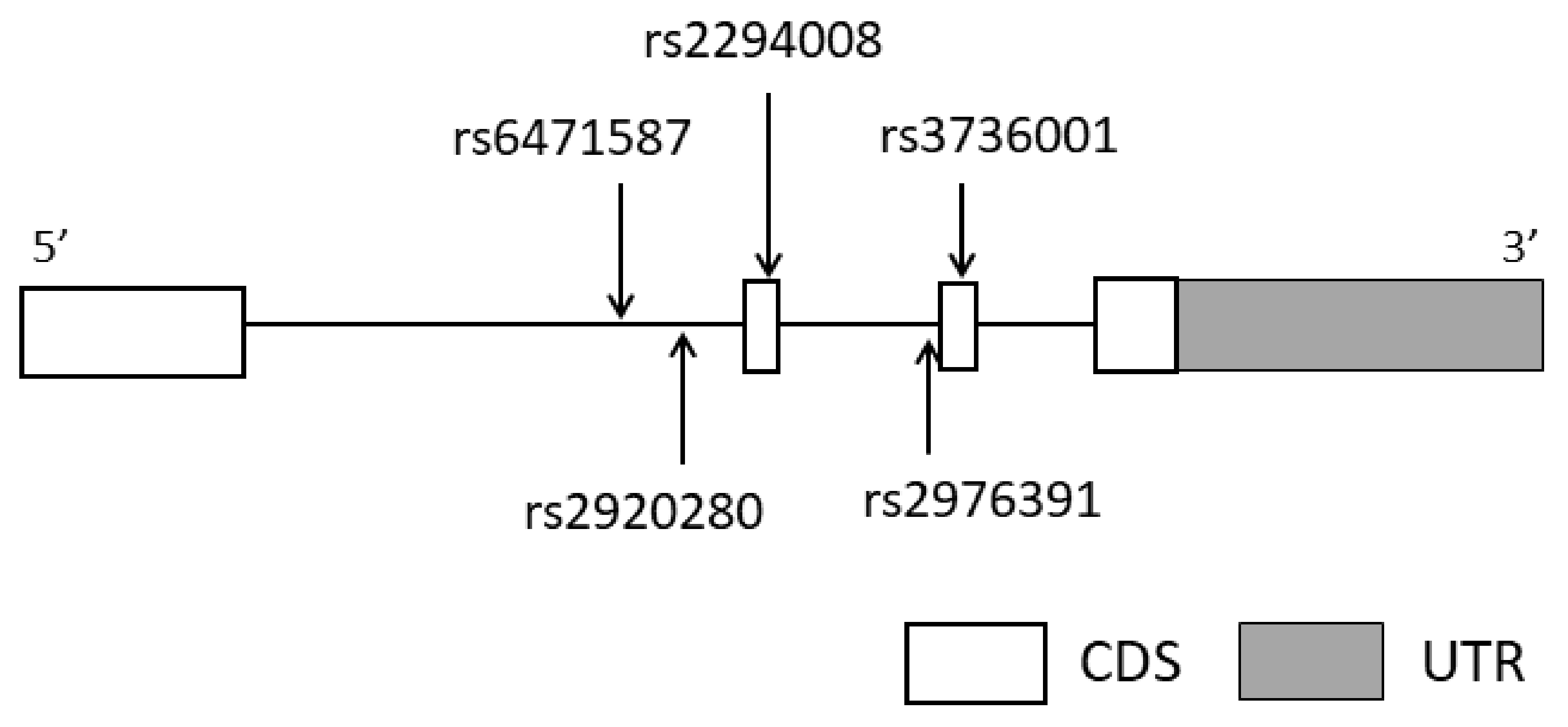
2.3. Selection of Tag SNPs in PSCA

PSCA (OMIM #602470, located at 8q24.3) was selected as the candidate gene for GMA susceptibility. Five tag SNPs (rs6471587, rs2920280, rs2294008, rs2976391, and rs3736001) were genotyped based on information concerning SNPs in PSCA according to previous methods [20,21]. In brief, we extracted the SNPs reported in Japanese people according to the International HapMap database in the region from the candidate gene and promoter to 2 kb upstream. Then, we extracted five SNPs with minor allele frequency over 0.05 and that met linkage disequilibrium (r2 > 0.8) using Haploview ver. 4.2 as tag SNPs. The gene structure and locations of the tag SNP sites in PSCA are shown in Figure 1.

2.4. SNP Genotyping

Each participant’s peripheral blood was stored in a blood collection tube with EDTA. Genomic DNA was extracted from the peripheral blood of each participant using Nucleo Spin® (Takara, Shiga, Japan) according to the manufacturer’s instructions. Genotyping of the five tag SNPs in PSCA was carried out by the probe-based high-resolution melting method [22,23] using the primers, probes, and annealing temperatures listed in Table 1.

2.5. Statistical Analyses

The mean participant age, presented as the mean ± standard deviation, was compared between the GMA and non-GMA groups using the Mann–Whitney U test. Likewise, sex was compared using the chi-square test. Statistical analyses were performed using SPSS 17 (SPSS Japan, Tokyo, Japan) or Prism 6 (GraphPad Software Inc., La Jolla, CA, USA).
A chi-square test was performed based on the expectation-maximization algorithm using the SNPAlyze® ver. 7.1 standard software package (DynaCom Inc., Yokohama, Japan) to determine whether each tag SNP was in Hardy–Weinberg equilibrium. The frequencies of alleles and genotypes of each tag SNP between the GMA and non-GMA groups were compared by chi-square or Fisher’s exact test with an odds ratio (OR) and 95% confidence interval in three different inheritance models: the minor allele, minor allele dominant, and minor allele recessive using the SNPAlyze® ver.7.1 standard software package and SPSS 17.
In addition to univariate analyses, multivariate logistic regression analysis was conducted to assess the interaction of genetic and environmental factors with GMA susceptibility using SPSS 17. p < 0.05 was considered to indicate significance.

3. Results

3.1. Association between PSCA Polymorphisms and GMA Susceptibility by the PG Method

The frequencies of alleles and genotypes of the five tag SNPs in PSCA showed no significant differences in the allele and genotype frequencies between the GMA (n = 92) and non-GMA (n = 103) groups in the three inheritance models: allele, dominant, and recessive models (Table 2).

3.2. Association between PSCA Polymorphisms and GMA Susceptibility Based on Endoscopic Findings According to the Kimura–Takemoto Classification

The frequency of the minor G allele of rs2920280 in PSCA in the minor allele model was significantly higher in the GMA group (n = 123, 50.8%) than in the non-GMA group (n = 72, 37.5%, p = 0.011, OR = 1.722; Table 3), thereby indicating a 1.7-fold increase in susceptibility to GMA. Likewise, the frequency of the minor homozygosity (G/G genotype of rs2920280 in PSCA) in the minor allele recessive model was significantly higher in the GMA group than in the non-GMA group (27.7% vs. 9.7%, p = 0.003, OR = 3.547; Table 3), showing that there was an approximately 3.5-fold increase in susceptibility to GMA. In addition, the frequency of the minor C allele of rs2294008 in PSCA in the minor allele model was significantly lower in the GMA group than in the non-GMA group (35.8% vs. 46.5%, p = 0.036, OR = 0.640; Table 3), indicating a 0.64-fold susceptibility to GMA. Thus, the presence of the C allele of rs2294008 in PSCA indicated an approximately 1.6-fold increased resistance to GMA development. Moreover, the frequency of the T/C or C/C genotype of rs2294008 in PSCA in the minor allele dominant model was significantly lower in the GMA group than in the non-GMA group (61.0% vs. 76.4%, p = 0.028, OR = 0.483; Table 3), indicating a 0.48-fold susceptibility to GMA. Thus, the possession of the T/C or C/C genotype of rs2294008 in PSCA indicated an approximately 2.1-fold increased resistance to GMA development. Conversely, the possession of the major homozygous T/T genotype of rs2294008 in PSCA indicated an approximately 2.1-fold increase in susceptibility to GMA.

3.3. Comparison of Clinical Characteristics between GMA and Non-GMA Groups Based on Endoscopic Findings According to Kimura–Takemoto Classification

According to the mean age, patients in the GMA group were approximately six years older than those in the non-GMA group (59.1 vs. 53.2, p = 0.0002; Table 3), indicating that the older participants showed greater GMA susceptibility. However, there was no significant difference in sex between the GMA and non-GMA groups (Table 3).

3.4. Contribution of Gene–Environment Interactions to GMA Susceptibility

The results of univariate analyses revealed that the G/G genotype of rs2920280 in PSCA, the T/T genotype of rs2294008 in PSCA, and age contributed to GMA susceptibility (Table 3). The results of ultivariate analysis indicated that three independent factors contributed to susceptibility to GMA with statistical significance: the G/G genotype of rs2920280 in PSCA, T/T genotype of rs2294008 in PSCA, and age (Table 4 and Table 5). Furthermore, genetic testing indicated that the G/G genotype of rs2920280 in PSCA, with a sensitivity of 27.6%, specificity of 90.3%, positive predictive value (PPV) of 82.9%, and negative predictive value of 42.2%, was more useful as a biomarker than the T/T genotype of rs2294008 in PSCA (Table 6).

4. Discussion

The present study suggests that PSCA polymorphisms contributed to the onset and/or development of GMA in H. pylori positive Japanese individuals who were endoscopically evaluated using the Kimura–Takemoto classification. Individuals with the G/G genotype of rs2920280 or the T/T genotype of rs2294008 in PSCA showed a 3.5- or 2.1-fold increase in susceptibility to GMA, respectively. The results of multivariate analysis also indicated that age is an independent factor that significantly contributed to susceptibility to GMA. This result agrees with that of a previous study, in which over 30% of patients’ gastric mucosa worsened during the atrophic and intestinal metaplasia phases with increasing age [24].
PSCA is upregulated in bladder cancer, renal carcinoma, and pancreatic cancer, and is downregulated in gastric, esophagus, and gallbladder cancer [25,26]. In the field of gastric and esophagus cancer, the downregulation of PSCA promotes the proliferation of cancer cells, and patients whose PSCA is downregulated have poor prognoses [26,27]. These data suggest that PSCA has a tumor-suppressing effect in gastric and esophagus cancer development. Zhang et al. reported that PSCA arrests cell cycle progression and promotes cell differentiation in esophageal squamous cell carcinoma by binding to retinoblastoma 1-inducible coiled-coil 1 (RB1CC1) [26]. Although the precise function of PSCA in gastric cancer remains unknown [28], the fact that PSCA protein is mainly expressed in the isthmus, which contains stem cells and precursors of cells, and in a variety of cell lineage and differentiation stage, indicates that PSCA is involved in the differentiation [7]. Moreover, a slower growth rate of the PSCA expression cells than that without PSCA expression suggests that PSCA arrests the cell cycle without cell death [7].
rs2294008T reduces PSCA mRNA expression rather than rs2294008C in bladder cancer [29]. The T allele of rs2294008, which is considered a risk allele in gastric cancer, upregulated the expression of PSCA mRNA compared with the C allele of rs22904008 both in normal gastric and gastric cancer tissues, and the prognosis worsened with increased mRNA expression [30]. These contradictory results may be due to the qualitative change in traits in PSCA [30]. rs2294008T produces a long PSCA, which is nine amino acids longer than the short PSCA produced by rs2294008C [8]. Long PSCA is located on the membrane and has a growth-promoting ability, whereas short PSCA is in the cytoplasm and easily disassembles [8]. The signal analysis of long PSCA is not fully elucidated. Further investigation is required.
SNP rs2920280 may be more useful as a prognostic biomarker for detecting individuals at risk of developing GMA among H. pylori infected individuals than SNP rs2294008. The G/G genotype of rs2920280 in PSCA showed a higher OR, lower p-value, higher specificity, and higher PPV than the T/T genotype of rs2294008, indicating that the G/G genotype is a more useful biomarker despite its lower sensitivity (Table 6).
The lower sensitivity of the biomarker rs2294008 implies the detection of only one-quarter of individuals developing GMA. Nevertheless, the higher PPV implies that approximately 83% of H. pylori positive individuals with the G/G genotype of rs2920280 in PSCA develop GMA. These high-risk individuals may require careful observation and/or occasional treatment for developing GMA and gastric cancer because early initiation of eradication treatment may delay the progression toward advanced GMA [31,32,33].
SNP rs2920280 is located within intron 1 of PSCA (Figure 1). The results of GWAS analysis revealed that nine PSCA SNPs, including rs2294008 and rs2920283, are identified as risk SNPs for gastric atrophy and severe gastric atrophy [34]. Sophie K.M. Heinrichs et al. conducted eQTL analysis using 143 subjects of German descent with intestinal metaplasia [35]. They reported that the lead eQTL for PSCA is rs2920283, and an upregulated PSCA mRNA expression in gastric tissue was observed in gastric cancer risk allele carriers. Although we do not have data indicating the frequency of the genotype at rs2920283, rs2920280, which has linkage distribution (D’ = 0.971, r2 = 0.600) to rs2920283 according to Ensembl (https://asia.ensembl.org/Homo_sapiens/Info/Index accessed on 29 July 2022), may increase PSCA mRNA. However, the localization and function of PSCA with rs2920280G are not fully elucidated. Further experiments are needed.
Although early detection and eradication of H. pylori infection in adolescents is important, the benefit and harm of the eradication therapy should be always considered. In addition, these two SNP biomarkers would be useful if combined with the ABC methods, which are used to evaluate the conditions of the stomach based on the noninvasive methods of measuring pepsinogen and H. pylori antibody [36]. Stratifying the patients between the high- and the low-risk groups would be helpful for deciding whether the patients should take medicine or encourage frequent endoscopic surveillance for gastric cancer identification.
Our study had some limitations. First, the sample size was quite small. The minor allele frequencies of associated SNPs ranged from 10% to 20%, the OR was 2 to 3.5, and the p-values ranged from 0.03 to 0.003 in this study. Further prospective studies with larger sample sizes and higher statistical power are warranted to confirm the significant positive association between PSCA SNPs and the development to GMA. Second, the PSCA SNPs that met linkage disequilibrium (r2 > 0.8) were selected as tag SNPs. We could not deny that rs2294008 and rs2920280 were completely in linkage disequilibrium because these two SNPs were located within a short distance from each other. We separately included these two SNPs in the analysis in association with environmental factors and age. Third, this study did not include a correlation analysis between the role of PSCA genetic variations in GMA and gastric cancer, and there was no follow-up survey of the enrolled subjects. In addition, the CagA virulence factor of the bacterium was not examined, although almost all Japanese people harbor the East-Asia-type virulent H. pylori strain [37]. Nevertheless, such genetic testing may provide future directions for precision medicine for individuals more likely to develop GMA and encourage frequent endoscopic surveillance for gastric cancer identification or its prevention by eradication therapy. Again, adolescents with risky SNPs of PSCA would potentially be at risk of GMA development, linking to GC decades later.

5. Conclusions

Our study findings indicate that PSCA SNPs rs2920280 might be associated with GMA in Japanese individuals with H. pylori infection. PSCA SNPs consisting of the well-known T/T genotype of rs2294008 and the newly identified G/G genotype of rs2920280 can be a useful biomarker for predicting GMA development. Accordingly, we encourage H. pylori infected individuals, who cannot or did not eradicate H. pylori and possess these two SNPs, to undergo timely endoscopic screening for early-stage gastric cancer. Further investigations regarding PSCA polymorphisms and their functions are required to provide pathophysiological findings and establish reliable biomarkers, finally leading to the development of new therapeutic GMA drugs to reduce gastric cancer risk.

Author Contributions

Conceptualization, K.T. and H.I.; methodology, T.I., S.T., K.T., D.F. and K.O.; software, T.I. and K.T.; validation, T.S. (Takuki Sakaguchi), T.I. and K.T.; formal analysis, T.S. (Takuki Sakaguchi), T.I. and K.T.; investigation, T.S. (Takuki Sakaguchi), S.T., D.F., K.O., T.S. (Takaaki Sugihara), K.M., T.K., T.H. (Tetsuro Honda) and T.H. (Tatsuro Hirayama); data curation, K.T.; writing—original draft preparation, T.S. (Takuki Sakaguchi), S.T., H.I. and K.T.; writing—review and editing, H.I. and K.T.; supervision, K.N. and K.T.; project administration, H.I.; funding acquisition, K.T. All authors have read and agreed to the published version of the manuscript.

Funding

This work was supported by a research grant from the Non-Profit Organization Aimed at Supporting Community Medicine Research in Nagasaki, Japan (Kazuhiro Tsukamoto).

Institutional Review Board Statement

The study protocol was approved by the Ethics Committee for Human Genome and Gene Analysis at Nagasaki University (No. 120221, approved on 16 February 2012).

Informed Consent Statement

Written informed consent was obtained from all the participants.

Data Availability Statement

Not applicable.

Acknowledgments

We are grateful to the individuals who participated in this study.

Conflicts of Interest

The authors declare no conflict of interest. The funders had no role in the design of the study; in the collection, analyses, or interpretation of data; in the writing of the manuscript; or in the decision to publish the results.

References

  1. Yamaoka, Y. Mechanisms of disease: Helicobacter pylori virulence factors. Nat. Rev. Gastroenterol. Hepatol. 2010, 7, 629–641. [Google Scholar] [CrossRef]
  2. Malfertheiner, P.; Megraud, F.; O’Morain, C.A.; Atherton, J.; Axon, A.T.; Bazzoli, F.; Gensini, G.F.; Gisbert, J.P.; Graham, D.Y.; Rokkas, T.; et al. Management of Helicobacter pylori infection—The Maastricht IV/Florence Consensus Report. Gut 2012, 61, 646–664. [Google Scholar] [CrossRef] [PubMed]
  3. Correa, P. Human gastric carcinogenesis: A multistep and multifactorial process--First American Cancer Society Award Lecture on Cancer Epidemiology and Prevention. Cancer Res. 1992, 52, 6735–6740. [Google Scholar] [PubMed]
  4. Uemura, N.; Okamoto, S.; Yamamoto, S.; Matsumura, N.; Yamaguchi, S.; Yamakido, M.; Taniyama, K.; Sasaki, N.; Schlemper, R.J. Helicobacter pylori infection and the development of gastric cancer. N. Engl. J. Med. 2001, 345, 784–789. [Google Scholar] [CrossRef] [PubMed]
  5. Saeki, N.; Ono, H.; Sakamoto, H.; Yoshida, T. Genetic factors related to gastric cancer susceptibility identified using a genome-wide association study. Cancer Sci. 2013, 104, 1–8. [Google Scholar] [CrossRef]
  6. Reiter, R.E.; Gu, Z.; Watabe, T.; Thomas, G.; Szigeti, K.; Davis, E.; Wahl, M.; Nisitani, S.; Yamashiro, J.; Le Beau, M.M.; et al. Prostate stem cell antigen: A cell surface marker overexpressed in prostate cancer. Proc. Natl. Acad. Sci. USA 1998, 95, 1735–1740. [Google Scholar] [CrossRef]
  7. Sakamoto, H.; Yoshimura, K.; Saeki, N.; Katai, H.; Shimoda, T.; Matsuno, Y.; Saito, D.; Sugimura, H.; Tanioka, F.; Kato, S.; et al. Genetic variation in PSCA is associated with susceptibility to diffuse-type gastric cancer. Nat. Genet. 2008, 40, 730–740. [Google Scholar] [CrossRef]
  8. Tanikawa, C.; Urabe, Y.; Matsuo, K.; Kubo, M.; Takahashi, A.; Ito, H.; Tajima, K.; Kamatani, N.; Nakamura, Y.; Matsuda, K. A genome-wide association study identifies two susceptibility loci for duodenal ulcer in the Japanese population. Nat. Genet. 2012, 44, 430–434. [Google Scholar] [CrossRef]
  9. Lochhead, P.; Frank, B.; Hold, G.L.; Rabkin, C.S.; Ng, M.T.; Vaughan, T.L.; Risch, H.A.; Gammon, M.D.; Lissowska, J.; Weck, M.N.; et al. Genetic variation in the prostate stem cell antigen gene and upper gastrointestinal cancer in white individuals. Gastroenterology 2011, 140, 435–441. [Google Scholar] [CrossRef]
  10. Matsuo, K.; Tajima, K.; Suzuki, T.; Kawase, T.; Watanabe, M.; Shitara, K.; Misawa, K.; Ito, S.; Sawaki, A.; Muro, K.; et al. Association of prostate stem cell antigen gene polymorphisms with the risk of stomach cancer in Japanese. Int. J. Cancer 2009, 125, 1961–1964. [Google Scholar] [CrossRef]
  11. Lu, Y.; Chen, J.; Ding, Y.; Jin, G.; Wu, J.; Huang, H.; Deng, B.; Hua, Z.; Zhou, Y.; Shu, Y.; et al. Genetic variation of PSCA gene is associated with the risk of both diffuse- and intestinal-type gastric cancer in a Chinese population. Int. J. Cancer 2010, 127, 2183–2189. [Google Scholar] [CrossRef]
  12. Argani, P.; Rosty, C.; Reiter, R.E.; Wilentz, R.E.; Murugesan, S.R.; Leach, S.D.; Ryu, B.; Skinner, H.G.; Goggins, M.; Jaffee, E.M.; et al. Discovery of new markers of cancer through serial analysis of gene expression: Prostate stem cell antigen is overexpressed in pancreatic adenocarcinoma. Cancer Res. 2001, 61, 4320–4324. [Google Scholar]
  13. De Nooij-van Dalen, A.G.; van Dongen, G.A.; Smeets, S.J.; Nieuwenhuis, E.J.; Stigter-van Walsum, M.; Snow, G.B.; Brakenhoff, R.H. Characterization of the human Ly-6 antigens, the newly annotated member Ly-6K included, as molecular markers for head-and-neck squamous cell carcinoma. Int. J. Cancer 2003, 103, 768–774. [Google Scholar] [CrossRef]
  14. Wu, X.; Ye, Y.; Kiemeney, L.A.; Sulem, P.; Rafnar, T.; Matullo, G.; Seminara, D.; Yoshida, T.; Saeki, N.; Andrew, A.S.; et al. Genetic variation in the prostate stem cell antigen gene PSCA confers susceptibility to urinary bladder cancer. Nat. Genet. 2009, 41, 991–995. [Google Scholar] [CrossRef]
  15. Marra, E.; Uva, P.; Viti, V.; Simonelli, V.; Dogliotti, E.; De Rinaldis, E.; Lahm, A.; La Monica, N.; Nicosia, A.; Ciliberto, G.; et al. Growth delay of human bladder cancer cells by Prostate Stem Cell Antigen downregulation is associated with activation of immune signaling pathways. BMC Cancer 2010, 10, 129. [Google Scholar] [CrossRef]
  16. Zhao, Z.; Ma, W.; Zeng, G.; Qi, D.; Ou, L.; Liang, Y. Small interference RNA-mediated silencing of prostate stem cell antigen attenuates growth, reduces migration and invasion of human prostate cancer PC-3M cells. Urol. Oncol. 2013, 31, 343–351. [Google Scholar] [CrossRef]
  17. Rizzato, C.; Kato, I.; Plummer, M.; Muñoz, N.; Canzian, F. Genetic variation in PSCA and risk of gastric advanced preneoplastic lesions and cancer in relation to Helicobacter pylori infection. PLoS ONE 2013, 8, e73100. [Google Scholar] [CrossRef]
  18. Mukoubayashi, C.; Yanaoka, K.; Ohata, H.; Arii, K.; Tamai, H.; Oka, M.; Ichinose, M. Serum pepsinogen and gastric cancer screening. Intern. Med. 2007, 46, 261–266. [Google Scholar] [CrossRef]
  19. Kimura, K. An endoscopic recognition of the atrophic border and its significance in chronic gastritis. Endoscopy 1969, 13, 87–97. [Google Scholar] [CrossRef]
  20. Nanashima, K.; Mawatari, T.; Tahara, N.; Higuchi, N.; Nakaura, A.; Inamine, T.; Kondo, S.; Yanagihara, K.; Fukushima, K.; Suyama, N.; et al. Genetic variants in antioxidant pathway: Risk factors for hepatotoxicity in tuberculosis patients. Tuberculosis 2012, 92, 253–259. [Google Scholar] [CrossRef]
  21. Urabe, S.; Isomoto, H.; Ishida, T.; Maeda, K.; Inamine, T.; Kondo, S.; Higuchi, N.; Sato, K.; Uehara, R.; Yajima, H.; et al. Genetic Polymorphisms of IL-17F and TRAF3IP2 Could Be Predictive Factors of the Long-Term Effect of Infliximab against Crohn’s Disease. Biomed. Res. Int. 2015, 2015, 416838. [Google Scholar] [CrossRef] [PubMed]
  22. Araki, C.; Yoshimura, M.; Fukumitsu, Y.; Ma, S.; Ishida, T.; Urabe, S.; Matsushima, K.; Honda, T.; Uehara, R.; Fukuda, Y.; et al. The evidence of genetic polymorphisms of genes involved in the P2RX7 signaling pathway as predictive biomarkers for response and loss of response to infliximab against Crohn’s disease. Integr. Mol. Med. 2016, 3, 1–15. [Google Scholar] [CrossRef]
  23. Yamashita, A.; Inamine, T.; Suzuki, S.; Fukuda, S.; Unoike, M.; Kawafuchi, Y.; Machida, H.; Isomoto, H.; Nakao, K.; Tsukamoto, K. Genetic variants of SMAD2/3/4/7 are associated with susceptibility to ulcerative colitis in a Japanese genetic background. Immunol. Lett. 2019, 207, 64–72. [Google Scholar] [CrossRef] [PubMed]
  24. Chooi, E.Y.; Chen, H.M.; Miao, Q.; Weng, Y.R.; Chen, X.Y.; Ge, Z.Z.; Xiao, S.D.; Fang, J.Y. Chronic atrophic gastritis is a progressive disease: Analysis of medical reports from Shanghai (1985–2009). Singap. Med. J. 2012, 53, 318–324. [Google Scholar]
  25. Toyoshima, O.; Tanikawa, C.; Yamamoto, R.; Watanabe, H.; Yamashita, H.; Sakitani, K.; Yoshida, S.; Kubo, M.; Matsuo, K.; Ito, H.; et al. Decrease in PSCA expression caused by Helicobacter pylori infection may promote progression to severe gastritis. Oncotarget 2018, 9, 3936–3945. [Google Scholar] [CrossRef]
  26. Zhang, L.Y.; Wu, J.L.; Qiu, H.B.; Dong, S.S.; Zhu, Y.H.; Lee, V.H.; Qin, Y.R.; Li, Y.; Chen, J.; Liu, H.B.; et al. PSCA acts as a tumor suppressor by facilitating the nuclear translocation of RB1CC1 in esophageal squamous cell carcinoma. Carcinogenesis 2016, 37, 320–332. [Google Scholar] [CrossRef]
  27. Xu, L.P.; Qiu, H.B.; Yuan, S.Q.; Chen, Y.M.; Zhou, Z.W.; Chen, Y.B. Downregulation of PSCA promotes gastric cancer proliferation and is related to poor prognosis. J. Cancer 2020, 11, 2708–2715. [Google Scholar] [CrossRef]
  28. Saeki, N.; Gu, J.; Yoshida, T.; Wu, X. Prostate stem cell antigen: A Jekyll and Hyde molecule? Clin. Cancer Res. 2010, 16, 3533–3538. [Google Scholar] [CrossRef]
  29. Yang, J.; Li, W.; Zhang, Z.; Shen, J.; Zhang, N.; Yang, M.; Yang, M.; Yu, Y. PSCArs2294008 T polymorphism increases the risk of bladder cancer in Bai, Dai, and Han ethnicity in China and a potential mechanism. Genes Genom. 2018, 40, 531–541. [Google Scholar] [CrossRef]
  30. Sung, H.; Hu, N.; Yang, H.H.; Giffen, C.A.; Zhu, B.; Song, L.; Su, H.; Wang, C.; Parisi, D.M.; Goldstein, A.M.; et al. Association of high-evidence gastric cancer susceptibility loci and somatic gene expression levels with survival. Carcinogenesis 2017, 38, 1119–1128. [Google Scholar] [CrossRef]
  31. Choi, I.J.; Kook, M.C.; Kim, Y.I.; Cho, S.J.; Lee, J.Y.; Kim, C.G.; Park, B.; Nam, B.H. Helicobacter pylori Therapy for the Prevention of Metachronous Gastric Cancer. N. Engl. J. Med. 2018, 378, 1085–1095. [Google Scholar] [CrossRef]
  32. Wong, B.C.; Lam, S.K.; Wong, W.M.; Chen, J.S.; Zheng, T.T.; Feng, R.E.; Lai, K.C.; Hu, W.H.; Yuen, S.T.; Leung, S.Y.; et al. Helicobacter pylori eradication to prevent gastric cancer in a high-risk region of China: A randomized controlled trial. JAMA 2004, 291, 187–194. [Google Scholar] [CrossRef]
  33. Leung, W.K.; Lin, S.R.; Ching, J.Y.; To, K.F.; Ng, E.K.; Chan, F.K.; Lau, J.Y.; Sung, J.J. Factors predicting progression of gastric intestinal metaplasia: Results of a randomised trial on Helicobacter pylori eradication. Gut 2004, 53, 1244–1249. [Google Scholar] [CrossRef]
  34. Hishida, A.; Ugai, T.; Fujii, R.; Nakatochi, M.; Wu, M.C.; Ito, H.; Oze, I.; Tajika, M.; Niwa, Y.; Nishiyama, T.; et al. GWAS analysis reveals a significant contribution of PSCA to the risk of Heliobacter pylori-induced gastric atrophy. Carcinogenesis 2019, 40, 661–668. [Google Scholar] [CrossRef]
  35. Heinrichs, S.K.M.; Hess, T.; Becker, J.; Hamann, L.; Vashist, Y.K.; Butterbach, K.; Schmidt, T.; Alakus, H.; Krasniuk, I.; Höblinger, A.; et al. Evidence for PTGER4, PSCA, and MBOAT7 as risk genes for gastric cancer on the genome and transcriptome level. Cancer Med. 2018, 7, 5057–5065. [Google Scholar] [CrossRef]
  36. Yamaguchi, Y.; Nagata, Y.; Hiratsuka, R.; Kawase, Y.; Tominaga, T.; Takeuchi, S.; Sakagami, S.; Ishida, S. Gastric Cancer Screening by Combined Assay for Serum Anti-Helicobacter pylori IgG Antibody and Serum Pepsinogen Levels—The ABC Method. Digestion 2016, 93, 13–18. [Google Scholar] [CrossRef]
  37. Hatakeyama, M. Helicobacter pylori CagA and gastric cancer: A paradigm for hit-and-run carcinogenesis. Cell Host Microbe 2014, 15, 306–316. [Google Scholar] [CrossRef]
Figure 1. Gene structure and locations of the genotyped tag single-nucleotide polymorphism (SNP) sites in prostate stem cell antigen gene (PSCA). The horizontal bar indicates the genomic sequence of PSCA. Open boxes and lines represent exons and introns, respectively, with the widths not representative of the base-pair length. The dark gray box shows the untranslated region (UTR). Vertical arrows indicate genotyped tag SNP sites in this study.
Diagnostics 12 01988 g001
Table 1. Information regarding genotyping of tag SNPs in PSCA.
SNPPrimer Sequences
(5′ to 3′)
Annealing Temperature (°C)
forwardTCAGTGCACAGCTTGATGGT
rs6471587reverseCCCCGTATAACCCAGACATT56
probeCTGGTTGGCAATCGCTTGCAAGAGTTATC
forwardGGAACAGCCAGCGTTAACAT
rs2920280reverseCATCTTCTGGTTGGCAATCG55
probeGTCTCCACAACCCGGTATAACCCAGA
forwardCCTCACTGGCTCCAGGAAAC
rs2294008reverseAAGCCTGCCATCAACAGGG58
probeCCACCAGTGACCATGAAGGCTGTGCT
forwardGGAGGAGACATTGAGGGTGA
rs2976391reverseTTTGCAGGAGTAGCACAGCA57
probeCAGCCTCTGAGGCCCCTCCCACTCCA
forwardTGCTGTGCTACTCCTGCAAA
rs3736001reverseAGGTGGAAGGAAAACAGCAC56
probeTGCCTGCAGGTGGAGAACTGCACCC
Abbreviations: SNP, single-nucleotide polymorphism; PSCA, prostate stem cell antigen.
Table 2. Allele and genotype frequencies of tag SNPs in PSCA for the GMA and non-GMA groups at the selected serum PG I level and the specified PG I/II ratio.
SNPGenotypeGMA (%) Non-GMA (%)Inheritance ModelOR (95% CI)p-Value
PG I ≤ 70 and PG I/II ≤ 3Others
Clinical
characteristics
Number92103
Age, mean ± SD59.0 ± 9.655.0 ± 11.0 0.0055
Male/female37/5549/54 0.302
rs6471587G allele *24 (13.0)32 (15.5)Allele0.816 (0.456–1.467)0.484
C/C70 (76.1)74 (71.9)
C/G20 (21.7)26 (25.2)Dominant0.802 (0.432–1.520)0.501
G/G2 (2.2)3 (2.9)Recessive0.741 (0.129–3.700)1.000
rs2920280G allele *92 (50.0)87 (42.2)Allele1.368 (0.922–2.037)0.124
C/C24 (26.1)33 (32.0)
C/G44 (47.8)53 (51.5)Dominant1.336 (0.732–2.502)0.362
G/G24 (26.1)17 (16.5)Recessive1.785 (0.900–3.481)0.101
rs2294008C allele *69 (37.5)86 (41.7)Allele0.837 (0.553–1.259)0.392
T/T34 (37.0)31 (30.1)
T/C47 (51.1)58 (56.3)Dominant0.735 (0.398–1.342)0.310
C/C11 (11.9)14 (13.6)Recessive0.863 (0.3588–1.997)0.733
rs2976391A allele *33 (17.9)39 (18.9)Allele0.936 (0.551–1.563)0.800
C/C61 (65.2)67 (65.1)
C/A29 (32.6)33 (32.0)Dominant0.946 (0.517–1.711)0.854
A/A2 (2.2)3 (2.9)Recessive0.7461 (0.129–3.700)1.000
rs3736001A allele *15 (8.2)24 (11.7)Allele0.673 (0.350–1.346)0.250
G/G77 (83.7)80 (77.7)
G/A15 (16.3)22 (21.3)Dominant0.678 (0.326–1.400)0.289
A/A0 (0)1 (1.0)Recessive0.000 (0.099–10.080)1.000
Asterisks indicate the number of minor alleles of each SNP but not genotypes. Abbreviations: SNP, single-nucleotide polymorphism; PSCA, prostate stem cell antigen; GMA, gastric mucosal atrophy; PG, pepsinogen; OR, odds ratio; CI, confidence interval; SD, standard deviation.
Table 3. Frequencies of alleles and genotypes of tag SNPs in PSCA for the GMA and non-GMA groups under the Kimura–Takemoto classification.
SNPGenotypeGMA (%) Non-GMA (%)Inheritance ModelOR (95% CI)p-Value
C3 or O1-3C1 or C2
Clinical
characteristics
Number12372
Age, mean ± SD59.1 ± 9.153.2 ± 11.6 0.0002
Male/female55/6831/41 0.822
rs6471587G allele *33 (13.4)23 (16.0)Allele0.815 (0.466–1.485)0.487
C/C93 (75.6)51 (70.8)
C/G27 (22.0)19 (26.4)Dominant0.783 (0.406–1.480)0.464
G/G3 (2.4)2 (2.8)Recessive0.875 (0.175–5.028)1.000
rs2920280G allele *125 (50.8)54 (37.5)Allele1.722 (1.122–2.629)0.011
C/C32 (26.0)25 (34.7)
C/G57 (46.3)40 (55.6)Dominant1.513 (0.801–2.808)0.197
G/G34 (27.7)7 (9.7)Recessive3.547 (1.545–9.006)0.003
rs2294008C allele *88 (35.8)67 (46.5)Allele0.640 (0.424–0.966)0.036
T/T48 (39.0)17 (23.6)
T/C62 (50.4)43 (59.7)Dominant0.483 (0.246–0.905)0.028
C/C13 (10.6)12 (16.7)Recessive0.591 (0.249–1.375)0.219
rs2976391A allele *39 (15.9)33 (22.9)Allele0.634 (0.376–1.046)0.083
C/C86 (69.9)42 (58.3)
C/A35 (28.5)27 (37.5)Dominant0.602 (0.322–1.131)0.100
A/A2 (1.6)3 (4.2)Recessive0.380 (0.067–1.909)0.360
rs3736001A allele *22 (8.9)17 (11.8)Allele0.734 (0.388–1.4361)0.363
G/G102 (82.9)55 (76.4)
G/A20 (16.3)17 (23.6)Dominant0.666 (0.318–1.344)0.266
A/A1 (0.8)0 (0)Recessive0.000 (0.065–15.380)1.000
Asterisks indicate the number of minor alleles of each SNP but not genotypes. Abbreviations: SNP, single-nucleotide polymorphism; PSCA, prostate stem cell antigen; GMA, gastric mucosal atrophy; PG, pepsinogen; OR, odds ratio; CI, confidence interval; SD, standard deviation.
Table 4. Contribution of gene–environment interaction to GMA with regard to rs2920280 in PSCA.
FactorFactor Comparison
OR (95% CI)p-Value
Age1.060 (1.028–1.093)<0.001
G/G genotype of rs2920280 in PSCA3.665 (1.499–8.911)0.004
Abbreviations: GMA, gastric mucosal atrophy; PSCA, prostate stem cell antigen; OR, odds ratio; CI, confidence interval.
Table 5. Contribution of gene–environment interaction to GMA with regard to rs2294008 in PSCA.
FactorFactor Comparison
OR (95% CI)p-Value
Age1.058 (1.026–1.091)<0.001
T/T genotype of rs2294008 in PSCA1.992 (1.016–3.906)0.045
Abbreviations: GMA, gastric mucosal atrophy; PSCA, prostate stem cell antigen; OR, odds ratio; CI, confidence interval.
Table 6. Evaluation of a genetic test using the associated tag SNPs as a biomarker for susceptibility to GMA.
BiomarkerOR (95% CI)p-ValueSensitivity (%)Specificity (%)PPV (%)NPV (%)
G/G genotype of rs29202803.547 (1.545–9.006)0.00327.690.3 82.942.2
T/T genotype of rs22940082.070 (1.105–4.065)0.0283976.473.842.3
Abbreviations: SNPs, single-nucleotide polymorphisms; GMA, gastric mucosal atrophy; PSCA, prostate stem cell antigen; OR, odds ratio; CI, confidence interval; PPV, positive predictive value; NPV, negative predictive value.
Publisher’s Note: MDPI stays neutral with regard to jurisdictional claims in published maps and institutional affiliations.

