1. Introduction
Glaucoma is an ophthalmic pathology frequently encountered in current medical practice, with a high prevalence of patients with high intraocular pressure (IOP) or glaucomatous progression despite maximum antiglaucoma drug treatment. Micropulsed (Iridex, Silicon Valley, CA, USA) or subliminal (Quantel Medical, Cournon-d’Auvergne, France) transscleral cyclophotocoagulation is a new, noninvasive and non-incisional technique that bridges the gap between medical and surgical antiglaucomatous therapy.
There are at least three important mechanisms for lowering IOP using this technique: inflammation, decreased production of aqueous humor and increased uveoscleral elimination. The uveoscleral pathway represents 10% to 20% of the aqueous humor drainage pathway, and it is independent of intraocular pressure. State-of-the-art enhanced depth imaging optical coherence tomography (EDI-OCT) and swept source optical coherence tomography (SS-OCT) have made it possible to clearly visualize the entire choroidal structure and to measure its thickness [
1,
2,
3].
Using SS-OCT technology, we sought to estimate the success rate of subliminal transscleral cyclophotocoagulation for glaucoma and to determine the correlation between the decrease in intraocular pressure and the variation in choroidal thickness.
2. Materials and Methods
We conducted a prospective non-randomized pre–post study over a period of 3 years (2018–2021) including eyes with various types of drug-refractory glaucoma that were not amenable to surgery. Patients with significant cardiovascular issues and congenital/juvenile forms of glaucoma were excluded. This study was approved by the Ethics Committee of Ponderas Academic Hospital, Bucharest, Romania and was conducted in accordance with all principles of the World Medical Association’s Declaration of Helsinki. Written informed consent was obtained from all patients.
The study included 81 eyes from 67 patients with a one year follow-up. In each case, we performed subliminal transscleral cyclophotocoagulation (Quantel laser, SubCyclo laser procedure, subliminal transscleral cyclophotocoagulation, Lumibird, Lannion, France). The following data were obtained from each patient: best-corrected visual acuity (BCVA), IOP and choroidal thickness as measured by SS-OCT (TOPCON, Tokyo, Japan). IOP was measured using the same Goldmann tonometer. Choroidal thickness measurements were performed at the level of the foveal depression. Depending on the OCT appearance, the thickness of the choroid was measured from Bruch’s membrane to the choroid-sclera interface, to the suprachoroidal space or to the external limit of the large choroidal vessels (
Figure 1).
We performed these measurements in all 81 patients preoperatively and postoperatively at 1 month, 3 months, 6 months and 1 year. Numerous studies [
1,
2,
4] have described variations in choroidal thickness depending on the circadian rhythm and vascular factors, which is why all patients included in the study were within normal cardiovascular parameters and were examined in the morning to eliminate errors related to these variations.
The cyclophotocoagulation procedure was performed with parabulbar anesthesia (lidocaine 4% and ropivacaine) and by moving the probe 360° at approximately 3 mm from the sclerocorneal limbus, avoiding the 3 and 9 o’clock meridians. The total exposure time was 240 s (120 s × 2). The default characteristics of the laser were as follows: duty cycle of 31.3% and power of 2000 mW.
After cyclophotocoagulation, all patients received topical nonsteroidal anti-inflammatory drops and continued previous antiglaucomatous therapy until the first check-up at one month. Statistical analyses were conducted using SPSS 23.0 (IBM Corp., Armonk, NY, USA); statistical significance was set at p < 0.05. IOP, and choroidal thickness at different time points of follow-up are described using the mean and standard deviation. To assess the correlations between the difference in IOP and the difference in choroidal thickness, the Pearson correlation coefficient was used.
3. Results
For this study, 81 eyes from 67 patients with glaucoma refractory to maximum drug treatment were selected. All patients exhibited well-compensated cardiovascular status. The distribution by sex was approximately equal (49% women vs. 51% men), and there were no significant differences in the distributions of glaucoma by sex (chi-square = 2.33;
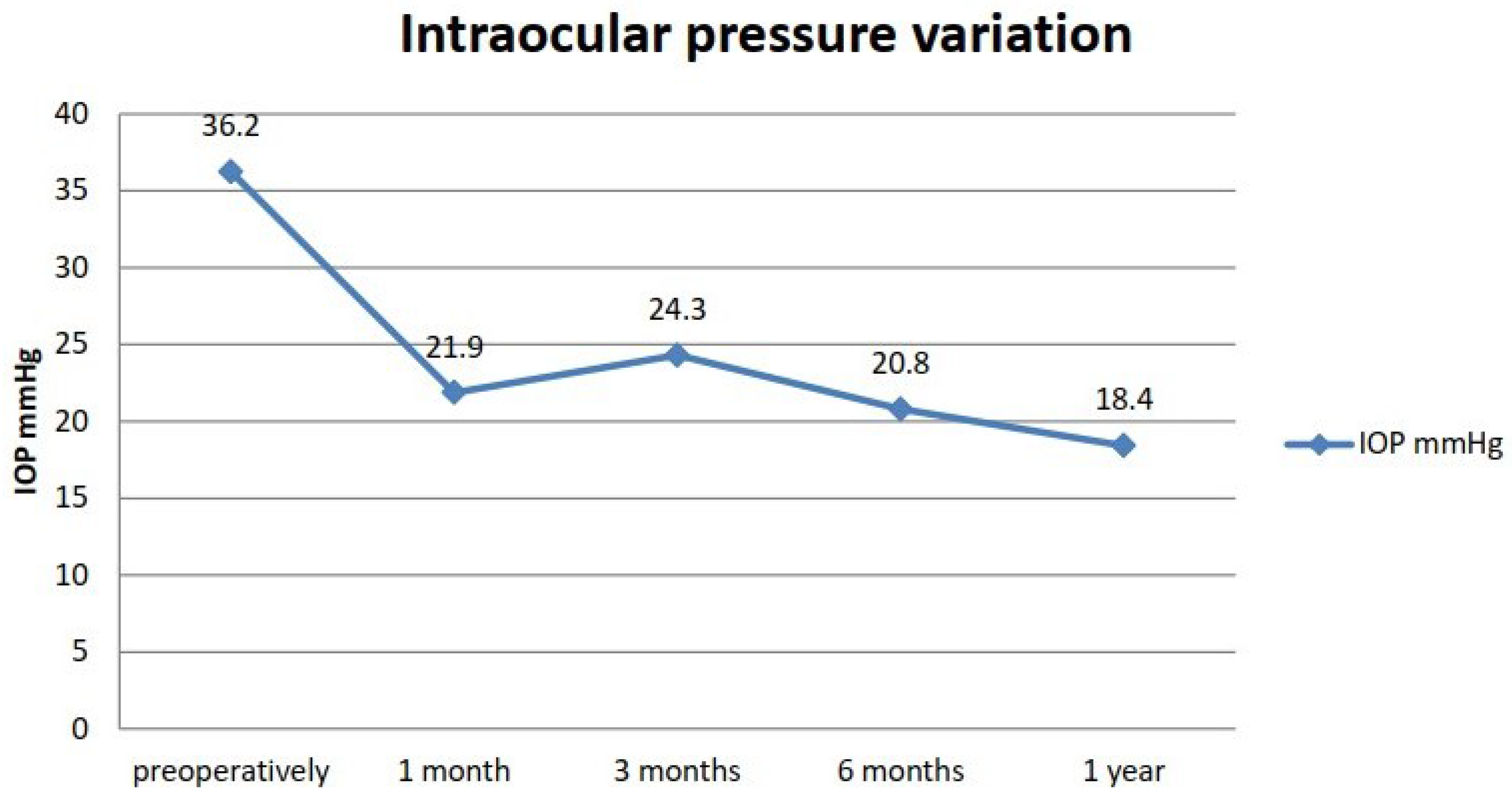
p = 0.675). No significant sex-related differences in BCVA were observed at any given time point before or after the SubCyclo laser procedure. A large difference was observed in the IOP before and after treatment, even at one month (
Figure 2). At one year, the average IOP was half the average preoperative IOP.
The distribution of cases according to type of glaucoma was assessed. The results were as follows: 35 patients (43.2%) had primary open-angle glaucoma, 21 patients (25.9%) had secondary neovascular glaucoma, 17 patients (21.0%) had glaucoma secondary to vitreoretinal surgery, seven patients (8.6%) had secondary inflammatory glaucoma, and only one patient (1.2%) had post-traumatic glaucoma. (
Table 1).
We defined the success rate at 1 month, 3 months, 6 months and 1 year as the percentage of patients with an IOP measurement below 21 mmHg. The success rate was 80% at 1 month (65 patients), 74% at 3 months (60 patients), 64% at 6 months (52 patients) and 50.6% at 1 year (41 patients). Regarding choroidal thickness, an increase from the average preoperative value (291.78 µm) was observed at 1 year (318.42 µm). Even though the increase observed at 1 month postoperatively did not remain at the same values until the last assessment, when compared to the preoperative baseline value, an increase in choroidal thickness was still observed.
A correlation was observed between the success rate and an increase in choroidal thickness. Overall, 88% of the patients responsive at 3 months demonstrated an increase in choroidal thickness at 1 month. The same percentage was observed for the patients responsive at 6 months. On the other hand, only 57% of unresponsive patients showed an increase in choroidal thickness at 1 month. Moreover, 67% of patients with success at 3 months, but not maintaining success at 6 months, showed a decrease in choroidal thickness (
Table 2).
The decrease in IOP due to SubCyclo is also supported by the decrease in the average daily number of antiglaucomatous drops used (3.69 preoperatively, 3.61 at 1 month, 3.35 at 3 months, 3.26 at 6 months and 2.7 at 1 year). With the same trend, the average daily number of acetazolamide tablets used (250 mg each) decreased gradually postoperatively (1.197 preoperatively, 1.197 at one month, 0.605 at 3 months, 0.45 at 6 months and 0.106 at 1 year).
4. Discussion
The aim of this study was to determine the correlation between the decrease in IOP and the variations in choroidal thickness in patients with initially uncompensated glaucoma, in whom the SubCyclo procedure was performed. The IOP results presented above emphasize the effectiveness of the procedure both in the short and long term. Similar results in all types of glaucoma indicate that this technique could be an effective solution for more patients. Given the inherent bias of this study towards recalcitrant glaucoma, the SubCyclo laser procedure allowed us to reach our therapeutic goal in cases where other procedures are deemed inadequate.
The significant increase in choroidal thickness after treatment supports the hypothesis that increased uveoscleral outflow may indeed be a key IOP-lowering mechanism of the SubCyclo procedure. The correlation of increased choroidal thickness at 1-month with the success rate of the procedure could be used clinically as a predictive factor for the final outcome of each case. The effectiveness of subliminal [
5] or micropulsed transscleral cyclophotocoagulation [
6,
7,
8,
9,
10] has been demonstrated in numerous studies. It is an undemanding intervention for both doctors and patients.
Thus far, there are several papers that describe an increase in choroidal thickness after trabeculoplasty [
11,
12]. We do not know exactly whether this is determined by the same pathophysiological mechanism as described in this article. The choroid is one of the eye structures that has been insufficiently studied thus far, although SS-OCT has been used extensively to gauge ocular anatomy and its response to therapy [
3]. We previously performed and published several studies showing the correlation between choroid thickness and prostaglandin analogue administration and the performance of micropulsed transscleral cyclophotocoagulation [
13].
The aim of our study was to demonstrate the correlation between increased choroidal thickness and decreased IOP after the SubCyclo procedure, confirming the mechanism of increased uveoscleral drainage of aqueous humor after SubCyclo. New imaging techniques, such as OCT angiography or adaptive optics, have allowed the imaging of retinal vessels and choroidal vessels [
14]. We intend to continue this study to establish the success rate at 2 years and to conduct further studies using OCT angiography and adaptive optics.
In conclusion, transscleral cyclophotocoagulation is a procedure with rapid post-intervention recovery, a satisfactory success rate, minimal possible complications and the possibility of repeated treatments.
Author Contributions
Conceptualization, F.B. and R.I.B.; methodology, V.D.; software, G.B.; validation, V.D., R.I.B. and F.B.; formal analysis, S.S. and M.Z.; investigation, R.I.B.; resources, F.B.; data curation, C.D.M.D.; writing—original draft preparation, R.I.B. and G.B.; writing—review and editing, R.I.B.; visualization, A.D.B.; supervision, F.B.; and project administration, F.B. All authors have read and agreed to the published version of the manuscript.
Funding
This research received no external funding.
Institutional Review Board Statement
The study was retrospectively approved by the Institutional Review Board and the Ethics Committee of Ponderas Academic Hospital.
Informed Consent Statement
All procedures conformed to the tenets of the World Medical Association’s Declaration of Helsinki, and written informed consent was obtained from all patients.
Data Availability Statement
The first author and the corresponding author have full access to all the data and materials in the study.
Conflicts of Interest
The authors declare no conflict of interest.
Abbreviations
The following abbreviations are used in this manuscript:
| IOP | Intraocular pressure |
| EDI-OCT | Enhanced depth imaging optical coherence tomography |
| SS-OCT | Swept source optical coherence tomography |
| BCVA | Best-corrected visual acuity |
References
- Usui, S.; Ikuno, Y.; Akiba, M.; Maruko, I.; Sekiryu, T.; Nishida, K.; Iida, T. Circadian Changes in Subfoveal Choroidal Thickness and the Relationship with Circulatory Factors in Healthy Subjects. Investig. Opthalmol. Vis. Sci. 2012, 53, 2300. [Google Scholar] [CrossRef] [PubMed]
- Chung, S.E.; Kang, S.W.; Lee, J.H.; Kim, Y.T. Choroidal Thickness in Polypoidal Choroidal Vasculopathy and Exudative Age-related Macular Degeneration. Ophthalmology 2011, 118, 840–845. [Google Scholar] [CrossRef] [PubMed]
- Iglicki, M.; Busch, C.; Zur, D.; Okada, M.; Mariussi, M.; Chhablani, J.K.; Cebeci, Z.; Fraser-Bell, S.; Chaikitmongkol, V.; Couturier, A.; et al. Dexamethasone implant for diabetic macular edema in naive compared with refractory eyes: The International Retina Group Real-Life 24-Month Multicenter Study. The IRGREL-DEX Study. Retina 2019, 39, 44–51. [Google Scholar] [CrossRef] [PubMed]
- Tan, C.S.; Ouyang, Y.; Ruiz, H.; Sadda, S.R. Diurnal Variation of Choroidal Thickness in Normal, Healthy Subjects Measured by Spectral Domain Optical Coherence Tomography. Investig. Ophthalmol. Vis. Sci. 2012, 53, 261. [Google Scholar] [CrossRef] [PubMed]
- Lutic, I.; Dragne, C.; Filip, M.; Filip, A.; Nicolae, M.; Moisescu, R.; Ungureanu, I.; Triantafyllidis, G.; Antonescu, C. Subcyclo laser procedure results in patients with glaucoma. Rom. J. Ophthalmol. 2018, 62, 296–299. [Google Scholar] [CrossRef] [PubMed]
- Emanuel, M.E.; Grover, D.S.; Fellman, R.L.; Godfrey, D.G.; Smith, O.; Butler, M.R.; Kornmann, H.L.; Feuer, W.J.; Goyal, S. Micropulse Cyclophotocoagulation. J. Glaucoma 2017, 26, 726–729. [Google Scholar] [CrossRef] [PubMed]
- Souissi, S.; Baudouin, C.; Labbé, A.; Hamard, P. Micropulse transscleral cyclophotocoagulation using a standard protocol in patients with refractory glaucoma naive of cyclodestruction. Eur. J. Ophthalmol. 2021, 31, 112–119. [Google Scholar] [CrossRef] [PubMed]
- Tan, A.M.; Chockalingam, M.; Aquino, M.C.; Lim, Z.I.; See, J.L.; Chew, P.T. Micropulse transscleral diode laser cyclophotocoagulation in the treatment of refractory glaucoma. Clin. Exp. Ophthalmol. 2010, 38, 266–272. [Google Scholar] [CrossRef] [PubMed]
- Toyos, M.M.; Toyos, R. Clinical Outcomes of Micropulsed Transcleral Cyclophotocoagulation in Moderate to Severe Glaucoma. J. Clin. Exp. Ophthalmol. 2016, 7, 2. [Google Scholar] [CrossRef] [Green Version]
- Ndulue, J.; Rahmatnejad, K.; Sanvicente, C.; Wizov, S.; Moster, M. Evolution of cyclophotocoagulation. J. Ophthalmic Vis. Res. 2018, 13, 55. [Google Scholar] [CrossRef] [PubMed] [Green Version]
- Stanca, H.T.; Munteanu, M.; Jianu, D.C.; Motoc, A.G.M.; Tăbăcaru, B.; Stanca, S.; Ungureanu, E.; Boruga, V.M.; Preda, M.A. New perspectives in the use of laser diode transscleral cyclophotocoagulation. A prospective single center observational cohort study. Rom. J. Morphol. Embryol. Rev. Roum. Morphol. Embryol. 2018, 59, 869–872. [Google Scholar]
- Kara, N.; Baz, O.; Altan, C.; Satana, B.; Kurt, T.; Demirok, A. Changes in choroidal thickness, axial length, and ocular perfusion pressure accompanying successful glaucoma filtration surgery. Eye 2013, 27, 940–945. [Google Scholar] [CrossRef] [PubMed] [Green Version]
- Barac, R.; Vuzitas, M.; Balta, F. Choroidal thickness increase after micropulse transscleral cyclophotocoagulation. Rom. J. Ophthalmol. 2018, 62, 144–148. [Google Scholar] [CrossRef] [PubMed]
- Ochinciuc, R.; Ochinciuc, U.; Stanca, H.T.; Barac, R.; Darabus, D.; Şuţă, M.; Baltă, F.; Burcea, M. Photoreceptor assessment in focal laser-treated central serous chorioretinopathy using adaptive optics and fundus autofluorescence. Medicine 2020, 99, e19536. [Google Scholar] [CrossRef] [PubMed]
| Publisher’s Note: MDPI stays neutral with regard to jurisdictional claims in published maps and institutional affiliations. |
© 2022 by the authors. Licensee MDPI, Basel, Switzerland. This article is an open access article distributed under the terms and conditions of the Creative Commons Attribution (CC BY) license (https://creativecommons.org/licenses/by/4.0/).


 ,
, 
