
Superpixel-Based Optic Nerve Head Segmentation Method of Fundus Images for Glaucoma Assessment

Abstract
:1. Introduction
2. Materials and Methods
2.1. Retinal Fundus Dataset
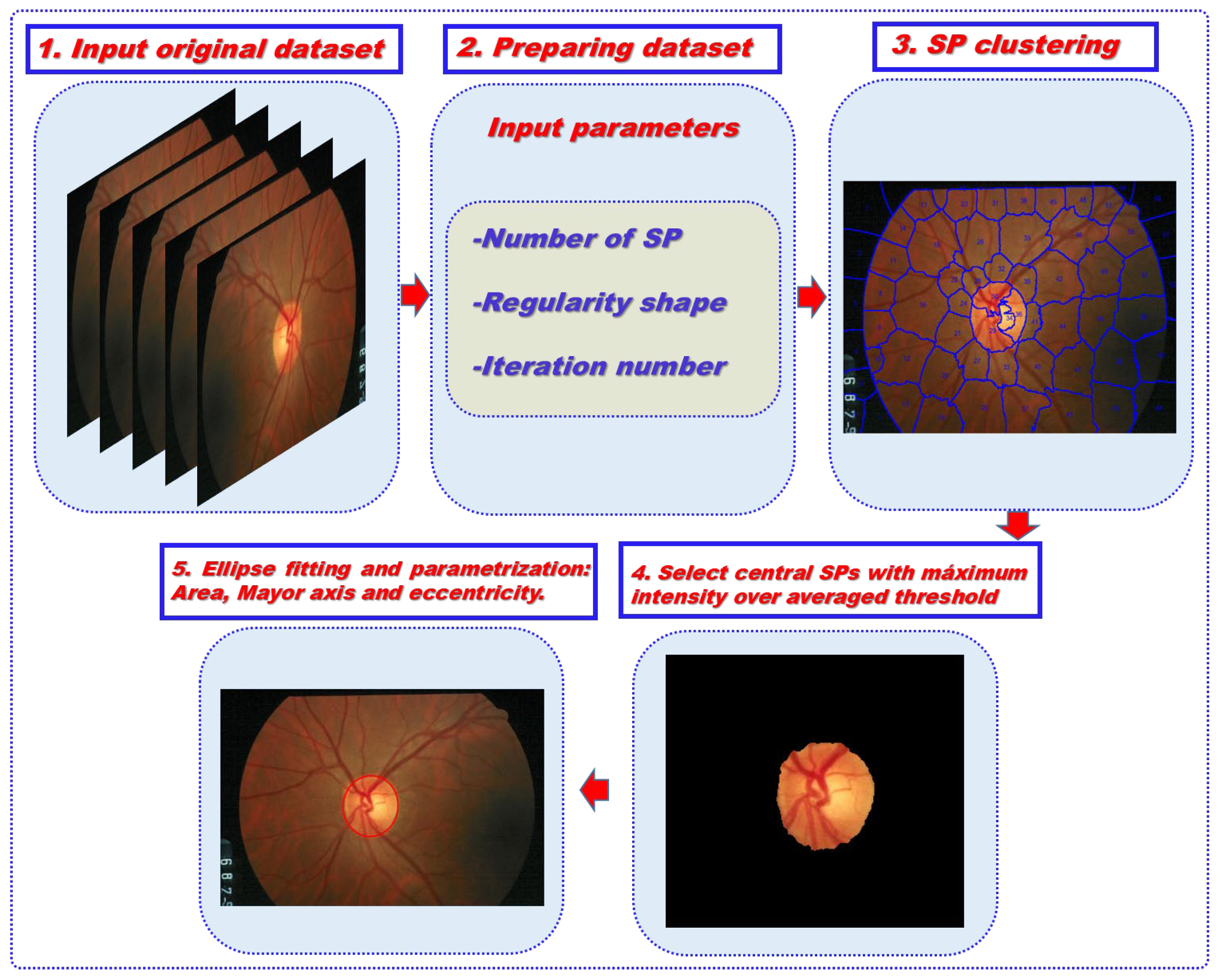
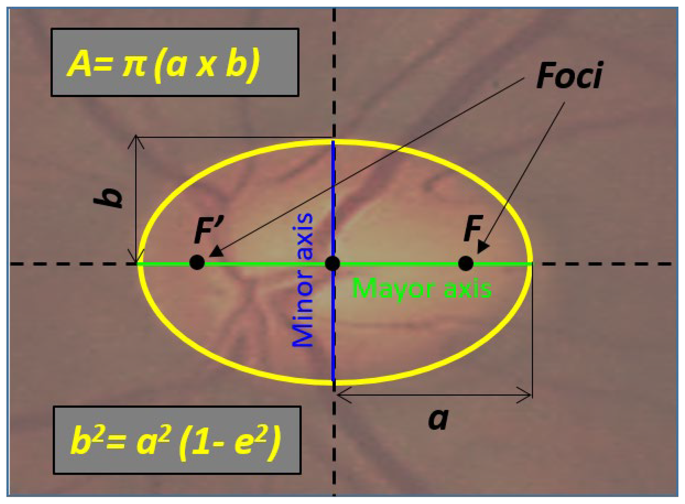
2.2. Algorithm Description and Image Processing
2.3. Data Analysis
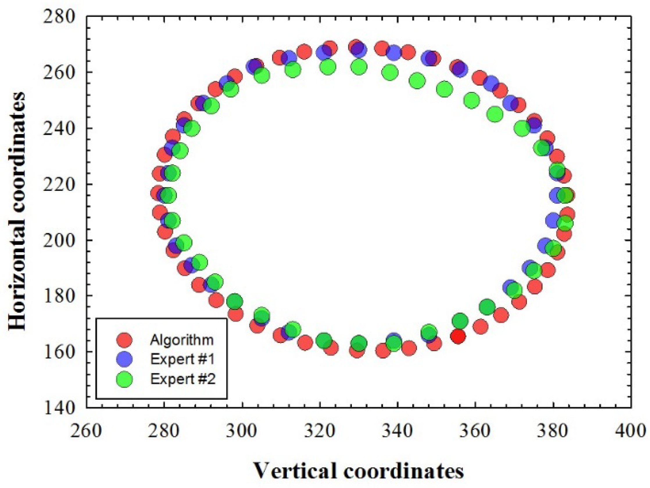
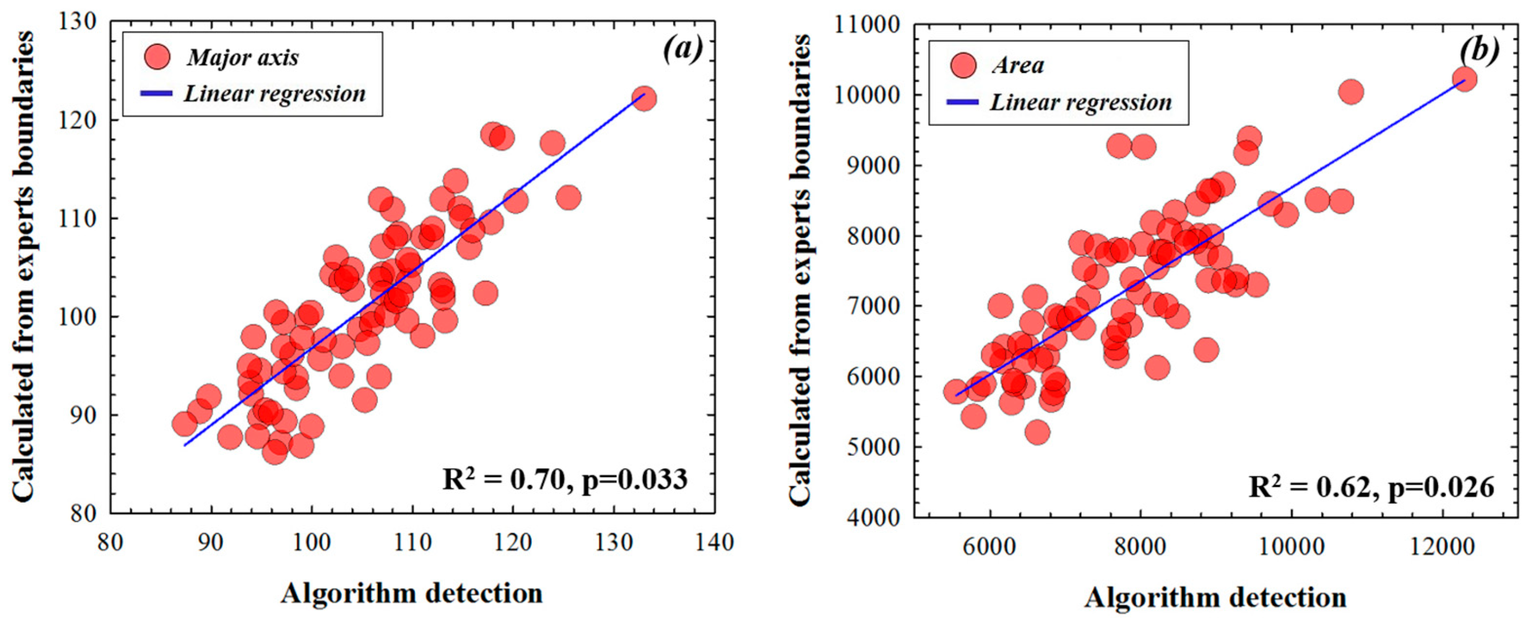
3. Results


4. Discussion and Conclusions
Author Contributions
Funding
Data Availability Statement
Conflicts of Interest
References
- Remo, S.; De Moraes, C.G.; Cioffi, G.A.; Ritch, R. Why do people (still) go blind from Glaucoma? Trans. Vis. Sci. Technol. 2015, 4, 1. [Google Scholar]
- Bussel, I.; Wollstein, G.; Schuman, J. OCT for glaucoma diagnosis, screening and detection of glaucoma progression. Br. J. Ophthalmol. 2013, 98, 15–19. [Google Scholar] [CrossRef] [Green Version]
- Lemij, H.G.; Reus, N.J. New developments in scanning laser polarimetry for glaucoma. Curr. Opin. Opthalmol. 2008, 19, 136–140. [Google Scholar] [CrossRef]
- Yaghoubi, M.; Moradi-Lakeh, M.; Mokhtari-Payam, M.; Fakhraie, G.; Shokraneh, F. Confocal scan laser ophthalmoscope for diagnosing glaucoma: A systematic review and meta-analysis. Asia Pac. J. Ophthalmol. 2015, 4, 32–39. [Google Scholar] [CrossRef]
- Chakrabarti, L.; Joshi, G.D.; Chakrabarti, A.; Raman, G.V.; Krishnadas, S.R.; Sivaswamy, J. Automated Detection of Glaucoma from Topographic Features of the Optic Nerve Head in Color Fundus Photographs. J. Glaucoma 2016, 25, 590–597. [Google Scholar] [CrossRef] [Green Version]
- Walter, T.; Klein, J.C. Segmentation of color fundus images of the human retina: Detection of the optic disc and the vascular tree using morphological techniques. In Proceedings of the Second International Symposium on Medical Data Analysis; Springer: Madrid, Spain, 2001; pp. 282–287. [Google Scholar]
- Haleem, M.S.; Han, L.; Hemert, J.V.; Li, B.; Fleming, A.; Pasquale, L.R.; Song, B.J. A novel adaptive deformable model for automated optic disc and cup segmentation to aid glaucoma diagnosis. J. Med. Syst. 2018, 42, 20. [Google Scholar] [CrossRef] [Green Version]
- Zhu, X.; Rangayyan, R.M. Detection of the optic disc in images of the retina using the hough transform. In Proceedings of the International Conference of the IEEE Engineering in Medicine and Biology Society, Vancouver, BC, Canada, 20–25 August 2008; pp. 3546–3549. [Google Scholar]
- Aquino, A.; Gegúndez-Arias, M.E.; Marín, D. Detecting the optic disc boundary in digital fundus images using morphological, edge detection, and feature extraction techniques. IEEE Trans. Med. Imaging 2010, 29, 1860–1869. [Google Scholar] [CrossRef] [Green Version]
- Chan, T.; Vese, L. An active contour model without edges. IEEE Trans. Image Process. 2002, 10, 266–277. [Google Scholar] [CrossRef] [Green Version]
- Tang, L.; Garvin, M.K.; Kwon, Y.H.; Abramoff, M.D. Segmentation of optic nerve head rim in color fundus photographs by probability based active shape model. Investig. Ophthalmol. Vis. Sci. 2012, 53, 2144. [Google Scholar]
- Gao, Y.; Yu, X.; Wu, C.; Zhou, W.; Lei, X.; Zhuang, Y. Automatic optic disc segmentation based on modified local image fitting model with shape prior information. J. Healthc. Eng. 2019, 2019, 2745183. [Google Scholar] [CrossRef]
- Xu, J.; Chutatape, O.; Sung, E.; Zheng, C.; Kuan, P.C.T. Optic disk feature extraction via modified deformable model technique for glaucoma analysis. Pattern Recognit. 2007, 40, 2063–2076. [Google Scholar] [CrossRef]
- Ayub, J.; Ahmad, J.; Muhammad, J.; Aziz, L.; Ayub, S.; Akram, U.; Basit, I. Glaucoma detection through optic disc and cup segmentation using K-mean clustering. In Proceedings of the 2016 International Conference on Computing, Electronic and Electrical Engineering (ICE Cube), Quetta, Pakistan, 11–12 April 2016. [Google Scholar]
- Zhang, K.; Zhang, L.; Lam, K.; Zhang, D. A level set approach to image segmentation with intensity inhomogeneity. IEEE Trans. Cybern. 2016, 46, 546–557. [Google Scholar] [CrossRef]
- Fu, H.; Cheng, J.; Xu, Y.; Liu, J. Glaucoma Detection Based on Deep Learning Network in Fundus Image. In Deep Learning and Convolutional Neural Networks for Medical Imaging and Clinical Informatics; Lu, L., Wang, X., Carneiro, G., Yang, L., Eds.; Advances in Computer Vision and Pattern Recognition; Springer: Berlin/Heidelberg, Germany, 2019; pp. 119–137. [Google Scholar]
- Park, K.; Kim, J.; Lee, J. Automatic optic nerve head localization and cup-to-disc ratio detection using state-of-the-art deep-learning architectures. Sci. Rep. 2020, 10, 5025. [Google Scholar] [CrossRef] [Green Version]
- Sreng, S.; Maneerat, N.; Hamamoto, K.; Win, K. Deep Learning for Optic Disc Segmentation andGlaucoma Diagnosis on Retinal Images. Appl. Sci. 2020, 10, 4916. [Google Scholar] [CrossRef]
- Gheisari, S.; Shariflou, S.; Phu, J.; Kennedy, P.J.; Agar, A.; Kalloniatis, M.; Golzan, S.M. A combined convolutional and recurrent neural network for enhanced glaucoma detection. Sci. Rep. 2021, 11, 1945. [Google Scholar] [CrossRef]
- Mohamed, N.A.; Zulkifley, M.A.; Zaki, W.M.; Hussain, A. An automated glaucoma screening system using cup-to-disc ratio via Simple Linear Iterative Clustering superpixel approach. Biomed. Signal Process. Control. 2019, 53, 101454. [Google Scholar] [CrossRef]
- Carmona, E.J.; Rincón, M.; García-Feijoo, J.; Martínez-de-la-Casa, J.M. Identification of the optic nerve head with genetic algorithms. Artif. Intell. Med. 2008, 43, 243–259. [Google Scholar] [CrossRef]
- Zhang, Z.; Yin, F.; Liu, J.; Wong, W.K.; Tan, N.M.; Lee, B.H.; Cheng, J.; Wong, T.Y. ORIGA(-light): An online retinal fundus image database for glaucoma analysis and research. Annu. Int. Conf. IEEE Engin. Med. Biol. Soc. 2010, 2010, 3065–3068. [Google Scholar]
- Ren, X.; Malik, J. Learning a classification model for segmentation. In Proceedings of the Ninth IEEE International Conference on Computer Vision, Nice, France, 13–16 October 2003; pp. 10–17. [Google Scholar]
- Masumoto, H.; Tabuchi, H.; Nakakura, S.; Naofumi, I. Deep-learning Classifier with an Ultrawide-field Scanning Laser Ophthalmoscope Detects Glaucoma Visual Field Severity. J. Glaucoma 2018, 27, 647–652. [Google Scholar] [CrossRef]
- Ran, A.; Tham, C.; Chan, P.; Cheng, C.-Y.; Tham, Y.-C.; Rim, T.H.; Cheung, C.Y. Deep learning in glaucoma with optical coherence tomography: A review. Eye 2021, 35, 188–201. [Google Scholar] [CrossRef]
- Yin, F.; Liu, J.; Ong, S.; Sun, Y.; Wong, D.W.K.; Tan, N.M.; Cheung, C.; Baskaran, M.; Aung, T.; Wong, T.Y. Model-based optic nerve head segmentation on retinal fundus images. IEEE Int. Conf. Eng. Med. Biol. Soc. 2011, 2011, 2626–2629. [Google Scholar]
- Wong, D.; Lim, J.; Tan, N.; Tan, N.M.; Zhang, Z.; Lu, S.; Li, H.; Teo, M.H.; Chan, K.L.; Wong, T.Y. Intelligent fusing of cup-to-disc ratio determination methods for glaucoma diagnosis. Int. Conf. Engin. Med. Biol. Soc. 2009, 2009, 5777–5780. [Google Scholar]
- Xu, Y.; Xu, D.; Lin, S.; Liu, J.; Cheng, J.; Cheung, C.; Aung, T.; Wong, T.Y. Sliding window and regression based cup detection in digital fundus images for glaucoma diagnosis. Med. Image Comput. Comput. Assist. Interv. 2011, 14, 1–8. [Google Scholar]
- Tan, N.; Xu, Y.; Goh, W.; Liu, J. Robust multi-scale superpixel classification for optic cup localization. Comput. Med. Imaging Graph 2015, 40, 182–193. [Google Scholar] [CrossRef]
- Xu, Y.; Liu, J.; Lin, S.; Xu, D.; Cheung, C.Y.; Aung, T.; Wong, T.Y. Efficient Optic Cup Detection from Intra-image Learning with Retinal Structure Prior. In Medical Image Computing and Computer-Assisted Intervention—MICCAI 2012; Ayache, N., Delingette, H., Golland, P., Mori, K., Eds.; Lecture Notes in Computer Science; Springer: Berlin/Heidelberg, Germany, 2012; Volume 7510, pp. 58–65. [Google Scholar]
- Xu, Y.; Duan, L.; Lin, S.; Chen, X.; Wong, D.W.K.; Wong, T.Y.; Liu, J. Optic cup segmentation for glaucoma detection using low-rank superpixel representation. Med. Image Comput. Comput. Assist. Interv. 2014, 17, 788–795. [Google Scholar]
- Cheng, J.; Liu, J.; Xu, J.; Yin, F.; Wong, D.W.K.; Tan, N.-M.; Tao, D.; Cheng, C.-Y.; Aung, T.; Wong, T.Y. Superpixel classification based optic disc and optic cup segmentation for glaucoma screening. IEEE Trans. Med. Imaging 2013, 32, 1019–1032. [Google Scholar] [CrossRef]
- Weismann, R.L.; Asseff, C.F.; Phelps, C.D.; Podos, S.M.; Becker, B. Vertical elongation of the optic cup in glaucoma. Trans. Am. Acad. Ophthalmol. Otolaryngol. 1973, 77, OP157–OP161. [Google Scholar]
- Mohammadzadeh, V.; Rabiolo, A.; Fu, Q.; Morales, E.; Coleman, A.L.; Law, S.K.; Caprioli, J.; Nouri-Mahdavi, K. Longitudinal macular structure-function relationship in glaucoma. Ophtalmology 2020, 127, 888–900. [Google Scholar] [CrossRef]
- Lee, S.; Han, S.; Young, M.; Beg, M.F.; Sarunic, M.V.; MacKenzie, P.J. Optic Nerve Head and Peripapillary Morphometrics in Myopic Glaucoma. Glaucoma. Investig. Opthalmol. Vis. Sci. 2014, 55, 4378–4393. [Google Scholar] [CrossRef]


Publisher’s Note: MDPI stays neutral with regard to jurisdictional claims in published maps and institutional affiliations. |
© 2022 by the authors. Licensee MDPI, Basel, Switzerland. This article is an open access article distributed under the terms and conditions of the Creative Commons Attribution (CC BY) license (https://creativecommons.org/licenses/by/4.0/).
Share and Cite
Ávila, F.J.; Bueno, J.M.; Remón, L. Superpixel-Based Optic Nerve Head Segmentation Method of Fundus Images for Glaucoma Assessment. Diagnostics 2022, 12, 3210. https://doi.org/10.3390/diagnostics12123210
Ávila FJ, Bueno JM, Remón L. Superpixel-Based Optic Nerve Head Segmentation Method of Fundus Images for Glaucoma Assessment. Diagnostics. 2022; 12(12):3210. https://doi.org/10.3390/diagnostics12123210
Chicago/Turabian StyleÁvila, Francisco J., Juan M. Bueno, and Laura Remón. 2022. "Superpixel-Based Optic Nerve Head Segmentation Method of Fundus Images for Glaucoma Assessment" Diagnostics 12, no. 12: 3210. https://doi.org/10.3390/diagnostics12123210