Left Ventricle Wall Motion Analysis with Real-Time MRI Feature Tracking in Heart Failure Patients: A Pilot Study
Abstract
1. Introduction
2. Materials and Methods
2.1. Experimental Study
2.1.1. Study Design
2.1.2. Data Acquisition
2.1.3. Image Reconstruction
2.2. Image Analysis and Measurements
2.2.1. Volumetric Measurements with Retrospective Cine and Real-Time Images
2.2.2. Velocity Measurements with Retrospective Cine and Real-Time Images
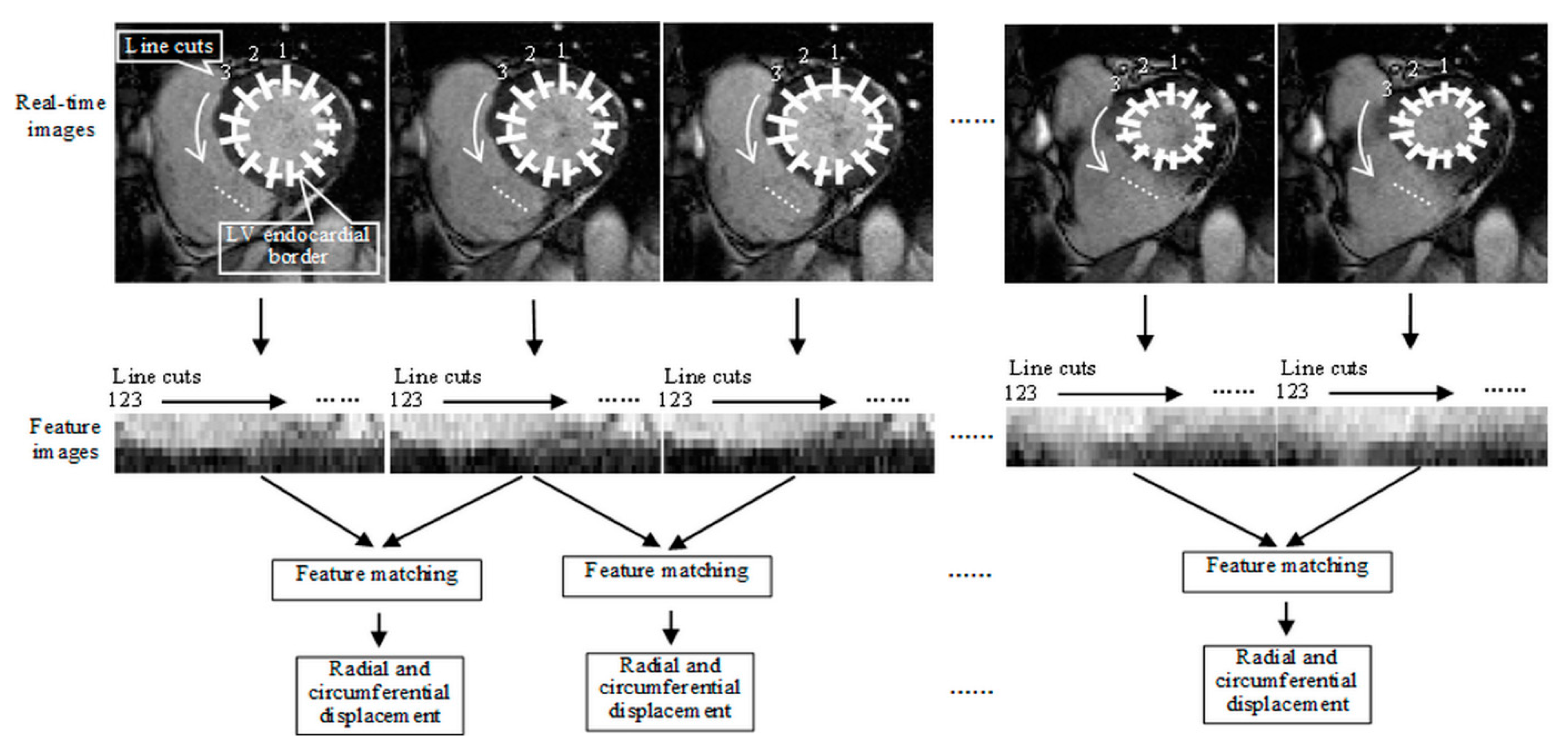
2.2.3. LV Wall Motion Analysis for HF Evaluation
2.3. Statistical Analysis
3. Results
3.1. Patient Characteristics
3.2. Collected Images and Volumetric Measurements
3.3. Velocity Measurements with Feature Tracking
3.4. Ventricular Function Assessment
4. Discussion
4.1. Main Findings
- (1)
- Torsion correlation was statistically different between the healthy volunteers and the patients with HFpEF (one-way ANOVA, p < 0.001) while volumetric indices were comparable, indicating correlation analysis provided diagnostic information complementary to volumetric measurements.
- (2)
- In the scatter plots of EF against torsion correlation, the HF patients with EF < 50%, the patients with HFpEF, and the healthy volunteers were well differentiated, indicating the potential of correlation analysis for improved HF evaluation.
4.2. Rationales for Correlation Analysis between Radial and Circumferential Motion
4.3. Retrospective Cine vs. Real-Time Imaging
4.4. Study Limitation
4.5. Clinical Signficiance
5. Conclusions
Author Contributions
Funding
Institutional Review Board Statement
Informed Consent Statement
Data Availability Statement
Acknowledgments
Conflicts of Interest
References
- Yancy, C.W.; Jessup, M.; Bozkurt, B.; Butler, J.; Casey, D.E., Jr.; Colvin, M.M.; Drazner, M.H.; Filippatos, G.S.; Fonarow, G.C.; Givertz, M.M.; et al. 2017 ACC/AHA/HFSA Focused Update of the 2013 ACCF/AHA Guideline for the Management of Heart Failure: A Report of the American College of Cardiology/American Heart Association Task Force on Clinical Practice Guidelines and the Heart Failure Society of America. Circulation 2017, 136, e137–e161. [Google Scholar] [CrossRef]
- Ponikowski, P.; Voors, A.A.; Anker, S.D.; Bueno, H.; Cleland, J.G.F.; Coats, A.J.S.; Falk, V.; Gonzalez-Juanatey, J.R.; Harjola, V.P.; Jankowska, E.A.; et al. 2016 ESC Guidelines for the diagnosis and treatment of acute and chronic heart failure: The Task Force for the diagnosis and treatment of acute and chronic heart failure of the European Society of Cardiology (ESC)Developed with the special contribution of the Heart Failure Association (HFA) of the ESC. Eur. Heart J. 2016, 37, 2129–2200. [Google Scholar] [CrossRef]
- King, M.; Kingery, J.E.; Casey, B. Diagnosis and evaluation of heart failure. Am. Fam. Physician 2012, 85, 1161–1168. [Google Scholar]
- Leong, D.P.; De Pasquale, C.G.; Selvanayagam, J.B. Heart failure with normal ejection fraction: The complementary roles of echocardiography and CMR imaging. JACC Cardiovasc. Imaging 2010, 3, 409–420. [Google Scholar] [CrossRef]
- Schulz-Menger, J.; Bluemke, D.A.; Bremerich, J.; Flamm, S.D.; Fogel, M.A.; Friedrich, M.G.; Kim, R.J.; von Knobelsdorff-Brenkenhoff, F.; Kramer, C.M.; Pennell, D.J.; et al. Standardized image interpretation and post-processing in cardiovascular magnetic resonance-2020 update: Society for Cardiovascular Magnetic Resonance (SCMR): Board of Trustees Task Force on Standardized Post-Processing. J. Cardiovasc. Magn. Reson. 2020, 22, 19. [Google Scholar] [CrossRef]
- Manning, W.J.; Pennell, D.J. Cardiovascular Magnetic Resonance; Elsevier Health Sciences: Amsterdam, The Netherlands, 2010. [Google Scholar]
- Tersalvi, G.; Gasperetti, A.; Schiavone, M.; Dauw, J.; Gobbi, C.; Denora, M.; Krul, J.D.; Cioffi, G.M.; Mitacchione, G.; Forleo, G.B. Acute heart failure in elderly patients: A review of invasive and non-invasive management. J. Geriatr. Cardiol. 2021, 18, 560–576. [Google Scholar] [CrossRef]
- Pedrizzetti, G.; Claus, P.; Kilner, P.J.; Nagel, E. Principles of cardiovascular magnetic resonance feature tracking and echocardiographic speckle tracking for informed clinical use. J. Cardiovasc. Magn. Reson. 2016, 18, 51. [Google Scholar] [CrossRef]
- Schuster, A.; Hor, K.N.; Kowallick, J.T.; Beerbaum, P.; Kutty, S. Cardiovascular Magnetic Resonance Myocardial Feature Tracking: Concepts and Clinical Applications. Circ. Cardiovasc. Imaging 2016, 9, e004077. [Google Scholar] [CrossRef]
- Hor, K.N.; Baumann, R.; Pedrizzetti, G.; Tonti, G.; Gottliebson, W.M.; Taylor, M.; Benson, D.W.; Mazur, W. Magnetic resonance derived myocardial strain assessment using feature tracking. J. Vis. Exp. 2011, 48, e2356. [Google Scholar] [CrossRef]
- Hor, K.N.; Gottliebson, W.M.; Carson, C.; Wash, E.; Cnota, J.; Fleck, R.; Wansapura, J.; Klimeczek, P.; Al-Khalidi, H.R.; Chung, E.S.; et al. Comparison of magnetic resonance feature tracking for strain calculation with harmonic phase imaging analysis. JACC Cardiovasc. Imaging 2010, 3, 144–151. [Google Scholar] [CrossRef]
- Bluemke, D.A.; Boxerman, J.L.; Atalar, E.; McVeigh, E.R. Segmented K-space cine breath-hold cardiovascular MR imaging: Part 1. Principles and technique. Am. J. Roentgenol. 1997, 169, 395–400. [Google Scholar] [CrossRef]
- Feng, L.; Grimm, R.; Block, K.T.; Chandarana, H.; Kim, S.; Xu, J.; Axel, L.; Sodickson, D.K.; Otazo, R. Golden-angle radial sparse parallel MRI: Combination of compressed sensing, parallel imaging, and golden-angle radial sampling for fast and flexible dynamic volumetric MRI. Magn. Reson. Med. 2014, 72, 707–717. [Google Scholar] [CrossRef] [PubMed]
- Li, Y.Y.; Zhang, P.; Rashid, S.; Cheng, Y.J.; Li, W.; Schapiro, W.; Gliganic, K.; Yamashita, A.-M.; Grgas, M.; Haag, E.; et al. Real-time exercise stress cardiac MRI with Fourier-series reconstruction from golden-angle radial data. Magn. Reson. Imaging 2021, 75, 89–99. [Google Scholar] [CrossRef] [PubMed]
- Zhang, S.; Uecker, M.; Voit, D.; Merboldt, K.-D.; Frahm, J. Real-time cardiovascular magnetic resonance at high temporal resolution: Radial FLASH with nonlinear inverse reconstruction. J. Cardiovasc. Magn. Reson. 2010, 12, 39. [Google Scholar] [CrossRef]
- Kramer, C.M.; Barkhausen, J.; Bucciarelli-Ducci, C.; Flamm, S.D.; Kim, R.J.; Nagel, E. Standardized cardiovascular magnetic resonance imaging (CMR) protocols: 2020 update. J. Cardiovasc. Magn. Reson. 2020, 22, 1–18. [Google Scholar] [CrossRef]
- Santelli, C.; Kozerke, S. L1 k-t ESPIRiT: Accelerating Dynamic MRI Using Efficient Auto-Calibrated Parallel Imaging and Compressed Sensing Reconstruction. J. Cardiovasc. Magn. Reson. 2016, 18, P302. [Google Scholar] [CrossRef]
- Sakuma, H.; Fujita, N.; Foo, T.K.; Caputo, G.R.; Nelson, S.J.; Hartiala, J.; Shimakawa, A.; Higgins, C.B. Evaluation of Left Ventricular Volume and Mass with Breath-hold Cine MR Imaging. Radiology 1993, 188, 377–380. [Google Scholar] [CrossRef]
- Mooij, C.F.; de Wit, C.J.; Graham, D.A.; Powell, A.J.; Geva, T. Reproducibility of MRI Measurements of Right Ventricular Size and Function in Patients with Normal and Dilated Ventricles. J. Magn. Reson. Imaging 2008, 28, 67–73. [Google Scholar] [CrossRef]
- Attili, A.K.; Schuster, A.; Nagel, E.; Reiber, J.H.C.; Geest, R.J.v.d. Quantification in cardiac MRI: Advances in image acquisition and processing. Int. J. Cardiovasc. Imaging 2010, 26, 27–49. [Google Scholar] [CrossRef]
- Pluempitiwiriyawej, C.; Moura, J.M.; Wu, Y.-J.L.; Ho, C. STACS: New active contour scheme for cardiac MR image segmentation. IEEE Trans. Med. Imaging 2005, 24, 593–603. [Google Scholar] [CrossRef]
- Hayes, M.H. Statistical Digital Signal Processing and Modeling; John Wiley & Sons: Hoboken, NJ, USA, 2009. [Google Scholar]
- Codreanu, I.; Pegg, T.; Selvanayagam, J.; Robson, M.; Rider, O. Comprehensive Assessment of Left Ventricular Wall Motion Abnormalities in Coronary Artery Disease Using Cardiac Magnetic Resonance. J. Cardiol. Neuro-Cardiovasc. Dis. 2015, 2, 2. [Google Scholar]
- Lindley, D. Regression and correlation analysis. In Time Series and Statistics; Springer: Berlin/Heidelberg, Germany, 1990; pp. 237–243. [Google Scholar]
- Miller, R.G., Jr. Beyond ANOVA: Basics of Applied Statistics; CRC Press: Boca Raton, FL, USA, 1997. [Google Scholar]
- D’Agostino, R.B. Tests for the normal distribution. In Goodness-of-Fit Techniques; Routledge: Oxfordshire, UK, 2017; pp. 367–420. [Google Scholar]
- Buckberg, G.D. Basic science review: The helix and the heart. J. Thorac. Cardiovasc. Surg. 2002, 124, 863–883. [Google Scholar] [CrossRef]
- Zerhouni, E.A.; Parish, D.M.; Rogers, W.J.; Yang, A.; Shapiro, E.P. Human heart: Tagging with MR imaging--a method for noninvasive assessment of myocardial motion. Radiology 1988, 169, 59–63. [Google Scholar] [CrossRef]
- Korosoglou, G.; Giusca, S.; Hofmann, N.P.; Patel, A.R.; Lapinskas, T.; Pieske, B.; Steen, H.; Katus, H.A.; Kelle, S. Strain-encoded magnetic resonance: A method for the assessment of myocardial deformation. ESC Heart Fail. 2019, 6, 584–602. [Google Scholar] [CrossRef]
- Jung, B.; Foll, D.; Bottler, P.; Petersen, S.; Hennig, J.; Markl, M. Detailed analysis of myocardial motion in volunteers and patients using high-temporal-resolution MR tissue phase mapping. J. Magn. Reson. Imaging 2006, 24, 1033–1039. [Google Scholar] [CrossRef]
- Notomi, Y.; Lysyansky, P.; Setser, R.M.; Shiota, T.; Popovic, Z.B.; Martin-Miklovic, M.G.; Weaver, J.A.; Oryszak, S.J.; Greenberg, N.L.; White, R.D.; et al. Measurement of ventricular torsion by two-dimensional ultrasound speckle tracking imaging. J. Am. Coll. Cardiol. 2005, 45, 2034–2041. [Google Scholar] [CrossRef]
- Nottin, S.; Doucende, G.; Schuster, I.; Tanguy, S.; Dauzat, M.; Obert, P. Alteration in left ventricular strains and torsional mechanics after ultralong duration exercise in athletes. Circ. Cardiovasc. Imaging 2009, 2, 323–330. [Google Scholar] [CrossRef]
- Nakatani, S. Left ventricular rotation and twist: Why should we learn? J. Cardiovasc. Ultrasound 2011, 19, 1–6. [Google Scholar] [CrossRef]
- Dong, S.-J.; Hees, P.S.; Siu, C.O.; Weiss, J.L.; Shapiro, E.P. MRI assessment of LV relaxation by untwisting rate: A new isovolumic phase measure of t. Am. J. Physiol.-Heart Circ. Physiol. 2001, 281, H2002–H2009. [Google Scholar] [CrossRef]
- Sengupta, P.P.; Tajik, A.J.; Chandrasekaran, K.; Khandheria, B.K. Twist mechanics of the left ventricle: Principles and application. JACC Cardiovasc. Imaging 2008, 1, 366–376. [Google Scholar] [CrossRef]
- Westermann, D.; Kasner, M.; Steendijk, P.; Spillmann, F.; Riad, A.; Weitmann, K.; Hoffmann, W.; Poller, W.; Pauschinger, M.; Schultheiss, H.P.; et al. Role of left ventricular stiffness in heart failure with normal ejection fraction. Circulation 2008, 117, 2051–2060. [Google Scholar] [CrossRef]


| ID | NYHA Class of HF | Relevant Cardiovascular and Pulmonary Problems | Cardiac MRI Assessment | |||
|---|---|---|---|---|---|---|
| LV | RV | |||||
| Size | Systolic Function | Size | Systolic Function | |||
| 1 | II | Nonischemic cardiomyopathy, shortness of breath, palpitation | Severely dilated, EDV index = 154 mL/m2 | Moderately reduced, EF = 37% | Normal, EDV index = 90 mL/m2 | Normal, EF = 56% |
| 2 | II | Hypertension, diabetes mellitus, thyroid nodule | Moderately dilated, EDV index = 110 mL/m2 | Moderately reduced EF = 42% | Normal, EDV index = 92 mL/m2 | Normal, EF = 57% |
| 3 | II | Hypertension, cardiomyopathy, chronic obstructive pulmonary disease, internal cardiac defibrillator procedure, hyperkalemia, dizziness | Normal, EDV index = 73 mL/m2 | Severely reduced, EF = 27% | Normal, EDV index = 72 mL/m2 | Moderately reduced, EF = 37% |
| 4 | II | Atrial fibrillation, stroke, nonischemic cardiomyopathy, transient ischemic attack, sleep apnea, chest pain, palpitation | Normal, EDV index = 90 mL/m2 | Severely reduced, EF = 33% | Normal, EDV index = 76 mL/m2 | Mildly reduced, EF = 41% |
| 5 | II | Atrial fibrillation, nonischemic cardiomyopathy, transient ischemic attack, chest pain, palpitation | Normal, EDV index = 90 mL/m2 | Moderately reduced, EF = 39% | Normal, EDV index = 90 mL/m2 | Mildly reduced, EF = 40% |
| 6 | II | Shortness of breath | Severely dilated, EDV index = 293 mL/m2 | Severely reduced, EF = 10% | Normal, EDV index = 90 mL/m2 | Moderately reduced, EF = 32% |
| 7 | II | Restrictive cardiomyopathy, dyspnea on exertion | Moderately dilated, EDV index = 114 mL/m2 | Moderately reduced, EF = 40% | Normal, EDV index = 67 mL/m2 | Normal, EF = 56% |
| 8 | II | Ventricular tachycardia, premature ventricular contraction, first degree atrioventricular block, non-sustained ventricular tachycardia, first degree heart block, left anterior fascicular block, hypertension, coronary artery disease, chest pain | Normal, EDV index = 74 mL/m2 | Mildly reduced, EF = 46% | Normal, EDV index = 55 mL/m2 | Normal, EF = 51% |
| ID | NYHA Class of HF | Relevant Cardiovascular and Pulmonary Problems | Cardiac MRI Assessment | |||
|---|---|---|---|---|---|---|
| LV | RV | |||||
| Size | Systolic Function | Size | Systolic Function | |||
| 1 | II | Pulmonary hypertension, cardiomyopathy, dilated cardiomyopathy, pulmonary embolism, atherosclerosis of arteries of extremities, chest pain, | Normal, EDV index = 75 mL/m2 | Normal, EF = 53% | Normal, EDV index = 79 mL/m2 | Normal, EF = 51% |
| 2 | II | Pulmonary hypertension, cardiomyopathy, dilated cardiomyopathy | Mildly dilated, EDV index = 111 mL/m2 | Normal EF = 59% | Mildly dilated, EDV index = 113 mL/m2 | Normal, EF = 59% |
| 3 | III | Hypertension, pulmonary hypertension, atrial fibrillation, obstructive sleep apnea | Mildly dilated, EDV index = 104 mL/m2 | Normal EF = 57% | Severely dilated, EDV index = 141 mL/m2 | Mildly reduced, EF = 42% |
| 4 | I | Cardiomyopathy, paroxysmal atrial fibrillation | Normal, EDV index = 72 mL/m2 | Normal, EF = 50% | Normal, EDV index = 66 mL/m2 | Low normal, EF = 49% |
| 5 | I-II | Nonischemic cardiomyopathy, atrial fibrillation | Normal, EDV index = 87 mL/m2 | Normal, EF = 54% | Normal, EDV index = 88 mL/m2 | Normal, EF = 50% |
| 6 | I-II | Nonischemic cardiomyopathy, atrial fibrillation | Normal, EDV index = 93 mL/m2 | Normal, 54% | Normal, EDV index = 87 mL/m2 | Normal, EF = 55% |
| 7 | I-II | Nonischemic cardiomyopathy, atrial fibrillation | Mildly dilated, EDV index = 101 mL/m2 | Normal, EF = 59% | Mildly dilated, EDV index = 104 mL/m2 | Normal, EF = 63% |
| 8 | III | Arteriosclerotic heart disease, pulmonary embolism on right, obstructive sleep apnea, hyperlipidemia, morbid obesity | Normal, EDV index = 86 mL/m2 | Normal, EF = 50% | Moderately dilated, EDV index = 120 mL/m2 | Mildly reduced, EF = 41% |
| 9 | III | Hypertension, pulmonary emboli, chronic obstructive pulmonary disease, asthma-COPD overlap syndrome, nonischemic cardiomyopathy, atrial fibrillation | Normal, EDV index = 66 mL/m2 | Normal, EF = 52% | Normal, EDV index = 69 mL/m2 | Normal, EF = 52% |
| 10 | II | Hypertension, coronary artery disease, non-ST elevated myocardial infarction, ventricular fibrillation, ventricular tachycardia, ischemic cardiomyopathy, ischemic heart disease, acute respiratory failure with hypoxia, hyperlipidemia, shortness of breath | Normal, EDV index = 95 mL/m2 | Normal, EF = 51% | Normal, EDV index = 80 mL/m2 | Normal, EF = 62% |
Publisher’s Note: MDPI stays neutral with regard to jurisdictional claims in published maps and institutional affiliations. |
© 2022 by the authors. Licensee MDPI, Basel, Switzerland. This article is an open access article distributed under the terms and conditions of the Creative Commons Attribution (CC BY) license (https://creativecommons.org/licenses/by/4.0/).
Share and Cite
Li, Y.; Craft, J.; Cheng, Y.; Gliganic, K.; Schapiro, W.; Cao, J. Left Ventricle Wall Motion Analysis with Real-Time MRI Feature Tracking in Heart Failure Patients: A Pilot Study. Diagnostics 2022, 12, 2946. https://doi.org/10.3390/diagnostics12122946
Li Y, Craft J, Cheng Y, Gliganic K, Schapiro W, Cao J. Left Ventricle Wall Motion Analysis with Real-Time MRI Feature Tracking in Heart Failure Patients: A Pilot Study. Diagnostics. 2022; 12(12):2946. https://doi.org/10.3390/diagnostics12122946
Chicago/Turabian StyleLi, Yu (Yulee), Jason Craft, Yang (Josh) Cheng, Kathleen Gliganic, William Schapiro, and Jie (Jane) Cao. 2022. "Left Ventricle Wall Motion Analysis with Real-Time MRI Feature Tracking in Heart Failure Patients: A Pilot Study" Diagnostics 12, no. 12: 2946. https://doi.org/10.3390/diagnostics12122946
APA StyleLi, Y., Craft, J., Cheng, Y., Gliganic, K., Schapiro, W., & Cao, J. (2022). Left Ventricle Wall Motion Analysis with Real-Time MRI Feature Tracking in Heart Failure Patients: A Pilot Study. Diagnostics, 12(12), 2946. https://doi.org/10.3390/diagnostics12122946


