Article

Risk-Based Colposcopy for Cervical Precancer Detection: A Cross-Sectional Multicenter Study in China

1
Department of Epidemiology and Biostatistics, School of Population Medicine and Public Health, Chinese Academy of Medical Sciences and Peking Union Medical College, Beijing 100730, China
2
Division of Health Research, Faculty of Health and Medicine, Lancaster University, Lancaster LA1 4YW, UK
3
Diagnosis and Treatment for Cervical Lesions Center, Shenzhen Maternity & Child Healthcare Hospital, Shenzhen 518028, China
*
Authors to whom correspondence should be addressed.
Diagnostics 2022, 12(11), 2585; https://doi.org/10.3390/diagnostics12112585
Submission received: 1 October 2022 / Accepted: 22 October 2022 / Published: 25 October 2022
(This article belongs to the Special Issue Recent Advances in Gynecologic Oncology: Diagnosis and Management)

Abstract

:
Recently published guidelines depend upon screening for cervical precancer risk stratification; however, colposcopy provides key information. There is no data from developing countries that could be used comparatively. Therefore, we assessed the potential benefits of intercalating colposcopic impressions with screening results to detect cervical precancers through a multicenter, cross-sectional study of a Chinese population. Anonymized data from 6012 women with cytologic assessment, human papillomavirus (HPV) testing, colposcopic impressions, and histological results were analyzed. Univariate and multivariate analysis showed that high-grade squamous intraepithelial lesion (HSIL) cytology, HPV16/18+, and/or high-grade colposcopic impressions markedly increased cervical precancer risk, while high-grade colposcopic impressions were associated with the highest risk. The risk of cervical intraepithelial neoplasia grade 3 or worse (CIN3+) ranged from 0% for normal/benign colposcopic impressions, <HSIL cytologies, and HPV negative to 63.61% for high-grade colposcopy, HSIL+ cytology, and HPV16/18+, across 18 subgroups. High-grade colposcopic impressions were associated with a >19% increased risk of CIN3+, even in participants without HSIL+ cytology and/or HPV16/18+. Regardless of screening outcomes, normal/benign colposcopic impressions were associated with the lowest risk of CIN3+ (<0.5%). Integrating colposcopic impressions into risk assessment may therefore provide key information for identifying cervical precancer cases. Adopting this approach may improve detection rates while also providing reassurance for women with a lower risk of developing cervical cancer.

1. Introduction

Cervical cancer is the fourth most prevalent cancer among women, and this presents a substantial problem for populations in low- and middle-income countries (LMICs). The World Health Organization has called on all nations to scale-up the implementation of proven, effective strategies to eradicate cervical cancer by 2030 [1]. These strategies include vaccinating 90% of all girls against human papillomavirus (HPV) by 15 years of age, screening 70% of all women, and identifying and treating 90% of all cases to manage precancers/cancers. Currently, potential cases are referred to a colposcopist for assessment to confirm the presence of precancers/cancers, which may then lead to biopsy or treatment [2,3,4]. Therefore, colposcopy is of vital importance in the diagnostic pathway and should be investigated for potential improvements.
The American Society of Colposcopy and Cervical Pathology (ASCCP) recently introduced quantitative risk estimates to adjust patient management techniques depending on precancer risk [5,6,7]. However, the guidelines depend solely upon screening results for cytology and/or HPV and are geared toward the detection of intraepithelial neoplasia grade 3 or worse (CIN3+). This is where colposcopy can provide key information for risk assessment and enhance the ability to identify cases. Several researchers have considered the feasibility of this approach using United-States–based datasets [8,9,10]. However, there is no comparative data available from LMICs, which face a greater burden related to cervical cancer. Therefore, we sought to assess the potential benefit of intercalating colposcopic impressions with screening test results (cytology and HPV testing) for cervical precancer detection in Chinese clinical settings using available registry data.

2. Materials and Methods

2.1. Study Participants

This study was conducted between January 2019 and October 2021 in six different colposcopy clinics across China. We collated registry data from women who visited a colposcopist. All participants were Chinese nationals. Patients older than 18 years of age and those who had been referred to a local colposcopist because of abnormal screening results or those with symptoms of suspected cervical disease were eligible to participate. Women who underwent a hysterectomy or previous treatment for cervical lesions and those who had undergone biopsies for cervical lesions without a definitive pathological diagnosis were excluded. Anonymized digital patient records, which included cytology, HPV testing, colposcopic impressions, and histological results, were collected.
This study was performed in accordance with the Declaration of Helsinki ethical standards and was approved by the Institutional Review Board (IRB) of the Chinese Academy of Medical Sciences and Peking Union Medical College (CAMS/PUMC). The need for informed consent was waived because this was a retrospective study of pre-existing clinical archives and because all data were completely anonymized.

2.2. Cytology and HPV Testing

Abnormal screening results from cytology or/and HPV testing were the main referral condition for colposcopy. In accordance with the Bethesda system [11], we categorized participants as either <HSIL or HSIL+: <HSIL included those negative for intraepithelial lesion or malignancies (NILM), atypical squamous cells of undetermined significance (ASC-US), and low-grade squamous intraepithelial lesion (LSIL); HSIL+ included those with atypical squamous cells that did not exclude high-grade squamous intraepithelial lesion (ASC-H), high-grade squamous intraepithelial lesion (HSIL), squamous cell carcinoma (SCC), adenocarcinoma in situ (AIS), or atypical glandular cells (AGC). The type of HPV test used was not recorded because it was not considered pertinent to this study. High-risk HPV types were defined as HPV16/18, non-16/18 hrHPV, or hrHPV-negative.

2.3. Colposcopy and Biopsy

Colposcopic examinations were performed by experienced colposcopists in hospitals; all colposcopists were accredited by the Chinese Society of Cervical Pathology and Colposcopy. International standards stipulate that qualified colposcopists should be trained and certified by a professional colposcopy training institution, have at least one year of experience and manage at least 100 newly diagnosed cases per year [12]. Colposcopic impressions were graded and distinguished following the classification of the International Federation of Cervical Pathology and Colposcopy (IFCPC) [13] as suspicious cancer, high-grade, low-grade, or normal/benign. Suspicious cases were also referred to as “high-grade” colposcopic impressions. All colposcopically detected abnormalities were directly biopsied. If no visible lesions were observed, a random biopsy was performed at the squamocolumnar junction in four quadrants, positionally at 2, 4, 8, or 10 o’clock. If necessary, an endocervical curettage was performed after cervical biopsies.

2.4. Histological Diagnosis

Histological reports of biopsy and excision specimens were reviewed by a panel of experienced pathologists from the six hospitals involved in the study. Disagreements were resolved through consultation with independent pathologists. Histological diagnoses were categorized as cervical intraepithelial neoplasia (CIN) by order of severity: normal; CIN grade 1, 2, 3, and adenocarcinoma; normal CIN with a negative biopsy; negative hrHPV results; and cytology showing either NILM or ASC-US. CIN2+ included patients with a histological diagnosis of CIN grade 2, 3, or adenocarcinoma [14]. CIN3+ included patients with a histological diagnosis of CIN3 or adenocarcinoma. When analyzing biopsies, excision specimens, and/or endocervical curettage together, the final histological diagnosis was considered the worst grade of dysplasia present.

2.5. Statistical Analysis

Two cervical precancer endpoints (CIN2+ and CIN3+) were defined for risk assessment. CIN3+ was established as the main threshold. CIN2+ may be considered suboptimal for these purposes and generally has poor pathological reproducibility and greater regression even without any treatment [15,16]. We considered all possible risk stratifications based on combinations of colposcopic impressions and screening results (cytology and/or HPV status). There were six subgroups of colposcopic impressions with cytology, nine subgroups for colposcopic impressions with HPV status, and 18 subgroups of colposcopy impressions with cytology and HPV status. For each subgroup, the immediate risk of histological CIN2+ or CIN3+ was calculated by dividing the number of CIN2+ or CIN3+ cases in all women in each category. Risk was reported as a frequency percentage. Univariate and multivariate logistic regression models were developed to assess independent factors that were reported as odds ratios (OR), with 95% confidence intervals (CI) for CIN2+ and CIN3+. All analyses were conducted using SAS software, version 9.4. A p value < 0.05 was set as the threshold for statistical significance.

2.6. List of Risk-Based Strata Assessed

We assessed the following risk strata in all possible combinations:
  • Colposcopy and cytology (six subgroups)
    1a.
    High-grade colp and HSIL+
    1b.
    High-grade colp and <HSIL
    1c.
    Low-grade colp and HSIL+
    1d.
    Low-grade colp and <HSIL
    1e.
    Normal/benign colp and HSIL+
    1f.
    Normal/benign colp and <HSIL
  • Colposcopy and HPV status (nine subgroups)
    2a.
    High-grade colp and HPV16/18
    2b.
    High-grade colp and non-16/18 hrHPV
    2c.
    High-grade colp and hrHPV negative
    2d.
    Low-grade colp and HPV16/18
    2e.
    Low-grade colp and non-16/18 hrHPV
    2f.
    Low-grade colp and hrHPV negative
    2g.
    Normal/benign colp and HPV16/18
    2h.
    Normal/benign colp and non-16/18 hrHPV
    2i.
    Normal/benign colp and hrHPV negative
  • Colposcopy, cytology and HPV status (18 subgroups)
    3a.
    High-grade colp and HSIL+ and HPV16/18+
    3b.
    High-grade colp and HSIL+ and non-16/18 hrHPV
    3c.
    High-grade colp and HSIL+ and hrHPV negative
    3d.
    High-grade colp and <HSIL and HPV16/18+
    3e.
    High-grade colp and <HSIL and non-16/18 hrHPV
    3f.
    High-grade colp and <HSIL and hrHPV negative
    3g.
    Low-grade colp and HSIL+ and HPV16/18+
    3h.
    Low-grade colp and HSIL+ and non-16/18 hrHPV
    3i.
    Low-grade colp and HSIL+ and hrHPV negative
    3j.
    Low-grade colp and <HSIL and HPV16/18+
    3k.
    Low-grade colp and <HSIL and non-16/18 hrHPV
    3l.
    Low-grade colp and <HSIL and hrHPV negative
    3m.
    Normal/benign colp and HSIL+ and HPV16/18+
    3n.
    Normal/benign colp and HSIL+ and non-16/18 hrHPV
    3o.
    Normal/benign colp and HSIL+ and hrHPV negative
    3p.
    Normal/benign colp and <HSIL and HPV16/18+
    3q.
    Normal/benign colp and <HSIL and non-16/18 hrHPV
    3r.
    Normal/benign colp and <HSIL and hrHPV negative

3. Results

A total of 6012 women with abnormal screening results or clinical symptoms of suspected cervical diseases who were referred to colposcopy were included in this study. The demographics and clinical information of all participants are provided in Table 1. Approximately 65.42% (n = 3933) of participants were 30–49 years of age, which is within the recommended age range for cervical cancer screening. In the total group, 12.31% (n = 740) had HSIL+ cytology results, 34.08% (n = 2049) had HPV16/18+ infection, and 21.37% (n = 1285) had high-grade colposcopic impressions. Histological analysis revealed that 75.57% (n = 4543) of samples were <CIN2, that 14.65% (n = 881) were CIN2, and that 8.65% (n = 520) were CIN3; cancer was found in 1.13% (n = 68) of samples.

3.1. Associations between Colposcopy, Cytology HPV Status, and Precancer Diagnosis

The associations between colposcopy, cytology HPV status, and the histology diagnosis of CIN2+ and CIN3+ using univariate and multivariate logistical regression analysis are shown in Table 2. In both univariate and multivariate analysis, HSIL+ cytology, HPV 16/18+, and high-grade colposcopic impressions were significantly related to an increased risk of CIN2+. However, the risk associated with high-grade colposcopic impressions (OR: 133.71, 95% CI: 95.63–186.95, p < 0.001; OR: 70.03, 95% CI: 49.43–99.19, p < 0.001) was substantially higher than HSIL+ cytology (OR: 11.39, 95% CI: 9.57–13.55, p < 0.001) and OR: 3.54, 95% CI: 2.79–4.51, p < 0.001). HPV 16/18+ also had an OR: 10.54, 95% CI: 7.95–13.97 (p < 0.001) and OR: 4.91, 95% CI: 3.41–7.07 (p < 0.001). Similar results were observed in individuals at risk of CIN3+.

3.2. Cervical Precancer Risk Strata Based on Colposcopy and Cytology

When HPV genotyping was not available in clinical scenarios, we considered risk estimates using only colposcopic impressions (i.e., high-grade, low-grade, and normal/benign) and referral cytology (HSIL+ and <HSIL). Data from each participant were categorized into one of six subgroups. Table 3 and Figure S1 show the categories with the numbers of women and the number of both CIN2+ and CIN3+ outcomes ranked by risk. The risk of histological CIN2+ ranged from 0% in the lowest risk subgroup (normal/benign colposcopy, HSIL+ cytology) to 89.02% for the highest risk subgroups (high-grade colposcopy, HSIL+ cytology).
The risk of histological CIN3+ ranged from 0% for normal/benign colposcopy, HSIL+ cytology in the lowest subgroup to 53.76% for the highest risk subgroups (high-grade colposcopy, HSIL+ cytology). Women with high-grade colposcopic impressions were at the highest risk of CIN2+ (more than 72%), and the risk of CIN3+ was more than 30%, even in those with cytologic results that did not suggest the presence of HSIL+. Irrespective of cytological outcomes, women with normal/benign colposcopic impressions had a risk of <3% for CIN2+ and <1% for CIN3+.

3.3. Cervical Precancer Risk Strata Based on Colposcopy and HPV Status

When cytology was not available in clinical scenarios, we considered colposcopic impressions (high-grade, low-grade and normal/benign) and referral HPV genotyping testing (hrHPV negative, non-16/18 hrHPV and HPV16/18). Participants were then assigned to one of nine subgroups. Table 4 and Figure S2 show the categories with the numbers of women and the number of both CIN2+ and CIN3+ outcomes ranked by risk. The risk of histological CIN2+ ranged from 0.90% in the lowest-risk subgroup (normal/benign colposcopy, hrHPV negative) to up to 85.33% in the highest-risk subgroup (high-grade colposcopy, HPV16/18+). The risk of histological CIN3+ ranged from 0% (normal/benign colposcopy, hrHPV negative) in the lowest-risk subgroup to up to 48.23% in the highest-risk subgroup (high-grade colposcopy, HPV16/18+). Women with high-grade colposcopic impressions showed a higher risk of CIN2+, greater than 60%, and a risk for CIN3+ greater than 27%. This finding was consistent even if HPV genotyping did not suggest the presence of HPV16/18. Irrespective of HPV genotyping, women with normal/benign colposcopies were at low risk for CIN2+ (<6%) and CIN3+ (<1%).

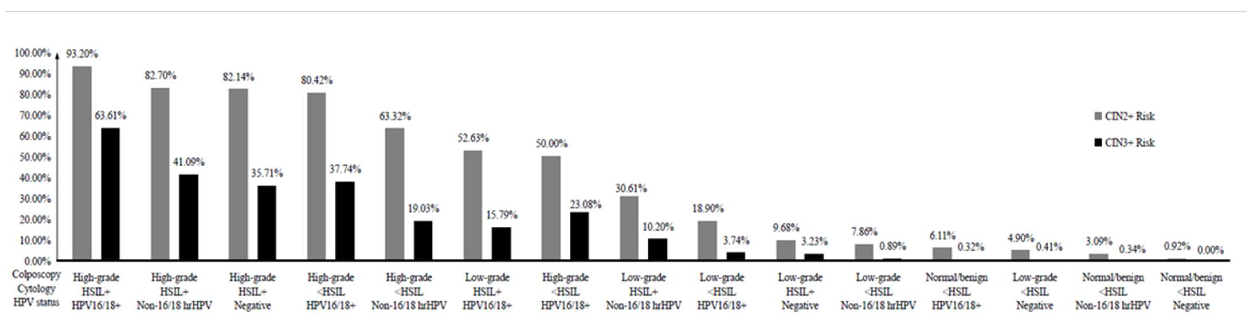
3.4. Cervical Precancer Risk Strata Based on Colposcopy, Cytology, and HPV Status

We assigned each participant to one of 18 subgroups on the basis of the findings of the three tests. Table 5 and Figure 1 show details on the categories with the number of women and the number of both CIN2+ and CIN3+ outcomes ranked by risk. The proportion of women in each subgroup varied considerably, ranging from 93.20% for those with high-grade colposcopic impressions, HSIL+ cytology, and HPV16/18+ to 0% for those with a normal/benign colposcopy, HSIL+ cytology, and hrHPV negative. Seven subgroups had pooled CIN2+ risk estimates above 50%: high-grade colposcopy impression, HSIL+ cytology, and HPV16/18+; high-grade colposcopy impression, HSIL+ cytology, and non-16/18 hrHPV; high-grade colposcopy impression, HSIL+ cytology, and hr-HPV negative; high-grade colposcopy impression, <HSIL cytology, and HPV16/18+; high-grade colposcopy impression, <HSIL cytology, and non-16/18 hrHPV; low-grade colposcopy impression, HSIL+ cytology, and HPV16/18+; and high-grade colposcopy impression, <HSIL cytology, and hr-HPV negative.
Pooled risk estimates for CIN3+ had similar patterns to CIN2+. There was also a correlation between high-risk subgroups and histological CIN2+ and CIN3+ risk: women with high-grade colposcopic impressions were more likely to have HSIL cytology and/or HPV16/18+. Women with high-grade colposcopic impressions showed a high risk for CIN2+ (50%) and CIN3+ (≥19%), even in those with screening results that did not suggest the presence of HPV16/18+ or HSIL+ cytology. Again, irrespective of the screening results, women with normal/benign colposcopies had a low risk for CIN2+ (<7%) and CIN3+ (<1%).

4. Discussion

Based on standard colposcopic practice in six centers across China, we found that HSIL cytology, HPV16/18+, and high-grade colposcopic impressions appear to be significantly related to both CIN2+ and CIN3+. These findings were consistent under both univariate and multivariate analyses and were increasingly evident when colposcopic impressions highly correlated. This suggests that colposcopy may be useful for CIN2+ and CIN3+ risk stratification in China. This is an important finding that advances our knowledge for clinical practice and education. In a previous systematic review, researchers studied the effectiveness of a pooled risk-based tool that combines three testing results (cytology, HPV16/18, and colposcopic impressions) for cervical precancer detection [17]. However, the data was limited because few researchers in the original studies examined the pooled risk of precancers. Therefore, few of the included studies intercalated all three tests, which influenced separate sample sizes. Additionally, many of the included studies chose a suboptimal CIN2+ endpoint as the threshold, which has poor pathological reproducibility and usually has a greater regression to the mean, even in those who did not receive treatment.
Herein, we conducted primary research and incorporated different thresholds that enabled us to analyze data from different perspectives. All possible risk stratifications were assessed using combinations of colposcopic impressions and cytology or/and HPV testing. We found that women with high-grade colposcopic impressions had a CIN2+ risk ranging from 50.0% to 93.20%. We also found that CIN3+ risk ranged from 19.03% to 63.19%, even among women who had an initial low-risk screening result, i.e., <HSIL cytology and/or non-16/18 hrHPV infection or hr-HPV negative. By contrast, normal/benign colposcopic impressions showed risks of CIN2+ ranging from 0% to 6.11% and CIN3+ risks ranging from 0% to less than 0.5%, even among women who were considered at high-risk, i.e., HPV16/18+. Our results suggest that there are similarities in low-risk patterns with normal/benign colposcopic impressions despite having different referral cytology and/or HPV testing results. Therefore, the low prevalence of CIN3+ in low-risk patients suggests that follow-up monitoring should be conducted in these women instead of biopsies, as previous reports have suggested [17,18].
Additionally, some combinations were not rare and were only occasionally detected, such as women with normal/benign colposcopic impressions, HSIL+ cytology, and HPV16/18+. This finding was again consistent with previous reports from clinical studies [19,20,21]. It is worth noting that our data come from experienced colposcopists in clinical settings in China. These findings therefore may not apply to other LMIC populations because training and skills may not be comparable. In China, our evidence appears to support the notion that cervical precancer risk assessment should intercalate colposcopic impressions. Additionally, women with at least two high-risk results from high-grade colposcopic impressions, HSIL+ cytology, and HPV16/18+, should be considered at highest risk because of the high prevalence of CIN2+ and CIN3+. Further research into 5- and 10-year recurrence rates following CIN2/3 treatment would be useful for follow-up scheduling using risk-based colposcopy. This may also provide more insight into the effectiveness of follow-up scheduling and could aid in the development of an artificial intelligence model that incorporates an initial screening step such as liquid-based cytology and HPV testing with lifestyle questionnaires before risk-based colposcopy for precancers [22].
The new ASCCP guidelines [5,6,7] suggest that there may be a need to expedite treatment without colposcopy-based biopsy. This is for women with HSIL and HPV16/18+ because the risk of CIN3+ is higher than 60%. The results from this study did not contradict these findings. We found that among Chinese women with high-grade colposcopic impressions (HSIL+ cytology and HPV16/18+) the prevalence of CIN3+ was 63.61%. In women with low-grade colposcopic impressions (HSIL+ cytology and HPV16/18+), the risk was considerably lower at 15.79%. This raises a number of questions around the immediacy of treatment that the ASCCP has tried to address. However, colposcopic assessment with biopsy should be the preferred strategy to ensure against the over-treatment of patients or causing unnecessary stress. The screening process impacts the emotions, self-esteem, and overall well-being of individuals. Various studies in the US, Germany, and Japan have highlighted the need for more psychosocial support throughout the cervical cancer screening process [23,24], and it is reasonable to assume that Chinese women encounter similar psychosocial issues. Therefore, reducing over-treatment can only be positive.
This study also found that, for women with HSIL+ cytology and HPV16/18+, the optimal treatment strategy may be different for women with high-grade colposcopic impressions compared with those thought to be at low-grade risk. This somewhat contradicts the “one-size-fits-all” approach to expediting treatments, as recommended in the most recent guidelines. The decision to intervene using invasive strategies should be modified by underlying risk. Women with normal/benign colposcopic impressions, <HSIL+ cytology, and non-hrHPV 16/18 negative or hr-HPV negative, were at lower risk of CIN2+, ranging from 0.92% to 3.09%, and CIN3+, ranging from 0% to 0.34%. This finding supports previous studies [9,10,17,18], which suggested that colposcopy-based biopsies are not recommended for underlying precancer identification. Therefore, adding colposcopy into standardized precancer risk assessment is likely to improve clinical practice. However, experienced colposcopists are not always readily available in LMICs. This also means that colposcopy training is not always standardized, and experience dramatically influences a physician’s performance.
In this study, we applied a risk assessment strategy recommended by National Cancer Institute (NCI) protocol [17]. However, risks vary on the basis of geographic regions [25]. Notably, we cannot determine risk based solely upon location because there are subtle differences between urban and rural risk. There are also a number of sub-cultures that may or may not be reported, adding risk and lifestyle elements that are not always considered. In this study, we attempted to gather data from a variety of sources that may provide more insight into the complexities of Chinese society. We included data from women visiting colposcopists in six different regions of mainland China. We hope this evidence will contribute to the development of risk-based management algorithms for Asian populations and may help initiate research into the subtle differences between and within global populations. Cervical cancer has heterogeneous variations in terms of metastatic spread, gene expression, and responses to treatments. Therefore, any well-designed research into cervical precancer screening and detection has the potential to provide valuable insights for population-specific disease prevention and health protection.
To the best of our knowledge, this is the first study to assess the potential benefit of intercalating colposcopic impressions with screening test results (cytology and HPV testing) for cervical precancer detection in Chinese population. However, the study has some limitations that must be considered. First, our findings only reflect the immediate risk of precancers. This study did not examine the course of development, which demands more attention. A longer follow-up period and screening histories and diagnosis will provide important information that will likely increase the precision of risk assessments. Second, we did not stratify patients by age. Several studies have suggested that refining risk-based assessment tools may also require factoring in age. We are conducting a related risk-stratification study to understand the effect of age on disease detection, and we hope to shed light on some of these issues.

5. Conclusions

This study suggests that colposcopy practice could play an important role in assessing patients for their risk of underlying cervical precancers. We found that adding colposcopic impressions to risk-stratification procedures improves case identification. This approach improves the detection of cervical precancers in individuals at highest risk and may also reduce over-treatment, which can have a negative impact on women’s health and well-being. This intercalated approach may also provide reassurance for patients receiving normal/benign colposcopy results who are at lower risk. Our findings support the recommendation against non-directed biopsies for women with normal/benign colposcopic impressions, <HSIL cytology, and hrHPV negative status and suggests that there is a need for directed biopsies for women with high-grade colposcopic impressions, regardless of screening outcomes.

Supplementary Materials

The following supporting information can be downloaded at: https://www.mdpi.com/article/10.3390/diagnostics12112585/s1, Figure S1: The sequence of cervical precancers risk strata based on colposcopy and cytology, Figure S2: The sequence of cervical precancers risk strata based on colposcopy and HPV status.

Author Contributions

P.X. planned and designed the study. P.X. and Q.L. collected the data, and P.X. analyzed and interpreted the data. P.X. performed the statistical analysis. P.X., S.S., Y.J. and Y.Q. provided conceptional assistance. P.X. and S.S. wrote the first draft, and P.X., S.S. and Y.Q. revised the manuscript. All co-authors reviewed the manuscript, made corrections, and approved this publication. All authors have read and agreed to the published version of the manuscript.

Funding

This research was funded by CAMS Innovation Fund for Medical Sciences [CAMS 2021-I2M-1-004].

Institutional Review Board Statement

Ethical approval was obtained from the ethics committee of the Chinese Academy of Medical Sciences and Peking Union Medical College (IEC-2022-022, Beijing, China, 14 March 2022). The need for informed consent was waived by the ethics committee of the Chinese Academy of Medical Sciences and Peking Union Medical College because of the retrospective nature of clinical archive datasets and fully anonymized personal information. All methods were carried out in accordance with relevant guidelines and regulations.

Informed Consent Statement

Patient consent was waived because this was a retrospective study of pre-existing clinical archives and because all data were completely anonymized.

Data Availability Statement

The datasets generated and/or analyzed during the current study are not publicly available because of personal information protection, patient privacy regulation, and medical institutional data regulatory policies but are available from the corresponding author on reasonable request and with permission.

Conflicts of Interest

The authors declare no conflict of interest.

References

  1. Ghebreyesus, T.A. WHO Director-General Calls for All Countries to Take Action to Help End the Suffering Caused by Cervical Cancer. Available online: https://www.who.int/news/item/18-05-2018-who-dg-calls-for-all-countries-to-take-action-to-help-end-the-suffering-caused-by-cervical-cancer (accessed on 13 January 2020).
  2. Xue, P.; Ng, M.T.A.; Qiao, Y. The challenges of colposcopy for cervical cancer screening in LMICs and solutions by artificial intelligence. BMC Med. 2020, 18, 169. [Google Scholar] [CrossRef] [PubMed]
  3. Farnsworth, A.; Roberts, J.M.; Garland, S.M.; Crescini, J.; Kaldor, J.M.; Machalek, D.A. Detection of high-grade cervical disease among women referred directly to colposcopy after a positive HPV screening test varies with age and cytology findings. Int. J. Cancer 2020, 147, 3068–3074. [Google Scholar] [CrossRef] [PubMed]
  4. Underwood, M.; Arbyn, M.; Parry-Smith, W.; De Bellis-Ayres, S.; Todd, R.; Redman, C.W.; Moss, E.L. Accuracy of colposcopy-directed punch biopsies: A systematic review and meta-analysis. BJOG Int. J. Obstet. Gynaecol. 2012, 119, 1293–1301. [Google Scholar] [CrossRef] [PubMed]
  5. Egemen, D.; Cheung, L.C.; Chen, X.; Demarco, M.; Perkins, R.B.; Kinney, W.; Poitras, N.; Befano, B.; Locke, A.; Guido, R.S.; et al. Risk Estimates Supporting the 2019 ASCCP Risk-Based Management Consensus Guidelines. J. Low. Genit. Tract. Dis. 2020, 24, 132–143. [Google Scholar] [CrossRef]
  6. Perkins, R.B.; Guido, R.S.; Castle, P.E.; Chelmow, D.; Einstein, M.H.; Garcia, F.; Huh, W.K.; Kim, J.J.; Moscicki, A.B.; Nayar, R.; et al. 2019 ASCCP Risk-Based Management Consensus Guidelines for Abnormal Cervical Cancer Screening Tests and Cancer Precursors. J. Low. Genit. Tract. Dis. 2020, 24, 102–131. [Google Scholar] [CrossRef] [Green Version]
  7. Schiffman, M.; Wentzensen, N.; Perkins, R.B.; Guido, R.S. An Introduction to the 2019 ASCCP Risk-Based Management Consensus Guidelines. J. Low. Genit. Tract. Dis. 2020, 24, 87–89. [Google Scholar] [CrossRef]
  8. Wentzensen, N.; Massad, L.S.; Mayeaux, E.J., Jr.; Khan, M.J.; Waxman, A.G.; Einstein, M.H.; Conageski, C.; Schiffman, M.H.; Gold, M.A.; Apgar, B.S.; et al. Evidence-Based Consensus Recommendations for Colposcopy Practice for Cervical Cancer Prevention in the United States. J. Low. Genit. Tract. Dis. 2017, 21, 216–222. [Google Scholar] [CrossRef]
  9. Wentzensen, N.; Schiffman, M.; Silver, M.I.; Khan, M.J.; Perkins, R.B.; Smith, K.M.; Gage, J.C.; Gold, M.A.; Conageski, C.; Einstein, M.H.; et al. ASCCP Colposcopy Standards: Risk-Based Colposcopy Practice. J. Low. Genit. Tract. Dis. 2017, 21, 230–234. [Google Scholar] [CrossRef]
  10. Wentzensen, N.; Walker, J.; Smith, K.; Gold, M.A.; Zuna, R.; Massad, L.S.; Liu, A.; Silver, M.I.; Dunn, S.T.; Schiffman, M. A prospective study of risk-based colposcopy demonstrates improved detection of cervical precancers. Am. J. Obs. Gynecol. 2018, 218, 604.e1–604.e8. [Google Scholar] [CrossRef]
  11. Solomon, D.; Davey, D.; Kurman, R.; Moriarty, A.; O’Connor, D.; Prey, M.; Raab, S.; Sherman, M.; Wilbur, D.; Wright, T., Jr.; et al. The 2001 Bethesda System: Terminology for reporting results of cervical cytology. JAMA 2002, 287, 2114–2119. [Google Scholar] [CrossRef]
  12. Luesley, D.; Leeson, S. Colposcopy and Programme Management: Guidelines for the NHS Cervical Screening Programme, 2nd ed.; Public Health England: London, UK, 2010.
  13. Bornstein, J.; Bentley, J.; Bösze, P.; Girardi, F.; Haefner, H.; Menton, M.; Perrotta, M.; Prendiville, W.; Russell, P.; Sideri, M.; et al. 2011 colposcopic terminology of the International Federation for Cervical Pathology and Colposcopy. Obs. Gynecol. 2012, 120, 166–172. [Google Scholar] [CrossRef] [PubMed] [Green Version]
  14. Cree, I.A.; White, V.A.; Indave, B.I.; Lokuhetty, D. Revising the WHO classification: Female genital tract tumours. Histopathology 2020, 76, 151–156. [Google Scholar] [CrossRef] [PubMed]
  15. Carreon, J.D.; Sherman, M.E.; Guillén, D.; Solomon, D.; Herrero, R.; Jerónimo, J.; Wacholder, S.; Rodríguez, A.C.; Morales, J.; Hutchinson, M.; et al. CIN2 is a much less reproducible and less valid diagnosis than CIN3: Results from a histological review of population-based cervical samples. Int. J. Gynecol. Pathol. 2007, 26, 441–446. [Google Scholar] [CrossRef]
  16. Herbert, A.; Arbyn, M.; Bergeron, C. Why CIN3 and CIN2 should be distinguished on histological reports. Cytopathology 2008, 19, 63–64. [Google Scholar] [CrossRef]
  17. Silver, M.I.; Andrews, J.; Cooper, C.K.; Gage, J.C.; Gold, M.A.; Khan, M.J.; Massad, L.S.; Parvu, V.; Perkins, R.B.; Schiffman, M.; et al. Risk of Cervical Intraepithelial Neoplasia 2 or Worse by Cytology, Human Papillomavirus 16/18, and Colposcopy Impression: A Systematic Review and Meta-analysis. Obs. Gynecol. 2018, 132, 725–735. [Google Scholar] [CrossRef] [PubMed]
  18. Del Pino, M.; Angeles, M.A.; Martí, C.; Henere, C.; Munmany, M.; Marimon, L.; Saco, A.; Rakislova, N.; Ordi, J.; Torné, A. Colposcopic Impression Has a Key Role in the Estimation of the Risk of HSIL/CIN3. Cancers 2021, 13, 1224. [Google Scholar] [CrossRef] [PubMed]
  19. Wentzensen, N.; Walker, J.L.; Gold, M.A.; Smith, K.M.; Zuna, R.E.; Mathews, C.; Dunn, S.T.; Zhang, R.; Moxley, K.; Bishop, E.; et al. Multiple biopsies and detection of cervical cancer precursors at colposcopy. J. Clin. Oncol. 2015, 33, 83–89. [Google Scholar] [CrossRef] [Green Version]
  20. Schiffman, M.; Adrianza, M.E. ASCUS-LSIL Triage Study. Design, methods and characteristics of trial participants. Acta Cytol. 2000, 44, 726–742. [Google Scholar] [CrossRef]
  21. Stoler, M.H.; Wright, T.C., Jr.; Parvu, V.; Vaughan, L.; Yanson, K.; Eckert, K.; Karchmer, T.; Kodsi, S.; Cooper, C.K. The Onclarity Human Papillomavirus Trial: Design, methods, and baseline results. Gynecol. Oncol. 2018, 149, 498–505. [Google Scholar] [CrossRef] [Green Version]
  22. Xue, P.; Tang, C.; Li, Q.; Li, Y.; Shen, Y.; Zhao, Y.; Chen, J.; Wu, J.; Li, L.; Wang, W.; et al. Development and validation of an artificial intelligence system for grading colposcopic impressions and guiding biopsies. BMC Med. 2020, 18, 406. [Google Scholar] [CrossRef]
  23. Jentschke, M.; Lehmann, R.; Drews, N.; Hansel, A.; Schmitz, M.; Hillemanns, P. Psychological distress in cervical cancer screening: Results from a German online survey. Arch. Gynecol. Obs. 2020, 302, 699–705. [Google Scholar] [CrossRef] [PubMed]
  24. Kobayashi, M.; Ohno, T.; Noguchi, W.; Matsuda, A.; Matsushima, E.; Kato, S.; Tsujii, H. Psychological distress and quality of life in cervical cancer survivors after radiotherapy: Do treatment modalities, disease stage, and self-esteem influence outcomes? Int. J. Gynecol. Cancer 2009, 19, 1264–1268. [Google Scholar] [CrossRef] [PubMed]
  25. Bray, F.; Ferlay, J.; Soerjomataram, I.; Siegel, R.L.; Torre, L.A.; Jemal, A. Global cancer statistics 2018: GLOBOCAN estimates of incidence and mortality worldwide for 36 cancers in 185 countries. CA Cancer J. Clin. 2018, 68, 394–424. [Google Scholar] [CrossRef] [PubMed]
Figure 1. The sequence of cervical precancer risk strata based on colposcopy cytology and HPV status. Abbreviations: CIN2+, cervical intraepithelial neoplasia grade 2 or worse; CIN3+, cervical intraepithelial neoplasia grade 3 or worse; <HSIL, includes negative for intraepithelial lesion or malignancy (NILM), atypical squamous cells of undetermined significance (ASC-US), low-grade squamous intraepithelial lesion (LSIL); HSIL+, high-grade squamous intraepithelial lesion or worse; HPV, human papillomavirus; hrHPV, high-risk HPV.
Diagnostics 12 02585 g001
Table 1. Baseline demographic and clinical information.
CharacteristicNumber (%)
Age groups, y
<30989 (16.45)
30–392261 (37.61)
40–491672 (27.81)
>501090 (18.13)
Menopause
No5065 (84.25)
Yes947 (15.75)
Gravidity
None584 (9.71)
11022 (17.00)
21685 (28.03)
31365 (22.70)
4+1356 (22.56)
Parity
None944 (15.70)
12588(43.05)
21935 (32.19)
3+545 (9.06)
Cytology
<HSIL5159 (85.81)
HSIL+740 (12.31)
Not performed113 (1.88)
HPV status
hrHPV negative889 (14.79)
Non-16/18 hrHPV3035 (50.48)
HPV16/18+2049 (34.08)
Not performed39 (0.65)
Colposcopy impression
Normal/benign1501 (24.97)
Low-grade3226 (53.66)
High-grade1285 (21.37)
Histological outcome
<CIN24543 (75.57)
CIN2881 (14.65)
CIN3520 (8.65)
Cancer68 (1.13)
Abbreviations: <HSIL, includes negative for intraepithelial lesion or malignancy (NILM), atypical squamous cells of undetermined significance (ASC-US), low-grade squamous intraepithelial lesion (LSIL); HSIL+, high-grade squamous intraepithelial lesion or worse; HPV, human papillomavirus; hrHPV, high-risk human papillomavirus; <CIN2, includes normal, cervical intraepithelial neoplasia grade 1; CIN2, cervical intraepithelial neoplasia grade 2; CIN3, cervical intraepithelial neoplasia grade 3.
Table 2. Association between colposcopy, cytology and HPV status and the diagnosis of cervical precancers.
Risk FactorsCIN2+CIN3+
Univariate AnalysisMultivariate AnalysisUnivariate AnalysisMultivariate Analysis
OR (95% CI)pOR (95% CI)pOR (95% CI)pOR (95% CI)p
Cytology
<HSIL1 (Reference) 1 (Reference) 1 (Reference) 1 (Reference)
HSIL+11.39 (9.57–13.55)<0.0013.54 (2.79–4.51)<0.00111.81 (9.77–14.29)<0.0012.87 (2.27–3.62)<0.001
HPV status
hrHPV negative1 (Reference) 1 (Reference) 1 (Reference) 1 (Reference)
Non-16/18 hrHPV3.12 (2.35–4.15)<0.0011.86 (1.29–2.67)0.0012.68 (1.63–4.38)<0.0011.27 (0.72–2.22)0.41
HPV16/18+10.54 (7.95–13.97)<0.0014.91 (3.41–7.07)<0.00111.74 (7.27–18.96)<0.0013.65 (2.11–6.33)<0.001
Colposcopy
Normal/benign1 (Reference) 1 (Reference) 1 (Reference) 1 (Reference)
Low-grade5.02 (3.63–6.93)<0.0014.00 (2.87–5.58)<0.00111.72 (3.69–37.24)<0.00110.14 (3.18–32.40)<0.001
High-grade133.71 (95.63–186.95)<0.00170.03 (49.43–99.19)<0.001329.66 (105.62–1028.94)<0.001162.15 (51.37–511.79)<0.001
Abbreviations: CIN2+, cervical intraepithelial neoplasia grade 2 or worse; CIN3+, cervical intraepithelial neoplasia grade 3 or worse; OR, odds ratio; CI, confidence interval; <HSIL, includes negative for intraepithelial lesion or malignancy (NILM), atypical squamous cells of undetermined significance (ASC-US), low-grade squamous intraepithelial lesion (LSIL); HSIL+, high-grade squamous intraepithelial lesion or worse; HPV, human papillomavirus; hrHPV, high-risk human papillomavirus.
Table 3. Cervical precancer risk strata based on colposcopy and cytology.
IDColposcopyCytologyTotal, NCIN2+, NRiskCIN3+, NRisk
1aHigh-gradeHSIL+5194620.89022790.5376
1bHigh-grade<HSIL7425400.72782240.3019
1cLow-gradeHSIL+187630.3369200.1070
1dLow-grade<HSIL29753290.1106530.0178
1fNormal/benign<HSIL1442420.029130.0021
1eNormal/benignHSIL+340000
Abbreviations: CIN2+, cervical intraepithelial neoplasia grade 2 or worse; CIN3+, cervical intraepithelial neoplasia grade 3 or worse; <HSIL, includes negative for intraepithelial lesion or malignancy (NILM), atypical squamous cells of undetermined significance (ASC-US), low-grade squamous intraepithelial lesion (LSIL); HSIL+, high-grade squamous intraepithelial lesion or worse. Risk is reported as a frequency percentage. The risk of histological CIN2+ or CIN3+ was calculated by dividing the number of CIN2+ or CIN3+ threshold over all women in each stratum.
Table 4. Cervical precancers risk in strata based on colposcopy and HPV status.
IDColposcopyHPV StatusTotal, NCIN2+, NRiskCIN3+, NRisk
2aHigh-gradeHPV16/187366280.85333550.4823
2bHigh-gradeNon-16/18 hrHPV4803410.71041310.2729
2cHigh-gradehrHPV negative54360.6667160.2963
2dLow-gradeHPV16/189932120.2135440.0443
2eLow-gradeNon-16/18 hrHPV19421760.0906260.0134
2gNormal/benignHPV16/18320190.059410.0031
2fLow-gradehrHPV negative277160.057820.0072
2hNormal/benignNon-16/18 hrHPV613180.029420.0033
2iNormal/benignhrHPV negative55850.009000
Abbreviations: CIN2+, cervical intraepithelial neoplasia grade2 or worse; CIN3+, cervical intraepithelial neoplasia grade3 or worse; HPV, human papillomavirus; hrHPV, high-risk human papillomavirus. Risk is reported as a frequency percentage. The risk of histological CIN2+ or CIN3+ was calculated by dividing the number of CIN2+ or CIN3+ threshold over all women in each stratum.
Table 5. Cervical precancers risk strata based on colposcopy, cytology, and HPV status.
IDColposcopyCytologyHPV StatusTotal, NCIN2+, NRiskCIN3+, NRisk
3aHigh-gradeHSIL+HPV16/18+2942740.93201870.6361
3bHigh-gradeHSIL+Non-16/18 hrHPV1851530.8270760.4109
3cHigh-gradeHSIL+hrHPV negative28230.8214100.3571
3dHigh-grade<HSILHPV16/18+4243410.80421600.3774
3eHigh-grade<HSILNon-16/18 hrHPV2891830.6332550.1903
3gLow-gradeHSIL+HPV16/18+57300.526390.1579
3fHigh-grade<HSILhrHPV negative26130.5060.2308
3hLow-gradeHSIL+Non-16/18 hrHPV98300.3061100.1020
3jLow-grade<HSILHPV16/18+9101720.1890340.0374
3iLow-gradeHSIL+hrHPV negative3130.096810.0323
3kLow-grade<HSILNon-16/18 hrHPV18071420.0786160.0089
3pNormal/benign<HSILHPV16/18+311190.061110.0032
3lLow-grade<HSILhrHPV negative245120.049010.0041
3qNormal/benign<HSILNon-16/18 hrHPV582180.030920.0034
3rNormal/benign<HSILhrHPV negative54150.009200
3nNormal/benignHSIL+Non-16/18 hrHPV170000
3oNormal/benignHSIL+hrHPV negative160000
3mNormal/benignHSIL+HPV16/18+None
Abbreviations: CIN2+, cervical intraepithelial neoplasia grade2 or worse; CIN3+, cervical intraepithelial neoplasia grade3 or worse; <HSIL, includes negative for intraepithelial lesion or malignancy (NILM), atypical squamous cells of undetermined significance (ASC-US), low-grade squamous intraepithelial lesion (LSIL); HSIL+, high-grade squamous intraepithelial lesion or worse; HPV, human papillomavirus; hrHPV, high-risk human papillomavirus. Risk is reported as a frequency percentage. The risk of histological CIN2+ or CIN3+ was calculated by dividing the number of CIN2+ or CIN3+ threshold over all women in each stratum.
Publisher’s Note: MDPI stays neutral with regard to jurisdictional claims in published maps and institutional affiliations.

