
Usefulness of Endoscopic Ultrasound with the Jelly-Filling Method for Esophageal Varices


 , , , ,
Abstract
1. Introduction
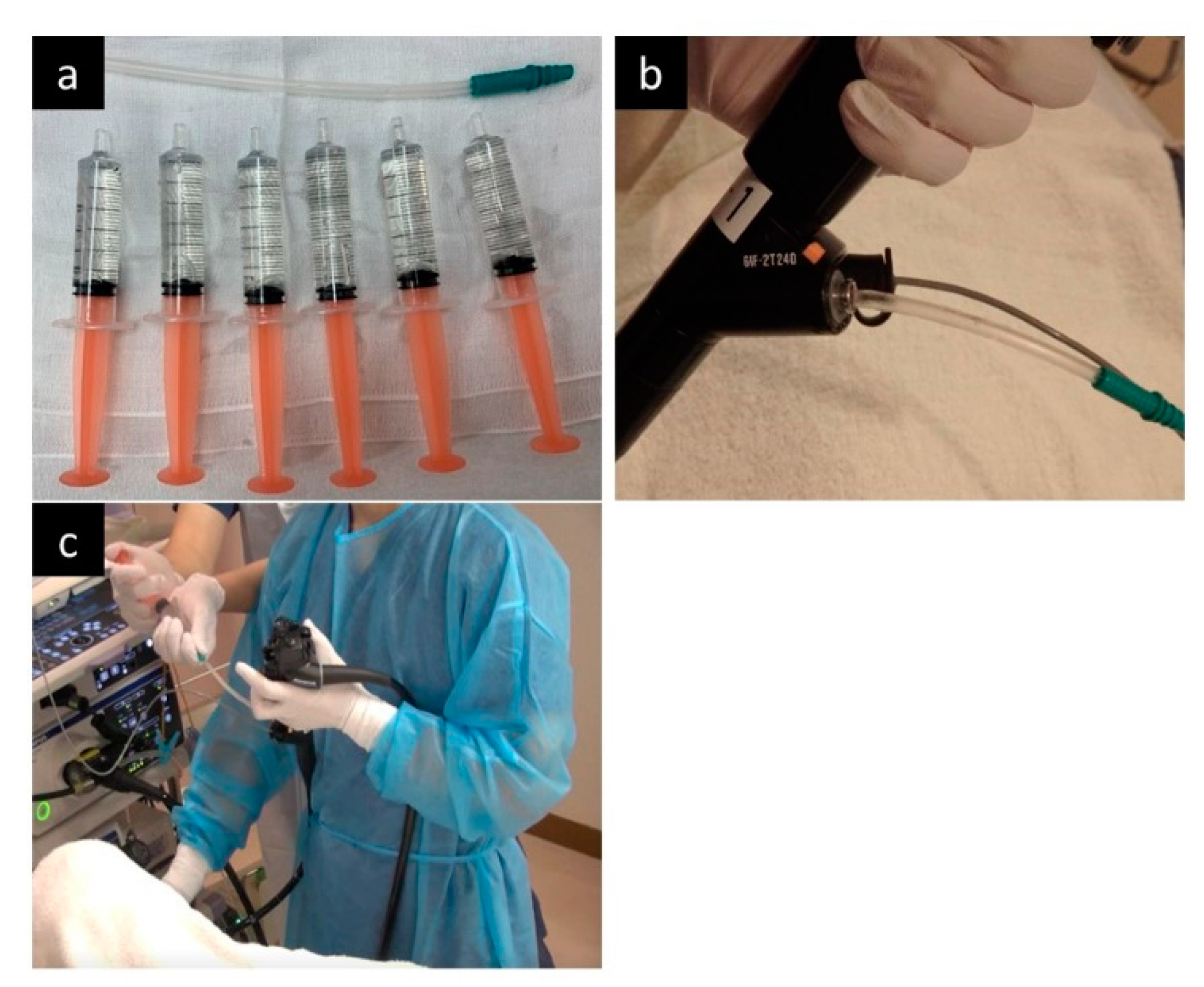
2. Methods
2.1. Study Setting
2.2. Study Design and Patients
2.3. Outcomes and Definitions
2.4. Statistical Analysis
3. Results
3.1. Patient Characteristics
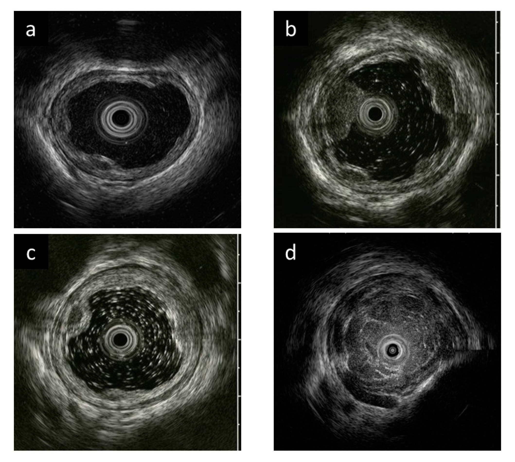
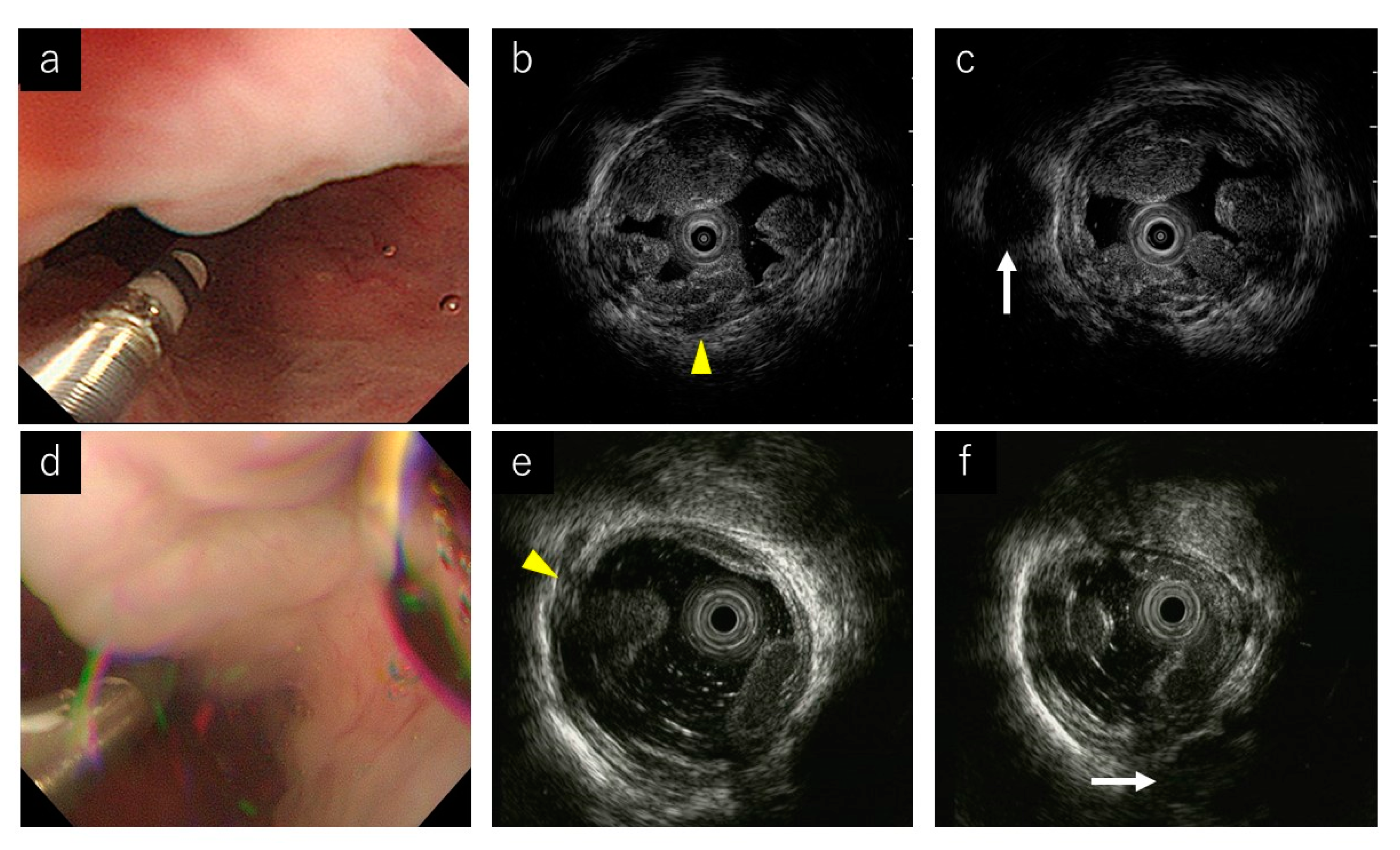
3.2. EUS Image Quality
3.3. Procedure Time and AEs
4. Discussion
Author Contributions
Funding
Institutional Review Board Statement
Informed Consent Statement
Data Availability Statement
Acknowledgments
Conflicts of Interest
References
- Jensen, D.M. Endoscopic screening for varices in cirrhosis: Findings, implications, and outcomes. Gastroenterology 2002, 122, 1620–1630. [Google Scholar] [CrossRef]
- Pagliaro, L.; D’Amico, G.; Sörensen, T.I.; Lebrec, D.; Burroughs, A.K.; Morabito, A.; Tiné, F.; Politi, F.; Traina, M. Prevention of first bleeding in cirrhosis. A meta-analysis of randomized clinical trials of nonsurgical treatment. Ann. Intern. Med. 1992, 117, 59–70. [Google Scholar] [CrossRef]
- D’Amico, G.; Pagliaro, L.; Bosch, J. The treatment of portal hypertension: A meta-analytic review. Hepatology 1995, 22, 332–354. [Google Scholar] [CrossRef] [PubMed]
- Triantos, C.; Kalafateli, M. Primary prevention of bleeding from esophageal varices in patients with liver cirrhosis. World J. Hepatol. 2014, 6, 363–369. [Google Scholar] [CrossRef] [PubMed]
- Van Stiegmann, G.; Cambre, T.; Sun, J.H. A new endoscopic elastic band ligating device. Gastrointest. Endosc. 1986, 32, 230–233. [Google Scholar] [CrossRef]
- Sarin, S.K.; Lamba, G.S.; Kumar, M.; Misra, A.; Murthy, N.S. Comparison of endoscopic ligation and propranolol for the primary prevention of variceal bleeding. N. Engl. J. Med. 1999, 340, 988–993. [Google Scholar] [CrossRef]
- Garbuzenko, D.V.; Arefyev, N.O. Primary prevention of bleeding from esophageal varices in patients with liver cirrhosis: An update and review of the literature. J. Evid. Based Med. 2020, 13, 313–324. [Google Scholar] [CrossRef]
- Trolle, E.; Trolle, D. Treatment of oesophageal varices by injections of sclerosing agents through oesophagoscope in splenectomized patient suffering from splenic phlebostenosis (splenic anemia); a case with autopsy. Acta Chir. Scand. 1946, 94, 385–396. [Google Scholar] [PubMed]
- Masaki, M.; Obara, K.; Suzuki, S.; Orikasa, K.; Mitsuhashi, H.; Iwasaki, K.; Sakamoto, H.; Morito, T.; Kasukawa, R. The destructive effects of sclerosant ethanolamine oleate on mammalian vessel endothelium. Gastroenterol. Jpn. 1990, 25, 230–235. [Google Scholar] [CrossRef] [PubMed]
- Miyaaki, H.; Ichikawa, T.; Taura, N.; Miuma, S.; Isomoto, H.; Nakao, K. Endoscopic management of esophagogastric varices in Japan. Ann. Transl. Med. 2014, 2, 42. [Google Scholar]
- Yuki, M.; Kazumori, H.; Yamamoto, S.; Shizuku, T.; Kinoshita, Y. Prognosis following endoscopic injection sclerotherapy for esophageal varices in adults: 20-year follow-up study. Scand. J. Gastroenterol. 2008, 43, 1269–1274. [Google Scholar] [CrossRef]
- Obara, K. Endoscopic Treatment of Esophagogastric Varices.; NARUNIA Inc.: Tokyo, Japan, 2016; pp. 1–77. [Google Scholar]
- Irisawa, A.; Obara, K.; Sato, Y.; Saito, A.; Takiguchi, F.; Shishido, H.; Sakamoto, H.; Kasukawa, R. EUS analysis of collateral veins inside and outside the esophageal wall in portal hypertension. Gastrointest. Endosc. 1999, 50, 374–380. [Google Scholar] [CrossRef]
- Irisawa, A.; Obara, K.; Saito, A.; Shibukawa, G.; Takagi, T.; Shishido, H.; Sakamoto, H.; Sato, Y.; Kasukawa, R. Endoscopic recurrence of esophageal varices is associated with the specific EUS abnormalities: Severe peri-esophageal collateral veins and large perforating veins. Gastrointest. Endosc. 2001, 53, 77–84. [Google Scholar] [CrossRef] [PubMed]
- Irisaw, A.; Saito, A.; Obara, K.; Shibukawa, G.; Takagi, T.; Yamamoto, G.; Sakamoto, H.; Takiguchi, F.; Shishido, H.; Hikichi, T.; et al. Usefulness of endoscopic ultrasonographic analysis of variceal hemodynamics for the treatment of esophageal varices. Fukushima J. Med. Sci. 2001, 47, 39–50. [Google Scholar] [CrossRef][Green Version]
- Irisawa, A.; Shibukawa, G.; Obara, K.; Saito, A.; Takagi, T.; Shishido, H.; Odajima, H.; Abe, M.; Sugino, T.; Suzuki, T.; et al. Collateral vessels around the esophageal wall in patients with portal hypertension: Comparison of EUS imaging and microscopic findings at autopsy. Gastrointest. Endosc. 2002, 56, 249–253. [Google Scholar] [CrossRef]
- Nagashima, K.; Irisawa, A.; Tominaga, K.; Kashima, K.; Kunogi, Y.; Minaguchi, T.; Izawa, N.; Yamamiya, A.; Yamabe, A.; Hoshi, K.; et al. The Role of Endoscopic Ultrasound for Esophageal Varices. Diagnostics 2020, 10, 1007. [Google Scholar] [CrossRef] [PubMed]
- Hino, S.; Kakutani, H.; Ikeda, K.; Uchiyama, Y.; Sumiyama, K.; Kuramochi, A.; Kitamura, Y.; Matsuda, K.; Arakawa, H.; Kawamura, M.; et al. Hemodynamic assessment of the left gastric vein in patients with esophageal varices with color Doppler EUS: Factors affecting development of esophageal varices. Gastrointest. Endosc. 2002, 55, 512–517. [Google Scholar] [CrossRef]
- Sato, T.; Yamazaki, K. Endoscopic color Doppler ultrasonography for esophagogastric varices. Diagn. Ther. Endosc. 2012, 2012, 859213. [Google Scholar] [CrossRef]
- Esaki, M.; Matsumoto, T.; Moriyama, T.; Hizawa, K.; Ohji, Y.; Nakamura, S.; Hirakawa, K.; Hirahashi, M.; Yao, T.; Iida, M. Probe EUS for the diagnosis of invasion depth in superficial esophageal cancer: A comparison between a jelly-filled method and a water-filled balloon method. Gastrointest. Endosc. 2006, 63, 389–395. [Google Scholar] [CrossRef] [PubMed]
- Hanaoka, N.; Ishihara, R.; Matsuura, N.; Uedo, N.; Takeuchi, Y.; Higashino, K.; Yamashina, T.; Aoi, K.; Iishi, H. Esophageal EUS by filling water-soluble lubricating jelly for diagnosis of depth of invasion in superficial esophageal cancer. Gastrointest. Endosc. 2015, 82, 164–165. [Google Scholar] [CrossRef]
- The Japan Society for Portal Hypertension. The General Rules for Study of Portal Hypertension, 3rd ed.; Kanehara: Tokyo, Japan, 2017. [Google Scholar]
- Chak, A.; Canto, M.; Stevens, P.D.; Lightdale, C.J.; van de Mierop, F.; Cooper, G.; Pollack, B.J.; Sivak, M.V., Jr. Clinical applications of a new through-the-scope ultrasound probe: Prospective comparison with an ultrasound endoscope. Gastrointest. Endosc. 1997, 45, 291–295. [Google Scholar] [CrossRef]
- Wallace, M.B.; Hoffman, B.J.; Sahai, A.S.; Inoue, H.; van Velse, A.; Hawes, R.H. Imaging of esophageal tumors with a water-filled condom and a catheter US probe. Gastrointest. Endosc. 2000, 51, 597–600. [Google Scholar] [CrossRef]
- Obara, K.; Irisawa, A.; Sato, Y. Usefulness of EUS in portal hypertension with esophageal varices. Dig. Endosc. 2004, 16, S168–S172. [Google Scholar] [CrossRef]
- Nagamine, N.; Ido, K.; Ueno, N.; Kimura, K.; Kawamata, T.; Kawada, H.; Hirasawa, T.; Suzuki, T.; Kubo, H.; Tokumaru, K.; et al. The usefulness of ultrasonic microprobe imaging for endoscopic variceal ligation. Am. J. Gastroenterol. 1996, 91, 523–529. [Google Scholar]
- Dhiman, R.K.; Choudhuri, G.; Saraswat, V.A.; Agarwal, D.K.; Naik, S.R. Role of paraoesophageal collateral and perforating veins on outcome of endoscopic sclerotherapy for oesophageal varices: An endosonographic study. Gut 1996, 38, 759–764. [Google Scholar] [CrossRef][Green Version]
- Miyamoto, S.; Takahashi, K.; Ohya, T.R. New method of esophageal endoscopic ultrasonography with injected gel: Endoscopic ultrasonography-gel filling method. Dig. Endosc 2021, 33, e49–e50. [Google Scholar] [CrossRef]
- Toyonaga, H.; Takahashi, K.; Kin, T.; Hayashi, T.; Katanuma, A. Gel immersion technique for the examination and treatment of an ampullary tumor. Endoscopy 2021. [Google Scholar] [CrossRef]
- Minoda, Y.; Nagatomo, S.; Fujimori, N.; Nishioka, K.S.; Teramatsu, K.; Takamatsu, Y.; Ogino, H.; Ihara, E. Usefulness of a gel immersion-assisted EUS-guided fine-needle aspiration/biopsy for ampullary lesions (with videos). Endosc. Ultrasound 2021. [Google Scholar] [CrossRef]
- Yano, T.; Nemoto, D.; Ono, K.; Miyata, Y.; Numao, N.; Iwashita, C.; Nagayama, M.; Takahashi, H.; Lefor, A.K.; Yamamoto, H. Gel immersion endoscopy: A novel method to secure the visual field during endoscopy in bleeding patients (with videos). Gastrointest. Endosc. 2016, 83, 809–811. [Google Scholar] [CrossRef] [PubMed]


| Evaluated Items | Jelly-Filling Method (n = 13) | Water-Filling Method (n = 17) | p-Value |
|---|---|---|---|
| Age (years), median (range) | 66 (40–77) | 66 (43–83) | 0.630 |
| Sex, n | 0.440 | ||
| Male | 10 | 10 | |
| Female | 3 | 7 | |
| Cause of liver cirrhosis, n | |||
| Alcoholic/PBC/NASH/virus/unknown | 5/3/3/1/1 | 6/4/2/3/2 | 0.205 |
| Indication of endoscopic treatment, n | |||
| Elective/prophylactic | 0/13 | 4/13 | 0.113 |
| Endoscopic findings of esophageal varices, n | |||
| Location | |||
| Li/Lm/Ls | 4/7/2 | 1/12/4 | 0.332 |
| Form | |||
| F1/F2/F3 | 1/12/0 | 2/15/0 | 0.717 |
| Color | |||
| Cb/Cw | 13/0 | 16/1 | 0.381 |
| Red color sign (RC) | |||
| RC0/≥RC1 | 4/9 | 10/7 | 0.133 |
| Evaluated Items | Jelly-Filling Method (n = 13) | Water-Filling Method (n = 17) | p-Value |
|---|---|---|---|
| EUS operator | 1.000 | ||
| Nonexpert | 6 | 8 | |
| Expert | 7 | 9 | |
| EUS image quality (points) * | 3 (3–4) | 2 (1–4) | <0.001 |
| Procedure time (min) * | 6 (3–10) | 4 (2–10) | 0.024 |
| Observation of Peri-v | 0.238 | ||
| Clear | 13 | 14 | |
| Unclear | 0 | 3 | |
| Observation of Para-v | 0.053 | ||
| Clear | 13 | 12 | |
| Unclear | 0 | 5 | |
| Observation of Pv | 0.004 | ||
| Clear | 13 | 9 | |
| Unclear | 0 | 8 | |
| Adverse events | 0 | 0 | – |
| Evaluated Items | Jelly-Filling Method (n = 13) | Water-Filling Method (n = 17) | p-Value |
|---|---|---|---|
| Expert, n | 7 | 9 | |
| EUS image quality (points) | 3 (3–4) | 2 (2–3) | 0.007 |
| Procedure time (min) | 5 (3–7) | 4 (2–7) | 0.329 |
| Nonexpert, n | 6 | 8 | |
| EUS image quality (points) | 3 (3–4) | 2 (1–3) | 0.003 |
| Procedure time (min) | 8 (6–10) | 4 (3–10) | 0.036 |
Publisher’s Note: MDPI stays neutral with regard to jurisdictional claims in published maps and institutional affiliations. |
© 2021 by the authors. Licensee MDPI, Basel, Switzerland. This article is an open access article distributed under the terms and conditions of the Creative Commons Attribution (CC BY) license (https://creativecommons.org/licenses/by/4.0/).
Share and Cite
Kato, T.; Hikichi, T.; Nakamura, J.; Takasumi, M.; Hashimoto, M.; Kobashi, R.; Yanagita, T.; Takagi, T.; Suzuki, R.; Sugimoto, M.; et al. Usefulness of Endoscopic Ultrasound with the Jelly-Filling Method for Esophageal Varices. Diagnostics 2021, 11, 1726. https://doi.org/10.3390/diagnostics11091726
Kato T, Hikichi T, Nakamura J, Takasumi M, Hashimoto M, Kobashi R, Yanagita T, Takagi T, Suzuki R, Sugimoto M, et al. Usefulness of Endoscopic Ultrasound with the Jelly-Filling Method for Esophageal Varices. Diagnostics. 2021; 11(9):1726. https://doi.org/10.3390/diagnostics11091726
Chicago/Turabian StyleKato, Tsunetaka, Takuto Hikichi, Jun Nakamura, Mika Takasumi, Minami Hashimoto, Ryoichiro Kobashi, Takumi Yanagita, Tadayuki Takagi, Rei Suzuki, Mitsuru Sugimoto, and et al. 2021. "Usefulness of Endoscopic Ultrasound with the Jelly-Filling Method for Esophageal Varices" Diagnostics 11, no. 9: 1726. https://doi.org/10.3390/diagnostics11091726
APA StyleKato, T., Hikichi, T., Nakamura, J., Takasumi, M., Hashimoto, M., Kobashi, R., Yanagita, T., Takagi, T., Suzuki, R., Sugimoto, M., Sato, Y., Irie, H., Okubo, Y., Kobayakawa, M., & Ohira, H. (2021). Usefulness of Endoscopic Ultrasound with the Jelly-Filling Method for Esophageal Varices. Diagnostics, 11(9), 1726. https://doi.org/10.3390/diagnostics11091726