
The Diagnostic Usefulness of 131I-SPECT/CT at Both Radioiodine Ablation and during Long-Term Follow-Up in Patients Thyroidectomized for Differentiated Thyroid Carcinoma: Analysis of Tissue Risk Factors Ascertained at Surgery and Correlated with Metastasis Appearance


 , , , and
Abstract
1. Introduction
2. Materials and Methods
2.1. Patients
2.2. Imaging
2.3. Image Analyses
2.4. Outcome
2.5. Statistics
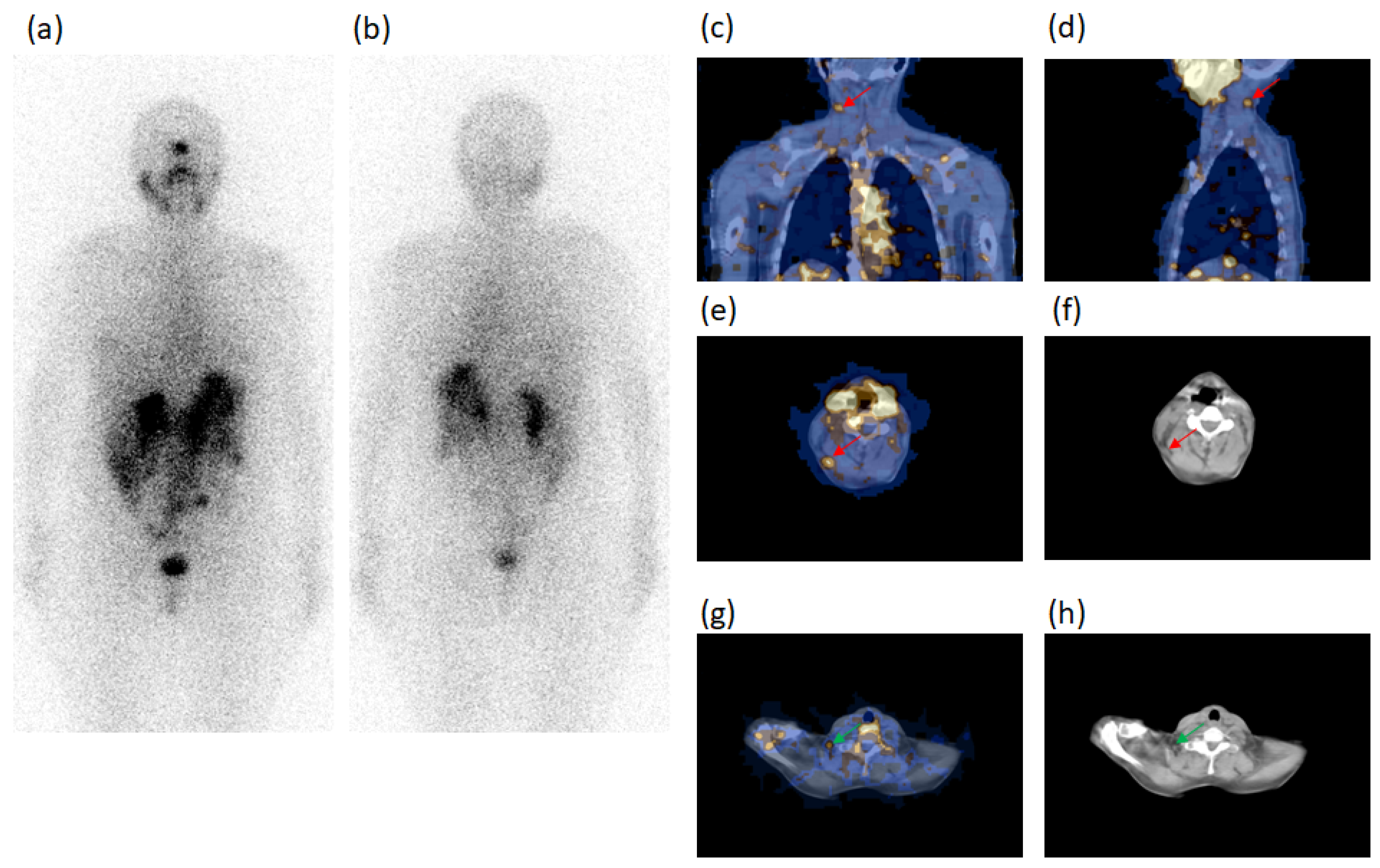
3. Results
Follow-Up
4. Discussion
5. Conclusions
Author Contributions
Funding
Institutional Review Board Statement
Informed Consent Statement
Data Availability Statement
Conflicts of Interest
References
- Beasley, N.J.P.; Lee, J.; Eski, S.; Walfish, P.; Witterick, I.; Freeman, J.L. Impact of Nodal Metastases on Prognosis in Patients with Well-Differentiated Thyroid Cancer. Arch. Otolaryngol. Head Neck Surg. 2002, 128, 825–828. [Google Scholar] [CrossRef]
- Enewold, L.; Zhu, K.; Ron, E.; Marrogi, A.J.; Stojadinovic, A.; Peoples, G.E.; Devesa, S.S. Rising Thyroid Cancer Incidence in the United States by Demographic and Tumor Characteristics, 1980–2005. Cancer Epidemiol. Biomark. Prev. 2009, 18, 784–791. [Google Scholar] [CrossRef] [PubMed]
- Avram, A.M. Radioiodine Scintigraphy with SPECT/CT: An Important Diagnostic Tool for Thyroid Cancer Staging and Risk Stratification. J. Nucl. Med. 2012, 53, 754–764. [Google Scholar] [CrossRef] [PubMed]
- Pellegriti, G.; Frasca, F.; Regalbuto, C.; Squatrito, S.; Vigneri, R. Worldwide Increasing Incidence of Thyroid Cancer: Update on Epidemiology and Risk Factors. J. Cancer Epidemiol. 2013, 2013, 965212. [Google Scholar] [CrossRef] [PubMed]
- Sellers, M.; Beenken, S.; Blankenship, A.; Soong, S.-J.; Turbat-Herrera, E.; Urist, M.; Maddox, W. Prognostic significance of cervical lymph node metastases in differentiated thyroid cancer. Am. J. Surg. 1992, 164, 578–581. [Google Scholar] [CrossRef]
- Hughes, C.J.; Shaha, A.R.; Shah, J.P.; Loree, T.R. Impact of lymph node metastasis in differentiated carcinoma of the thyroid: A matched-pair analysis. Head Neck 1996, 18, 127–132. [Google Scholar] [CrossRef]
- Mazzaferri, E.L.; Robyn, J. Postsurgical management of differentiated thyroid carcinoma. Otolaryngol. Clin. N. Am. 1996, 29, 637–662. [Google Scholar]
- Mercante, G.; Frasoldati, A.; Pedroni, C.; Formisano, D.; Renna, L.; Piana, S.; Gardini, G.; Valcavi, R.; Barbieri, V. Prognostic Factors Affecting Neck Lymph Node Recurrence and Distant Metastasis in Papillary Microcarcinoma of the Thyroid: Results of a Study in 445 Patients. Thyroid 2009, 19, 707–716. [Google Scholar] [CrossRef]
- Spanu, A.; Nuvoli, S.; Gelo, I.; Mele, L.; Piras, B.; Madeddu, G. Role of Diagnostic 131I SPECT/CT in Long-Term Follow-up of Patients with Papillary Thyroid Microcarcinoma. J. Nucl. Med. 2018, 59, 1510–1515. [Google Scholar] [CrossRef]
- Spanu, A.; Nuvoli, S.; Marongiu, A.; Gelo, I.; Mele, L.; Piras, B.; Madeddu, G. Neck lymph node metastasis detection in patients with differentiated thyroid carcinoma (DTC) in long-term follow-up: A 131I-SPECT/CT study. BMC Cancer 2020, 20, 239. [Google Scholar] [CrossRef]
- Simard, E.P.; Ward, E.M.; Siegel, R.; Jemal, A. Cancers with increasing incidence trends in the United States: 1999 through 2008. CA Cancer J. Clin. 2012, 62, 118–128. [Google Scholar] [CrossRef]
- Donohoe, K.J.; Aloff, J.; Avram, A.M.; Bennet, K.; Giovanella, L.; Greenspan, B.; Gulec, S.; Hassan, A.; Kloos, R.T.; Solórzano, C.C.; et al. Appropriate Use Criteria for Nuclear Medicine in the Evaluation and Treatment of Differentiated Thyroid Cancer. J. Nucl. Med. 2020, 61, 375–396. [Google Scholar] [CrossRef] [PubMed]
- Ito, Y.; Tomoda, C.; Uruno, T.; Takamura, Y.; Miya, A.; Kobayashi, K.; Matsuzuka, F.; Kuma, K.; Miyauchi, A. Prognostic Significance of Extrathyroid Extension of Papillary Thyroid Carcinoma: Massive but Not Minimal Extension Affects the Relapse-free Survival. World J. Surg. 2006, 30, 780–786. [Google Scholar] [CrossRef] [PubMed]
- Ito, Y.; Miyauchi, A.; Kihara, M.; Kobayashi, K.; Miya, A. Prognostic values of clinical lymph node metastasis and macroscopic extrathyroid extension in papillary thyroid carcinoma. Endocr. J. 2014, 61, 745–750. [Google Scholar] [CrossRef] [PubMed]
- Andersen, P.E.; Kinsella, J.; Loree, T.R.; Shaha, A.R.; Shah, J.P. Differentiated carcinoma of the thyroid with extrathyroidal extension. Am. J. Surg. 1995, 170, 467–470. [Google Scholar] [CrossRef]
- Ortiz, S.; Rodríguez, J.M.; Soria, T.; Pérez-Flores, D.; Piñero, A.; Moreno, J.; Parrilla, P. Extrathyroid Spread in Papillary Carcinoma of the Thyroid: Clinicopathological and Prognostic Study. Otolaryngol. Head Neck Surg. 2001, 124, 261–265. [Google Scholar] [CrossRef]
- Lombardi, C.P.; Bellantone, R.; De Crea, C.; Paladino, N.C.; Fadda, G.; Salvatori, M.; Raffaelli, M. Papillary Thyroid Microcarcinoma: Extrathyroidal Extension, Lymph Node Metastases, and Risk Factors for Recurrence in a High Prevalence of Goiter Area. World J. Surg. 2010, 34, 1214–1221. [Google Scholar] [CrossRef]
- Yin, D.-T.; Yu, K.; Lu, R.-Q.; Li, X.; Xu, J.; Lei, M. Prognostic impact of minimal extrathyroidal extension in papillary thyroid carcinoma. Medicine 2016, 95, e5794. [Google Scholar] [CrossRef]
- Youngwirth, L.M.; Adam, M.A.; Scheri, R.P.; Roman, S.A.; Sosa, J.A. Extrathyroidal Extension Is Associated with Compromised Survival in Patients with Thyroid Cancer. Thyroid 2017, 27, 626–631. [Google Scholar] [CrossRef]
- Liu, Z.; Huang, Y.; Chen, S.; Hu, D.; Wang, M.; Zhou, L.; Zhou, W.; Chen, D.; Feng, H.; Wei, W.; et al. Minimal extrathyroidal extension affects the prognosis of differentiated thyroid cancer: Is there a need for change in the AJCC classification system? PLoS ONE 2019, 14, e0218171. [Google Scholar] [CrossRef]
- Danilovic, D.L.; Castroneves, L.A.; Suemoto, C.K.; Elias, L.O.; Soares, I.C.; Camargo, R.Y.; Correa, F.D.A.; Hoff, A.O.; Marui, S. Is There a Difference Between Minimal and Gross Extension into the Strap Muscles for the Risk of Recurrence in Papillary Thyroid Carcinomas? Thyroid 2020, 30, 1008–1016. [Google Scholar] [CrossRef] [PubMed]
- Moon, H.J.; Kim, E.-K.; Chung, W.Y.; Yoon, J.H.; Kwak, J.Y. Minimal Extrathyroidal Extension in Patients with Papillary Thyroid Microcarcinoma: Is It a Real Prognostic Factor? Ann. Surg. Oncol. 2011, 18, 1916–1923. [Google Scholar] [CrossRef]
- Shin, J.H.; Ha, T.K.; Park, H.K.; Ahn, M.S.; Kim, K.H.; Bae, K.B.; Kim, T.H.; Choi, C.S.; Bae, S.K.; Kim, S.H. Implication of minimal extrathyroidal extension as a prognostic factor in papillary thyroid carcinoma. Int. J. Surg. 2013, 11, 944–947. [Google Scholar] [CrossRef] [PubMed][Green Version]
- Nixon, I.; Ganly, I.; Patel, S.; Palmer, F.L.; Whitcher, M.M.; Tuttle, R.M.; Shaha, A.R.; Shah, J.P. The impact of microscopic extrathyroid extension on outcome in patients with clinical T1 and T2 well-differentiated thyroid cancer. Surgery 2011, 150, 1242–1249. [Google Scholar] [CrossRef] [PubMed]
- Al-Qurayshi, Z.; Shama, M.A.; Randolph, G.W.; Kandil, E. Minimal extrathyroidal extension does not affect survival of well-differentiated thyroid cancer. Endocr.-Relat. Cancer 2017, 24, 221–226. [Google Scholar] [CrossRef] [PubMed]
- Tam, S.; Amit, M.; Boonsripitayanon, M.; Busaidy, N.L.; Cabanillas, M.E.; Waguespack, S.G.; Gross, N.D.; Grubbs, E.G.; Williams, M.D.; Lai, S.; et al. Effect of Tumor Size and Minimal Extrathyroidal Extension in Patients with Differentiated Thyroid Cancer. Thyroid 2018, 28, 982–990. [Google Scholar] [CrossRef]
- Castagna, M.G.; Forleo, R.; Maino, F.; Fralassi, N.; Barbato, F.; Palmitesta, P.; Pilli, T.; Capezzone, M.; Brilli, L.; Ciuoli, C.; et al. Small papillary thyroid carcinoma with minimal extrathyroidal extension should be managed as ATA low-risk tumor. J. Endocrinol. Investig. 2018, 41, 1029–1035. [Google Scholar] [CrossRef]
- Ahmaddy, F.; Wenter, V.; Ilhan, H.; Wacker, D.; Unterrainer, M.; Knösel, T.; Bartenstein, P.; Spitzweg, C.; Lehner, S.; Todica, A. Effects of the Minimal Extrathyroidal Extension on Early Response Rates after (Adjuvant) Initial Radioactive Iodine Therapy in PTC Patients. Cancers 2020, 12, 3357. [Google Scholar] [CrossRef]
- Hay, I.; Hutchinson, M.E.; Gonzalez-Losada, T.; McIver, B.; Reinalda, M.E.; Grant, C.S.; Thompson, G.B.; Sebo, T.J.; Goellner, J.R. Papillary thyroid microcarcinoma: A study of 900 cases observed in a 60-year period. Surgery 2008, 144, 980–988. [Google Scholar] [CrossRef]
- Wang, T.S.; Goffredo, P.; Sosa, J.A.; Roman, S.A. Papillary Thyroid Microcarcinoma: An Over-Treated Malignancy? World J. Surg. 2014, 38, 2297–2303. [Google Scholar] [CrossRef]
- Lubin, E.; Mechlis-Frish, S.; Zatz, S.; Shimoni, A.; Segal, K.; Avraham, A.; Levy, R.; Feinmesser, R. Serum thyroglobulin and iodine-131 whole-body scan in the diagnosis and assessment of treatment for metastatic differentiated thyroid carcinoma. J. Nucl. Med. 1994, 35, 257–262. [Google Scholar]
- Franceschi, M.; Kusić, Z.; Franceschi, D.; Lukinac, L.; Roncević, S. Thyroglobulin determination, neck ultrasonography and iodine-131 whole-body scintigraphy in differentiated thyroid carcinoma. J. Nucl. Med. 1996, 37, 446–451. [Google Scholar]
- Filesi, M.; Signore, A.; Ventroni, G.; Melacrinis, F.F.; Ronga, G. Role of initial iodine-131 whole-body scan and serum thyroglobulin in differentiated thyroid carcinoma metastases. J. Nucl. Med. 1998, 39, 1542–1546. [Google Scholar] [PubMed]
- Spies, W.G.; Wojtowicz, C.H.; Spies, S.M.; Shah, A.Y.; Zimmer, A.M. Value of Post-Therapy Whole-Body I-131 Imaging in the Evaluation of Patients with Thyroid Carcinoma Having Undergone High-Dose I-131 Therapy. Clin. Nucl. Med. 1989, 14, 793–800. [Google Scholar] [CrossRef] [PubMed]
- Sutter, C.W.; Masilungan, B.G.; Stadalnik, R.C. False-positive results of I-131 whole-body scans in patients with thyroid cancer. Semin. Nucl. Med. 1995, 25, 279–282. [Google Scholar] [CrossRef]
- Shapiro, B.; Rufini, V.; Jarwan, A.; Geatti, O.; Kearfott, K.J.; Fig, L.M.; Kirkwood, I.D.; Gross, M.D. Artifacts, anatomical and physiological variants, and unrelated diseases that might cause false-positive whole-body 131-I scans in patients with thyroid cancer. Semin. Nucl. Med. 2000, 30, 115–132. [Google Scholar] [CrossRef]
- Mitchell, G.; Pratt, B.E.; Vini, L.; McCready, V.R.; Harmer, C.L. False positive 131I whole body scans in thyroid cancer. Br. J. Radiol. 2000, 73, 627–635. [Google Scholar] [CrossRef]
- Avram, A.M.; Fig, L.M.; Frey, K.A.; Gross, M.D.; Wong, K.K. Preablation 131-I Scans With SPECT/CT in Postoperative Thyroid Cancer Patients: What Is the Impact on Staging? J. Clin. Endocrinol. Metab. 2013, 98, 1163–1171. [Google Scholar] [CrossRef]
- Avram, A.M.; Esfandiari, N.H.; Wong, K.K. Preablation 131-I Scans with SPECT/CT Contribute to Thyroid Cancer Risk Stratification and 131-I Therapy Planning. J. Clin. Endocrinol. Metab. 2015, 100, 1895–1902. [Google Scholar] [CrossRef] [PubMed]
- Kohlfuerst, S.; Igerc, I.; Lobnig, M.; Gallowitsch, H.J.; Gomez-Segovia, I.; Matschnig, S.; Mayr, J.; Mikosch, P.; Beheshti, M.; Lind, P. Posttherapeutic 131I SPECT-CT offers high diagnostic accuracy when the findings on conventional planar imaging are inconclusive and allows a tailored patient treatment regimen. Eur. J. Nucl. Med. Mol. Imaging 2009, 36, 886–893. [Google Scholar] [CrossRef]
- Schmidt, D.; Szikszai, A.; Linke, R.; Bautz, W.; Kuwert, T. Impact of 131I SPECT/Spiral CT on Nodal Staging of Differentiated Thyroid Carcinoma at the First Radioablation. J. Nucl. Med. 2009, 50, 18–23. [Google Scholar] [CrossRef][Green Version]
- Mustafa, M.; Kuwert, T.; Weber, K.; Knesewitsch, P.; Negele, T.; Haug, A.; Linke, R.; Bartenstein, P.; Schmidt, D. Regional lymph node involvement in T1 papillary thyroid carcinoma: A bicentric prospective SPECT/CT study. Eur. J. Nucl. Med. Mol. Imaging 2010, 37, 1462–1466. [Google Scholar] [CrossRef] [PubMed]
- Grewal, R.K.; Tuttle, R.M.; Fox, J.; Borkar, S.; Chou, J.F.; Gonen, M.; Strauss, H.W.; Larson, S.M.; Schöder, H. The Effect of Posttherapy 131I SPECT/CT on Risk Classification and Management of Patients with Differentiated Thyroid Cancer. J. Nucl. Med. 2010, 51, 1361–1367. [Google Scholar] [CrossRef] [PubMed]
- Ciappuccini, R.; Heutte, N.; Trzepla, G.; Rame, J.-P.; Vaur, D.; Aide, N.; Bardet, S. Postablation 131I scintigraphy with neck and thorax SPECT–CT and stimulated serum thyroglobulin level predict the outcome of patients with differentiated thyroid cancer. Eur. J. Endocrinol. 2011, 164, 961–969. [Google Scholar] [CrossRef] [PubMed]
- Maruoka, Y.; Abe, K.; Baba, S.; Isoda, T.; Sawamoto, H.; Tanabe, Y.; Sasaki, M.; Honda, H. Incremental Diagnostic Value of SPECT/CT with131I Scintigraphy after Radioiodine Therapy in Patients with Well-differentiated Thyroid Carcinoma. Radiology 2012, 265, 902–909. [Google Scholar] [CrossRef]
- Tharp, K.; Israel, O.; Hausmann, J.; Bettman, L.; Martin, W.H.; Daitzchman, M.; Sandler, M.P.; Delbeke, D. Impact of 131I-SPECT/CT images obtained with an integrated system in the follow-up of patients with thyroid carcinoma. Eur. J. Nucl. Med. Mol. Imaging 2004, 31, 1435–1442. [Google Scholar] [CrossRef]
- Spanu, A.; Solinas, M.E.; Chessa, F.; Sanna, D.; Nuvoli, S.; Madeddu, G. 131I SPECT/CT in the Follow-up of Differentiated Thyroid Carcinoma: Incremental Value Versus Planar Imaging. J. Nucl. Med. 2009, 50, 184–190. [Google Scholar] [CrossRef]
- Schmidt, D.; Linke, R.; Uder, M.; Kuwert, T. Five months’ follow-up of patients with and without iodine-positive lymph node metastases of thyroid carcinoma as disclosed by 131I-SPECT/CT at the first radioablation. Eur. J. Nucl. Med. Mol. Imaging 2010, 37, 699–705. [Google Scholar] [CrossRef]
- Menges, M.; Uder, M.; Kuwert, T.; Schmidt, D. 131I SPECT/CT in the Follow-Up of Patients with Differentiated Thyroid Carcinoma. Clin. Nucl. Med. 2012, 37, 555–560. [Google Scholar] [CrossRef]
- Xue, Y.-L.; Qiu, Z.-L.; Song, H.-J.; Luo, Q.-Y. Value of 131I SPECT/CT for the evaluation of differentiated thyroid cancer: A systematic review of the literature. Eur. J. Nucl. Med. Mol. Imaging 2012, 40, 768–778. [Google Scholar] [CrossRef]
- Kuker, R.; Sztejnberg, M.; Gulec, S. I-124 Imaging and Dosimetry. Mol. Imaging Radionucl. Ther. 2017, 26, 66–73. [Google Scholar] [CrossRef]
- Edge, S.B.; Compton, C.C. The American Joint Committee on Cancer: The 7th Edition of the AJCC Cancer Staging Manual and the Future of TNM. Ann. Surg. Oncol. 2010, 17, 1471–1474. [Google Scholar] [CrossRef]
- Pacini, F.; Schlumberger, M.; Dralle, H.; Elisei, R.; Smit, J.W.A.; Wiersinga, W. European consensus for the management of patients with differentiated thyroid carcinoma of the follicular epithelium. Eur. J. Endocrinol. 2006, 154, 787–803. [Google Scholar] [CrossRef] [PubMed]
- Grant, S.; Luttrell, B.; Reeve, T.; Wiseman, J.; Wilmshurst, E.; Stiel, J.; Donohoe, D.; Cooper, R.; Bridgman, M. Thyroglobulin may be undetectable in the serum of patients with metastatic disease secondary to differentiated thyroid carcinoma. Follow-up of differentiated thyroid carcinoma. Cancer 1984, 54, 1625–1628. [Google Scholar] [CrossRef]
- Miller, J.H.; Marcus, C.S. Metastatic Papillary Thyroid Carcinoma with Normal Serum Thyroglobulin Level. Clin. Nucl. Med. 1988, 13, 652–653. [Google Scholar] [CrossRef]
- Lee, S.-W. SPECT/CT in the Treatment of Differentiated Thyroid Cancer. Nucl. Med. Mol. Imaging 2017, 51, 297–303. [Google Scholar] [CrossRef] [PubMed]
- Szujo, S.; Sira, L.; Bajnok, L.; Bodis, B.; Gyory, F.; Nemes, O.; Rucz, K.; Kenyeres, P.; Valkusz, Z.; Sepp, K.; et al. The impact of post-radioiodine therapy SPECT/CT on early risk stratification in differentiated thyroid cancer; a bi-institutional study. Oncotarget 2017, 8, 79825–79834. [Google Scholar] [CrossRef] [PubMed]
- Sawyer, L.J.; Starritt, H.C.; Hiscock, S.C.; Evans, M.J. Effective doses to patients from CT acquisitions on the GE Infinia Hawkeye: A comparison of calculation methods. Nucl. Med. Commun. 2008, 29, 144–149. [Google Scholar] [CrossRef] [PubMed]
- Lee, H.S.; Park, C.; Kim, S.W.; Park, T.; Chun, B.K.; Hong, J.C.; Lee, K.D. Correlation of minimal extrathyroidal extension with pathologic features of lymph node metastasis in patients with papillary thyroid carcinoma. J. Surg. Oncol. 2015, 112, 592–596. [Google Scholar] [CrossRef] [PubMed]
- Belfiore, A.; Garofalo, M.R.; Giuffrida, D.; Runello, F.; Filetti, S.; Fiumara, A.; Ippolito, O.; Vigneri, R. Increased Aggressiveness of Thyroid Cancer in Patients with Graves’ Disease. J. Clin. Endocrinol. Metab. 1990, 70, 830–835. [Google Scholar] [CrossRef]


| Characteristics | Patients (n) | Characteristics | Patients (n) |
|---|---|---|---|
| Sex | Structural characteristics | ||
| Male | 29 | Unifocal carcinoma | 62 |
| Female | 77 | Multifocal carcinoma | 44 |
| Age | Minimal tumor extra-thyroid extension | 21 | |
| <45 y | 33 | Extended tumor extra-thyroid extension | 1 |
| ≥45 y | 73 | Lymph node metastasis | 14 |
| Histology | Distant metastasis | 2 | |
| Papillary carcinoma | 98 | Chronic thyroiditis | 20 |
| Follicular carcinoma | 3 | Graves/Basedow disease | 5 |
| Hürtle cell carcinoma | 5 | Risk stratification | |
| Size | High risk | 29 | |
| ≤10 mm | 42 | Low risk | 56 |
| >10 mm | 64 | Very low risk | 21 |
| Foci Concordantly Classified by WBS and SPECT/CT | Foci Unclear at WBS Characterized by SPECT/CT | Foci Occult (oc) at WBS Characterized by SPECT/CT | Foci Wrongly (W) Classified by WBS Correctly Characterized by SPECT/CT |
|---|---|---|---|
| n. 186 | n. 42 | n. 29 | n. 3 |
| 172 Residues 8 Lung metastases 5 Soft tissue metastases 1 Cutaneous contamination | 24 Residues 10 Neck lymph node metastases 6 Lymph node metastases outside the neck 1 Bone metastases 1 Physiologic uptake (stomach) | 12 Residues 12 Neck lymph node metastases 1 LN metastasis outside the neck 4 Lung metastases | 1 Neck lymph node metastasis 1 Thorax muscle metastasis 1 Benign vertebral disorder |
| AT SURGERY | AT RADIOIODINE ABLATION | AT FOLLOW-UP | |||||||||||||||||||
|---|---|---|---|---|---|---|---|---|---|---|---|---|---|---|---|---|---|---|---|---|---|
| Patients n. | Sex | Age | Histology | Size (mm) | Focality | ETE | Metastases | HT | GB | TNM | Risk Stratification | Thyroglobulin (ng/mL) | Planar WBS (n. foci) | SPECT/CT (n. foci) | TNM | Risk Stratification | Thyroglobulin (ng/mL) | Planar WBS (n. foci) | SPECT/CT (n. foci) | TNM | Risk Stratification |
| 1 | F | <45 | PC | ≤10 | UF | HT | T1aN0M0 | VL | und + AbTg | 1 unclear | 2 LTC (1 oc) | T1aN1M0 | H | und + AbTg | 0 | 3 LTC (oc) | T1aN1M0 | H | |||
| 2 | M | <45 | PC | >10 | UF | mETE | T3N0M0 | H | >10.0 | 2 unclear | 2 LTC | T3N1M0 | H | >10.0 | 0 | 3 LTC (oc) | T3N1M0 | H | |||
| 3 | F | ≥45 | PC | ≤10 | MF | T1aN0M0 | L | und | 1 unclear | 1 M | T1aN0M1 | H | >5.0–10.0 | 1 unclear | 1 M, 1 LTC (oc) | T1aN1M1 | H | ||||
| 4 | F | <45 | PC | >10 | UF | mETE | LN | T3N1M0 | H | >10.0 | 2 unclear | 2 LTC | T3N1M0 | H | >10.0 | 2 unclear | 2 LTC, 1 SM (oc), 1 lung (oc), 1 bone (oc) | T3N1M1 | H | ||
| 5 | F | ≥45 | PC | ≤10 | MF | T1aN0M0 | L | 2.5–5.0 | 1 unclear | 1 PT | T1aN1M0 | H | 2.5–5.0 | 0 | 1 PT (oc) | T1aN1M0 | H | ||||
| 6 | F | <45 | PC | >10 | MF | LN | T1bN1M0 | H | >10.0 | 0 | 1 PT (oc) | T1bN1M0 | H | >5.0–10.0 | 0 | 1 PT (oc) | T1bN1M0 | H | |||
| 7 | M | ≥45 | PC | >10 | UF | eETE | LN + DM | T4N1M1 | H | >10.0 | 6 unclear, 1 lung, 5 ST | 1 LTC, 1 bone, 5 AP (1 oc), 1 lung, 5 ST | T4N1M1 | H | >10.0 | 6 unclear, 1 lung, 5 ST | 1 LTC, 1 bone, 5 AP (1 oc), 1 lung, 5 ST | T4N1M1 | H | ||
| 8 | M | ≥45 | PCFV | >10 | UF | mETE | LN | T3N1M0 | H | >10.0 | 1 lung | 1 lung, 1 PT (oc) | T3N1M1 | H | >10.0 | 1 lung | 1 lung, 1 PT (oc) | T3N1M1 | H | ||
| 9 | F | <45 | PC | >10 | MF | HT | T1bN0M0 | L | >10.0 | 0 | 3 LTC (oc) | T1bN1M0 | H | >5.0–10.0 | 0 | 2 LTC (oc) | T1bN1M0 | H | |||
| 10 | F | <45 | PC | >10 | UF | mETE | LN | HT | T3N1M0 | H | >10.0 | 1 unclear | 2 LTC (1 oc) | T3N1M0 | H | <2.5 | 0 | 1 LTC (oc) | T3N1M0 | H | |
| 11 | F | ≥45 | PC | ≤10 | MF | HT | T1aN0M0 | L | <2.5 | 0 | 3 LTC (oc), 1 PT (oc) | T1aN1M0 | H | und | 0 | 1 PT (oc) | T1aN1M0 | H | |||
| 12 | M | ≥45 | PCFV | >10 | UF | DM | GB | T4N0M1 | H | >10.0 | 5 lung, 1 W | 5 lung, 1 TM (W) | T4N0M1 | H | >5.0–10.0 | 1 lung | 1 lung | T4N0M1 | H | ||
| 13 | F | ≥45 | PC | ≤10 | UF | T1aN0M0 | VL | >10.0 | 1 unclear | 1 SM | T1aN1M0 | H | und | 0 | 0 | T1aN0M0 | VL | ||||
| 14 | F | <45 | PC | ≤10 | MF | T1aN0M0 | L | <2.5 | 0 | 1 lung (oc) | T1aN0M1 | H | |||||||||
| 15 | M | ≥45 | PC | >10 | UF | mETE | LN | T3N1M0 | H | >10.0 | 1 lung, 1 unclear | 4 lung (3 oc), 1 M | T3N0M1 | H | |||||||
| 16 | F | ≥45 | PC | >10 | UF | T1bN0M0 | L | und | 1 unclear | 1 SC | T1bN1M0 | H | |||||||||
| 17 | M | ≥45 | PC | >10 | MF | mETE | LN | T3N1M0 | H | <2.5 | 1 W | 1 SM (W), 1 LTC (oc) | T3N1M0 | H | |||||||
| AT SURGERY | AT RADIOIODINE | AT FOLLOW-UP | |||||||||||||||
|---|---|---|---|---|---|---|---|---|---|---|---|---|---|---|---|---|---|
| Patient s n. | Sex | Age | Histology | Size (mm) | Focality | ETE | GB | TNM | Risk Stratification | Thyroglobulin (ng/mL) | Planar WBS (n. foci) | SPECT/CT (n. foci) | Thyroglobulin (ng/mL) | Planar WBS (n. foci) | SPECT/CT (n. foci) | TNM | Risk Stratification |
| 1 | F | <45 | PC | ≤10 | UF | T1aN0M0 | VL | und | 2 R | 2 R | <2.5 | 1 unclear | 1 SM | T1aN1M0 | H | ||
| 2 | F | ≥45 | PC | ≤10 | UF | T1aN0M0 | VL | und | 1 R | 1 R | <2.5 | 0 | 1 SM (oc) | T1aN1M0 | H | ||
| 3 | F | <45 | PC | ≤10 | UF | T1aN0M0 | VL | und + AbTg | 1 R | 1 R | und + AbTg | 0 | 1 SM (oc) | T1aN1M0 | H | ||
| 4 | F | <45 | PC | ≤10 | UF | mETE | GB | T3N0M0 | H | 2.5–5.0 | 2 R | 2 R | 2.5–5.0 | 0 | 1 SM (oc) | T3N1M0 | H |
| 5 | F | ≥45 | PC | >10 | UF | mETE | T3N0M0 | H | 2.5–5.0 | 1 R | 1 R | 2.5–5.0 | 0 | 1 SM (oc) | T3N1M0 | H | |
| 6 | M | ≥45 | PC | >10 | MF | mETE | T3N0M0 | H | 2.5–5.0 | 1 R | 1 R | 2.5–5.0 | 0 | 1 SM (oc) | T3N1M0 | H | |
| 7 | F | <45 | PC | >10 | UF | T1bN0M0 | L | und | 1 R | 1 R | <2.5 | 0 | 1 LTC (oc) | T1bN1M0 | H | ||
| 8 | F | ≥45 | PC | >10 | UF | T1bN0M0 | L | und + AbTg | 2 R | 2 R | und + AbTg | 1 unclear | 1 LTC | T1bN1M0 | H | ||
| 9 | F | <45 | PC | >10 | UF | T1bN0M0 | L | und + AbTg | 3 unclear | 3 R | und + AbTg | 0 | 1 LTC (oc) | T1bN1M0 | H | ||
| 10 | F | <45 | PC | >10 | UF | T1bN0M0 | L | <2.5 | 4 R | 4 R | >5.0–10.0 | 1 unclear | 1 LTC | T1bN1M0 | H | ||
| 11 | F | ≥45 | PC | >10 | UF | mETE | T3N0M0 | H | und | 3 R | 4 R | und | 0 | 2 LTC (oc) | T3N1M0 | H | |
| 12 | F | ≥45 | PC | >10 | MF | T1bN0M0 | L | und | 1 R | 2 R | <2.5 | 0 | 1 LTC (oc), 1 SM (oc) | T1bN1M0 | H | ||
| 13 | F | ≥45 | PC | >10 | MF | T1bN0M0 | L | 2.5–5.0 | 2 R | 2 R | >10.0 | 0 | 1 LTC (oc), 1 SC (oc) | T1bN1M0 | H | ||
Publisher’s Note: MDPI stays neutral with regard to jurisdictional claims in published maps and institutional affiliations. |
© 2021 by the authors. Licensee MDPI, Basel, Switzerland. This article is an open access article distributed under the terms and conditions of the Creative Commons Attribution (CC BY) license (https://creativecommons.org/licenses/by/4.0/).
Share and Cite
Spanu, A.; Nuvoli, S.; Marongiu, A.; Gelo, I.; Mele, L.; De Vito, A.; Rondini, M.; Madeddu, G. The Diagnostic Usefulness of 131I-SPECT/CT at Both Radioiodine Ablation and during Long-Term Follow-Up in Patients Thyroidectomized for Differentiated Thyroid Carcinoma: Analysis of Tissue Risk Factors Ascertained at Surgery and Correlated with Metastasis Appearance. Diagnostics 2021, 11, 1504. https://doi.org/10.3390/diagnostics11081504
Spanu A, Nuvoli S, Marongiu A, Gelo I, Mele L, De Vito A, Rondini M, Madeddu G. The Diagnostic Usefulness of 131I-SPECT/CT at Both Radioiodine Ablation and during Long-Term Follow-Up in Patients Thyroidectomized for Differentiated Thyroid Carcinoma: Analysis of Tissue Risk Factors Ascertained at Surgery and Correlated with Metastasis Appearance. Diagnostics. 2021; 11(8):1504. https://doi.org/10.3390/diagnostics11081504
Chicago/Turabian StyleSpanu, Angela, Susanna Nuvoli, Andrea Marongiu, Ilaria Gelo, Luciana Mele, Andrea De Vito, Maria Rondini, and Giuseppe Madeddu. 2021. "The Diagnostic Usefulness of 131I-SPECT/CT at Both Radioiodine Ablation and during Long-Term Follow-Up in Patients Thyroidectomized for Differentiated Thyroid Carcinoma: Analysis of Tissue Risk Factors Ascertained at Surgery and Correlated with Metastasis Appearance" Diagnostics 11, no. 8: 1504. https://doi.org/10.3390/diagnostics11081504
APA StyleSpanu, A., Nuvoli, S., Marongiu, A., Gelo, I., Mele, L., De Vito, A., Rondini, M., & Madeddu, G. (2021). The Diagnostic Usefulness of 131I-SPECT/CT at Both Radioiodine Ablation and during Long-Term Follow-Up in Patients Thyroidectomized for Differentiated Thyroid Carcinoma: Analysis of Tissue Risk Factors Ascertained at Surgery and Correlated with Metastasis Appearance. Diagnostics, 11(8), 1504. https://doi.org/10.3390/diagnostics11081504