Imaging Cardiovascular Inflammation in the COVID-19 Era
Abstract
1. Introduction
2. Inflammation and Cardiovascular Diseases
3. Cytokine Storm and Cardiac Injury
4. COVID-19—A Trigger for Systemic Thrombosis
4.1. ACS and COVID-19—A Challenging Combination
4.2. ACS Presentations in COVID-19 Era
5. Imaging Inflammation in COVID-19 Era
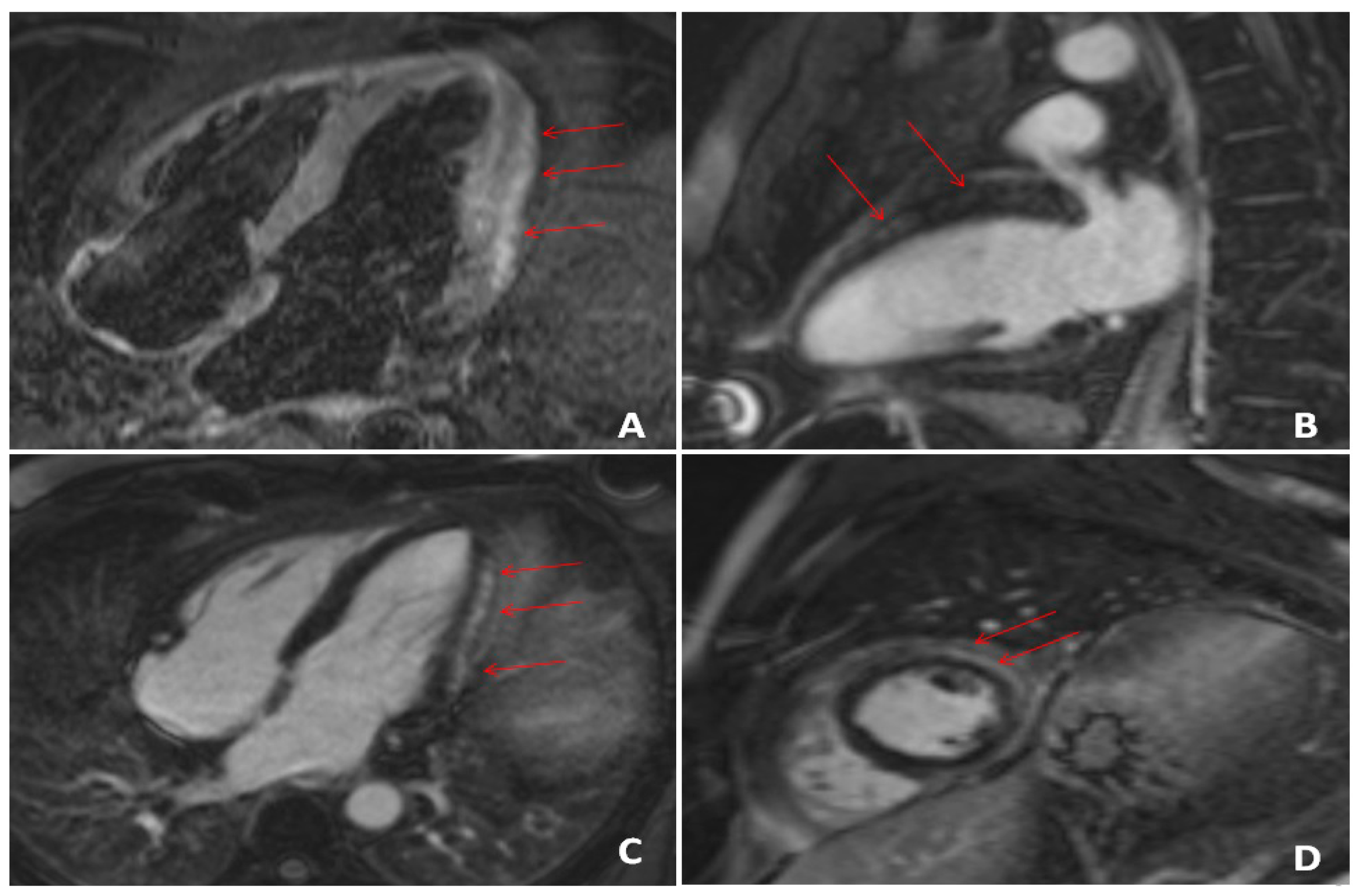
5.1. The Role of CMR for Detection of Cardiac Injury in COVID-19 Patients
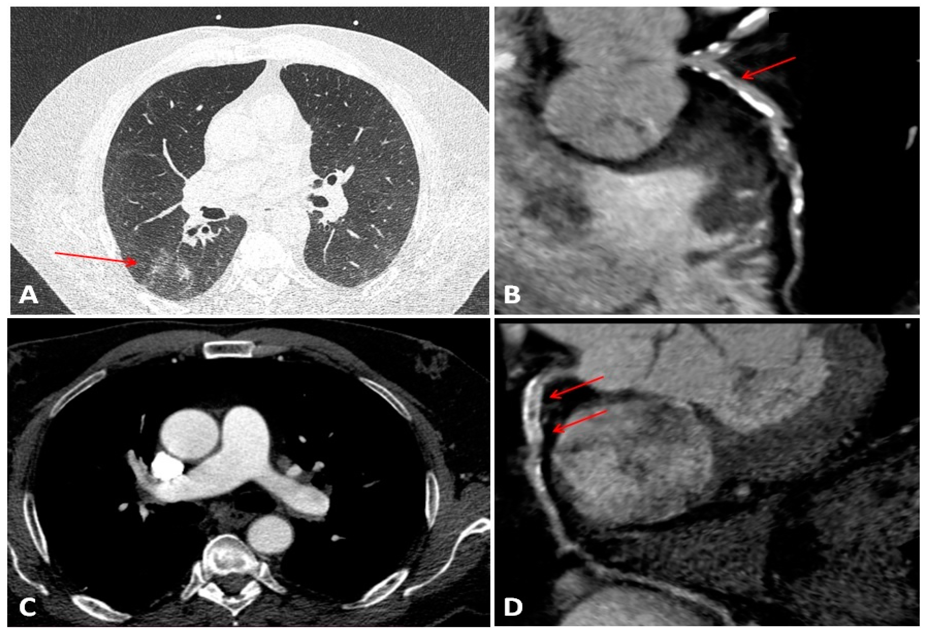
5.2. CTA and Multimodal Imaging in COVID-19 Patients
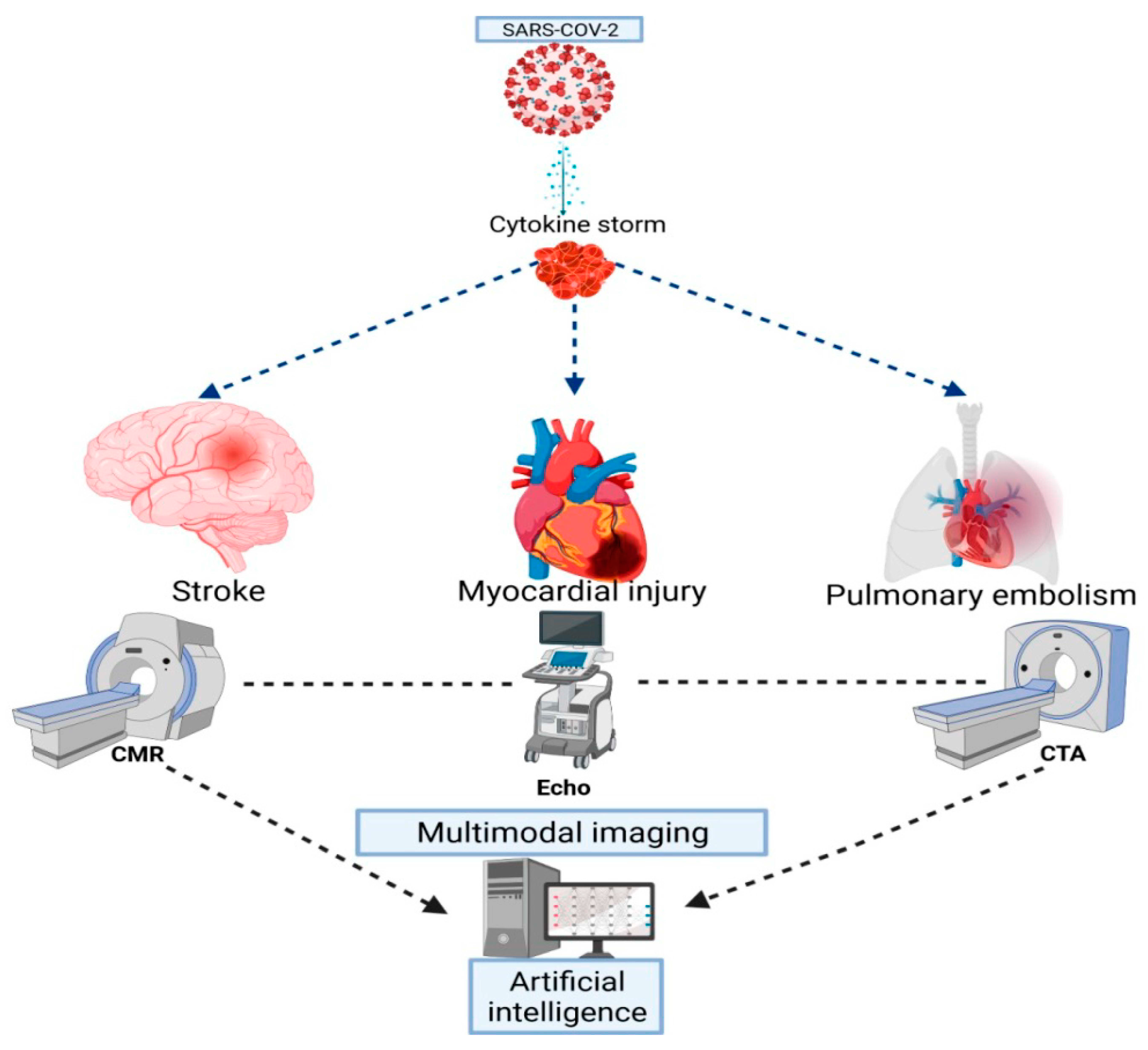
6. Future Perspectives—The Role of Artificial Intelligence in COVID-19 Patients
7. Conclusions
Author Contributions
Funding
Institutional Review Board Statement
Informed Consent Statement
Data Availability Statement
Acknowledgments
Conflicts of Interest
References
- Gupta, A.; Madhavan, M.V.; Sehgal, K.; Nair, N.; Mahajan, S.; Sehrawat, T.S.; Bikdeli, B.; Ahluwalia, N.; Ausiello, J.C.; Wan, E.Y.; et al. Extrapulmonary Manifestations of COVID-19. Nat. Med. 2020, 26, 1017–1032. [Google Scholar] [CrossRef] [PubMed]
- Bonow, R.O.; Fonarow, G.C.; O’Gara, P.T.; Yancy, C.W. Association of Coronavirus Disease 2019 (COVID-19) with Myocardial Injury and Mortality. JAMA Cardiol. 2020, 5, 751. [Google Scholar] [CrossRef]
- Inciardi, R.M.; Adamo, M.; Lupi, L.; Cani, D.S.; Di Pasquale, M.; Tomasoni, D.; Italia, L.; Zaccone, G.; Tedino, C.; Fabbricatore, D.; et al. Characteristics and Outcomes of Patients Hospitalized for COVID-19 and Cardiac Disease in Northern Italy. Eur. Heart J. 2020, 41, 1821–1829. [Google Scholar] [CrossRef]
- Ghio, S.; Baldi, E.; Vicentini, A.; Lenti, M.V.; Di Sabatino, A.; Di Matteo, A.; Zuccaro, V.; Piloni, D.; Corsico, A.; Gnecchi, M.; et al. Cardiac Involvement at Presentation in Patients Hospitalized with COVID-19 and Their Outcome in a Tertiary Referral Hospital in Northern Italy. Intern. Emerg. Med. 2020, 15, 1457–1465. [Google Scholar] [CrossRef] [PubMed]
- Guo, T.; Fan, Y.; Chen, M.; Wu, X.; Zhang, L.; He, T.; Wang, H.; Wan, J.; Wang, X.; Lu, Z. Cardiovascular Implications of Fatal Outcomes of Patients With Coronavirus Disease 2019 (COVID-19). JAMA Cardiol. 2020, 5, 811. [Google Scholar] [CrossRef] [PubMed]
- Shi, S.; Qin, M.; Shen, B.; Cai, Y.; Liu, T.; Yang, F.; Gong, W.; Liu, X.; Liang, J.; Zhao, Q.; et al. Association of Cardiac Injury With Mortality in Hospitalized Patients With COVID-19 in Wuhan, China. JAMA Cardiol. 2020, 5, 802. [Google Scholar] [CrossRef]
- Lippi, G.; Lavie, C.J.; Sanchis-Gomar, F. Cardiac Troponin I in Patients with Coronavirus Disease 2019 (COVID-19): Evidence from a Meta-Analysis. Prog. Cardiovasc. Dis. 2020, 63, 390–391. [Google Scholar] [CrossRef]
- Sheth, A.R.; Grewal, U.S.; Patel, H.P.; Thakkar, S.; Garikipati, S.; Gaddam, J.; Bawa, D. Possible Mechanisms Responsible for Acute Coronary Events in COVID-19. Med. Hypotheses 2020, 143. [Google Scholar] [CrossRef]
- Liu, P.P.; Blet, A.; Smyth, D.; Li, H. The Science Underlying COVID-19: Implications for the Cardiovascular System. Circulation 2020, 142, 68–78. [Google Scholar] [CrossRef]
- Akhmerov, A.; Marbán, E. COVID-19 and the Heart. Circ. Res. 2020, 126, 1443–1455. [Google Scholar] [CrossRef]
- Cau, R.; Bassareo, P.; Saba, L. Cardiac Involvement in COVID-19—Assessment with Echocardiography and Cardiac Magnetic Resonance Imaging. SN Compr. Clin. Med. 2020, 2, 845–851. [Google Scholar] [CrossRef] [PubMed]
- Tibaut, M.; Caprnda, M.; Kubatka, P.; Sinkovič, A.; Valentova, V.; Filipova, S.; Gazdikova, K.; Gaspar, L.; Mozos, I.; Egom, E.E.; et al. Markers of Atherosclerosis: Part 1—Serological Markers. Heart Lung Circ. 2019, 28, 667–677. [Google Scholar] [CrossRef] [PubMed]
- Knuuti, J.; Wijns, W.; Saraste, A.; Capodanno, D.; Barbato, E.; Funck-Brentano, C.; Prescott, E.; Storey, R.F.; Deaton, C.; Cuisset, T.; et al. 2019 ESC Guidelines for the Diagnosis and Management of Chronic Coronary Syndromes. Eur. Heart J. 2020, 41, 407–477. [Google Scholar] [CrossRef]
- Rusnak, J.; Fastner, C.; Behnes, M.; Mashayekhi, K.; Borggrefe, M.; Akin, I. Biomarkers in Stable Coronary Artery Disease. Curr. Pharm. Biotechnol. 2017, 18. [Google Scholar] [CrossRef]
- Held, C.; White, H.D.; Stewart, R.A.H.; Budaj, A.; Cannon, C.P.; Hochman, J.S.; Koenig, W.; Siegbahn, A.; Steg, P.G.; Soffer, J.; et al. on behalf of the STABILITY Investigators. Inflammatory Biomarkers Interleukin-6 and C-Reactive Protein and Outcomes in Stable Coronary Heart Disease: Experiences From the STABILITY (Stabilization of Atherosclerotic Plaque by Initiation of Darapladib Therapy) Trial. J. Am. Heart Assoc. 2017, 6. [Google Scholar] [CrossRef]
- Hemingway, H.; Philipson, P.; Chen, R.; Fitzpatrick, N.K.; Damant, J.; Shipley, M.; Abrams, K.R.; Moreno, S.; McAllister, K.S.L.; Palmer, S.; et al. Evaluating the Quality of Research into a Single Prognostic Biomarker: A Systematic Review and Meta-Analysis of 83 Studies of C-Reactive Protein in Stable Coronary Artery Disease. PLoS Med. 2010, 7, e1000286. [Google Scholar] [CrossRef] [PubMed]
- Hansson, G.K.; Libby, P.; Tabas, I. Inflammation and Plaque Vulnerability. J. Intern. Med. 2015, 278, 483–493. [Google Scholar] [CrossRef]
- Elnabawi, Y.A.; Dey, A.K.; Goyal, A.; Groenendyk, J.W.; Chung, J.H.; Belur, A.D.; Rodante, J.; Harrington, C.L.; Teague, H.L.; Baumer, Y.; et al. Coronary Artery Plaque Characteristics and Treatment with Biologic Therapy in Severe Psoriasis: Results from a Prospective Observational Study. Cardiovasc. Res. 2019, 115, 721–728. [Google Scholar] [CrossRef]
- Verhagen, S.N.; Visseren, F.L.J. Perivascular Adipose Tissue as a Cause of Atherosclerosis. Atherosclerosis 2011, 214, 3–10. [Google Scholar] [CrossRef]
- Opincariu, D.; Benedek, T.; Chițu, M.; Raț, N.; Benedek, I. From CT to Artificial Intelligence for Complex Assessment of Plaque-Associated Risk. Int. J. Cardiovasc. Imaging 2020, 36, 2403–2427. [Google Scholar] [CrossRef]
- Kim, H.W.; Shi, H.; Winkler, M.A.; Lee, R.; Weintraub, N.L. Perivascular Adipose Tissue and Vascular Perturbation/Atherosclerosis. Arterioscler. Thromb. Vasc. Biol. 2020, 40, 2569–2576. [Google Scholar] [CrossRef]
- Guzik, T.J.; Mohiddin, S.A.; Dimarco, A.; Patel, V.; Savvatis, K.; Marelli-Berg, F.M.; Madhur, M.S.; Tomaszewski, M.; Maffia, P.; D’Acquisto, F.; et al. COVID-19 and the Cardiovascular System: Implications for Risk Assessment, Diagnosis, and Treatment Options. Cardiovasc. Res. 2020, 116, 1666–1687. [Google Scholar] [CrossRef] [PubMed]
- Babapoor-Farrokhran, S.; Gill, D.; Walker, J.; Rasekhi, R.T.; Bozorgnia, B.; Amanullah, A. Myocardial Injury and COVID-19: Possible Mechanisms. Life Sci. 2020, 253. [Google Scholar] [CrossRef]
- Kwenandar, F.; Japar, K.V.; Damay, V.; Hariyanto, T.I.; Tanaka, M.; Lugito, N.P.H.; Kurniawan, A. Coronavirus Disease 2019 and Cardiovascular System: A Narrative Review. IJC Heart Vasc. 2020, 29. [Google Scholar] [CrossRef] [PubMed]
- Datta, P.K.; Liu, F.; Fischer, T.; Rappaport, J.; Qin, X. SARS-CoV-2 Pandemic and Research Gaps: Understanding SARS-CoV-2 Interaction with the ACE2 Receptor and Implications for Therapy. Theranostics 2020, 10, 7448–7464. [Google Scholar] [CrossRef]
- Cheng, H.; Wang, Y.; Wang, G. Organ-protective Effect of Angiotensin-converting Enzyme 2 and Its Effect on the Prognosis of COVID-19. J. Med. Virol. 2020, 92, 726–730. [Google Scholar] [CrossRef]
- Gheblawi, M.; Wang, K.; Viveiros, A.; Nguyen, Q.; Zhong, J.-C.; Turner, A.J.; Raizada, M.K.; Grant, M.B.; Oudit, G.Y. Angiotensin-Converting Enzyme 2: SARS-CoV-2 Receptor and Regulator of the Renin-Angiotensin System: Celebrating the 20th Anniversary of the Discovery of ACE2. Circ. Res. 2020, 126, 1456–1474. [Google Scholar] [CrossRef]
- Oudit, G.Y.; Kassiri, Z.; Jiang, C.; Liu, P.P.; Poutanen, S.M.; Penninger, J.M.; Butany, J. SARS-Coronavirus Modulation of Myocardial ACE2 Expression and Inflammation in Patients with SARS. Eur. J. Clin. Invest. 2009, 39, 618–625. [Google Scholar] [CrossRef]
- Boukhris, M.; Hillani, A.; Moroni, F.; Annabi, M.S.; Addad, F.; Ribeiro, M.H.; Mansour, S.; Zhao, X.; Ybarra, L.F.; Abbate, A.; et al. Cardiovascular Implications of the COVID-19 Pandemic: A Global Perspective. Can. J. Cardiol. 2020, 36, 1068–1080. [Google Scholar] [CrossRef] [PubMed]
- Wang, K.; Gheblawi, M.; Oudit, G.Y. Angiotensin Converting Enzyme 2: A Double-Edged Sword. Circulation 2020, 142, 426–428. [Google Scholar] [CrossRef] [PubMed]
- Rizzo, P.; Vieceli Dalla Sega, F.; Fortini, F.; Marracino, L.; Rapezzi, C.; Ferrari, R. COVID-19 in the Heart and the Lungs: Could We “Notch” the Inflammatory Storm? Basic Res. Cardiol. 2020, 115, 31. [Google Scholar] [CrossRef] [PubMed]
- Ragab, D.; Salah Eldin, H.; Taeimah, M.; Khattab, R.; Salem, R. The COVID-19 Cytokine Storm; What We Know So Far. Front. Immunol. 2020, 11. [Google Scholar] [CrossRef]
- Kang, Y.; Chen, T.; Mui, D.; Ferrari, V.; Jagasia, D.; Scherrer-Crosbie, M.; Chen, Y.; Han, Y. Cardiovascular Manifestations and Treatment Considerations in COVID-19. Heart 2020, 106, 1132–1141. [Google Scholar] [CrossRef] [PubMed]
- McGonagle, D.; Sharif, K.; O’Regan, A.; Bridgewood, C. The Role of Cytokines Including Interleukin-6 in COVID-19 Induced Pneumonia and Macrophage Activation Syndrome-Like Disease. Autoimmun. Rev. 2020, 19. [Google Scholar] [CrossRef]
- Liu, T.; Zhang, J.; Yang, Y.; Ma, H.; Li, Z.; Zhang, J.; Cheng, J.; Zhang, X.; Zhao, Y.; Xia, Z.; et al. The Role of Interleukin-6 in Monitoring Severe Case of Coronavirus Disease 2019. EMBO Mol. Med. 2020, 12. [Google Scholar] [CrossRef]
- El-Shabrawy, M.; Alsadik, M.E.; El-Shafei, M.; Abdelmoaty, A.A.; Alazzouni, A.S.; Esawy, M.M.; Shabana, M.A. Interleukin-6 and C-Reactive Protein/Albumin Ratio as Predictors of COVID-19 Severity and Mortality. Egypt. J. Bronchol. 2021, 15. [Google Scholar] [CrossRef]
- Chen, X.; Zhao, B.; Qu, Y.; Chen, Y.; Xiong, J.; Feng, Y.; Men, D.; Huang, Q.; Liu, Y.; Yang, B.; et al. Detectable Serum Severe Acute Respiratory Syndrome Coronavirus 2 Viral Load (RNAemia) Is Closely Correlated With Drastically Elevated Interleukin 6 Level in Critically Ill Patients With Coronavirus Disease 2019. Clin. Infect. Dis. 2020, 71, 1937–1942. [Google Scholar] [CrossRef]
- Lindner, D.; Fitzek, A.; Bräuninger, H.; Aleshcheva, G.; Edler, C.; Meissner, K.; Scherschel, K.; Kirchhof, P.; Escher, F.; Schultheiss, H.-P.; et al. Association of Cardiac Infection With SARS-CoV-2 in Confirmed COVID-19 Autopsy Cases. JAMA Cardiol. 2020, 5, 1281. [Google Scholar] [CrossRef]
- Lindner, D.; Bräuninger, H.; Stoffers, B.; Fitzek, A.; Meißner, K.; Aleshcheva, G.; Schweizer, M.; Weimann, J.; Rotter, B.; Warnke, S.; et al. Cardiac SARS-CoV-2 Infection Is Associated with Distinct Transcriptomic Changes within the Heart. Cardiovasc. Med. 2020. [Google Scholar] [CrossRef]
- Bearse, M.; Hung, Y.P.; Krauson, A.J.; Bonanno, L.; Boyraz, B.; Harris, C.K.; Helland, T.L.; Hilburn, C.F.; Hutchison, B.; Jobbagy, S.; et al. Factors Associated with Myocardial SARS-CoV-2 Infection, Myocarditis, and Cardiac Inflammation in Patients with COVID-19. Mod. Pathol. 2021. [Google Scholar] [CrossRef]
- Gauchotte, G.; Venard, V.; Segondy, M.; Cadoz, C.; Esposito-Fava, A.; Barraud, D.; Louis, G. SARS-Cov-2 Fulminant Myocarditis: An Autopsy and Histopathological Case Study. Int. J. Legal Med. 2021, 135, 577–581. [Google Scholar] [CrossRef] [PubMed]
- Albert, C.L.; Carmona-Rubio, A.E.; Weiss, A.J.; Procop, G.G.; Starling, R.C.; Rodriguez, E.R. The Enemy Within: Sudden-Onset Reversible Cardiogenic Shock With Biopsy-Proven Cardiac Myocyte Infection by Severe Acute Respiratory Syndrome Coronavirus 2. Circulation 2020, 142, 1865–1870. [Google Scholar] [CrossRef] [PubMed]
- Xiong, T.-Y.; Redwood, S.; Prendergast, B.; Chen, M. Coronaviruses and the Cardiovascular System: Acute and Long-Term Implications. Eur. Heart J. 2020, 41, 1798–1800. [Google Scholar] [CrossRef]
- Carsana, L.; Sonzogni, A.; Nasr, A.; Rossi, R.S.; Pellegrinelli, A.; Zerbi, P.; Rech, R.; Colombo, R.; Antinori, S.; Corbellino, M.; et al. Pulmonary Post-Mortem Findings in a Series of COVID-19 Cases from Northern Italy: A Two-Centre Descriptive Study. Lancet Infect. Dis. 2020, 20, 1135–1140. [Google Scholar] [CrossRef]
- Erdoğan, G.; Yenerçağ, M.; Arslan, U. The Relationship between Blood Viscosity and Acute Arterial Occlusion. J. Cardiovasc. Emergencies 2020, 6, 7–12. [Google Scholar] [CrossRef]
- Libby, P.; Lüscher, T. COVID-19 Is, in the End, an Endothelial Disease. Eur. Heart J. 2020, 41, 3038–3044. [Google Scholar] [CrossRef]
- Basso, C.; Leone, O.; Rizzo, S.; De Gaspari, M.; van der Wal, A.C.; Aubry, M.-C.; Bois, M.C.; Lin, P.T.; Maleszewski, J.J.; Stone, J.R. Pathological Features of COVID-19-Associated Myocardial Injury: A Multicentre Cardiovascular Pathology Study. Eur. Heart J. 2020, 41, 3827–3835. [Google Scholar] [CrossRef] [PubMed]
- Shah, R.M.; Shah, M.; Shah, S.; Li, A.; Jauhar, S. Takotsubo Syndrome and COVID-19: Associations and Implications. Curr. Probl. Cardiol. 2021, 46. [Google Scholar] [CrossRef]
- John, K.; Lal, A.; Mishra, A. A Review of the Presentation and Outcome of Takotsubo Cardiomyopathy in COVID-19. Monaldi Arch. Chest Dis. 2021. [Google Scholar] [CrossRef]
- Kariyanna, P.T.; Chandrakumar, H.P.; Jayarangaiah, A.; Khan, A.; Vulkanov, V.; Ashamalla, M.; Salifu, M.O.; McFarlane, S.I. Apical Takotsubo Cardiomyopathy in a COVID-19 Patient Presenting with Stroke: A Case Report and Pathophysiologic Insights. Am. J. Med. Case Rep. 2020, 8, 350–357. [Google Scholar] [CrossRef]
- Sharma, K.; Desai, H.D.; Patoliya, J.V.; Jadeja, D.M.; Gadhiya, D. Takotsubo Syndrome a Rare Entity in COVID-19: A Systemic Review—Focus on Biomarkers, Imaging, Treatment, and Outcome. SN Compr. Clin. Med. 2021, 3, 62–72. [Google Scholar] [CrossRef]
- Zhou, F.; Yu, T.; Du, R.; Fan, G.; Liu, Y.; Liu, Z.; Xiang, J.; Wang, Y.; Song, B.; Gu, X.; et al. Clinical Course and Risk Factors for Mortality of Adult Inpatients with COVID-19 in Wuhan, China: A Retrospective Cohort Study. Lancet 2020, 395, 1054–1062. [Google Scholar] [CrossRef]
- Middeldorp, S.; Coppens, M.; Haaps, T.F.; Foppen, M.; Vlaar, A.P.; Müller, M.C.A.; Bouman, C.C.S.; Beenen, L.F.M.; Kootte, R.S.; Heijmans, J.; et al. Incidence of Venous Thromboembolism in Hospitalized Patients with COVID-19. J. Thromb. Haemost. 2020, 18, 1995–2002. [Google Scholar] [CrossRef]
- Violi, F.; Pastori, D.; Cangemi, R.; Pignatelli, P.; Loffredo, L. Hypercoagulation and Antithrombotic Treatment in Coronavirus 2019: A New Challenge. Thromb. Haemost. 2020, 120, 949–956. [Google Scholar] [CrossRef]
- Harzallah, I.; Debliquis, A.; Drénou, B. Lupus Anticoagulant Is Frequent in Patients with Covid-19. J. Thromb. Haemost. 2020, 18, 2064–2065. [Google Scholar] [CrossRef]
- Bowles, L.; Platton, S.; Yartey, N.; Dave, M.; Lee, K.; Hart, D.P.; MacDonald, V.; Green, L.; Sivapalaratnam, S.; Pasi, K.J.; et al. Lupus Anticoagulant and Abnormal Coagulation Tests in Patients with Covid-19. N. Engl. J. Med. 2020, 383, 288–290. [Google Scholar] [CrossRef]
- Zhang, Y.; Xiao, M.; Zhang, S.; Xia, P.; Cao, W.; Jiang, W.; Chen, H.; Ding, X.; Zhao, H.; Zhang, H.; et al. Coagulopathy and Antiphospholipid Antibodies in Patients with Covid-19. N. Engl. J. Med. 2020, 382, e38. [Google Scholar] [CrossRef]
- Moriarty, P.M.; Gorby, L.K.; Stroes, E.S.; Kastelein, J.P.; Davidson, M.; Tsimikas, S. Lipoprotein(a) and Its Potential Association with Thrombosis and Inflammation in COVID-19: A Testable Hypothesis. Curr. Atheroscler. Rep. 2020, 22. [Google Scholar] [CrossRef] [PubMed]
- Anghel, L.; Prisacariu, C.; Sascău, R.; Macovei, L.; Cristea, E.-C.; Prisacariu, G.; Stătescu, C. Particularities of Acute Myocardial Infarction in Young Adults. J. Cardiovasc. Emerg. 2019, 5, 25–31. [Google Scholar] [CrossRef]
- Klok, F.A.; Kruip, M.J.H.A.; van der Meer, N.J.M.; Arbous, M.S.; Gommers, D.A.M.P.J.; Kant, K.M.; Kaptein, F.H.J.; van Paassen, J.; Stals, M.A.M.; Huisman, M.V.; et al. Incidence of Thrombotic Complications in Critically Ill ICU Patients with COVID-19. Thromb. Res. 2020, 191, 145–147. [Google Scholar] [CrossRef]
- Cantador, E.; Núñez, A.; Sobrino, P.; Espejo, V.; Fabia, L.; Vela, L.; de Benito, L.; Botas, J. Incidence and Consequences of Systemic Arterial Thrombotic Events in COVID-19 Patients. J. Thromb. Thrombolysis 2020, 50, 543–547. [Google Scholar] [CrossRef]
- Esenwa, C.; Cheng, N.T.; Lipsitz, E.; Hsu, K.; Zampolin, R.; Gersten, A.; Antoniello, D.; Soetanto, A.; Kirchoff, K.; Liberman, A.; et al. COVID-19-Associated Carotid Atherothrombosis and Stroke. Am. J. Neuroradiol. 2020, 41, 1993–1995. [Google Scholar] [CrossRef]
- Mohamud, A.Y.; Griffith, B.; Rehman, M.; Miller, D.; Chebl, A.; Patel, S.C.; Howell, B.; Kole, M.; Marin, H. Intraluminal Carotid Artery Thrombus in COVID-19: Another Danger of Cytokine Storm? Am. J. Neuroradiol. 2020. [Google Scholar] [CrossRef] [PubMed]
- Magro, C.; Mulvey, J.J.; Berlin, D.; Nuovo, G.; Salvatore, S.; Harp, J.; Baxter-Stoltzfus, A.; Laurence, J. Complement Associated Microvascular Injury and Thrombosis in the Pathogenesis of Severe COVID-19 Infection: A Report of Five Cases. Transl. Res. 2020, 220, 1–13. [Google Scholar] [CrossRef] [PubMed]
- Lodigiani, C.; Iapichino, G.; Carenzo, L.; Cecconi, M.; Ferrazzi, P.; Sebastian, T.; Kucher, N.; Studt, J.-D.; Sacco, C.; Bertuzzi, A.; et al. Venous and Arterial Thromboembolic Complications in COVID-19 Patients Admitted to an Academic Hospital in Milan, Italy. Thromb. Res. 2020, 191, 9–14. [Google Scholar] [CrossRef] [PubMed]
- Wichmann, D.; Sperhake, J.-P.; Lütgehetmann, M.; Steurer, S.; Edler, C.; Heinemann, A.; Heinrich, F.; Mushumba, H.; Kniep, I.; Schröder, A.S.; et al. Autopsy Findings and Venous Thromboembolism in Patients With COVID-19: A Prospective Cohort Study. Ann. Intern. Med. 2020, 173, 268–277. [Google Scholar] [CrossRef]
- Bangalore, S.; Sharma, A.; Slotwiner, A.; Yatskar, L.; Harari, R.; Shah, B.; Ibrahim, H.; Friedman, G.H. Thompson, C.; Alviar, C.L.; et al. ST-Segment Elevation in Patients with Covid-19—A Case Series. N. Engl. J. Med. 2020, 382, 2478–2480. [Google Scholar] [CrossRef]
- Stefanini, G.G.; Montorfano, M.; Trabattoni, D.; Andreini, D.; Ferrante, G.; Ancona, M.; Metra, M.; Curello, S.; Maffeo, D.; Pero, G. ST-Elevation Myocardial Infarction in Patients With COVID-19: Clinical and Angiographic Outcomes. Circulation 2020, 141, 2113–2116. [Google Scholar] [CrossRef] [PubMed]
- Guagliumi, G.; Sonzogni, A.; Pescetelli, I.; Pellegrini, D.; Finn, A.V. Microthrombi and ST-Segment–Elevation Myocardial Infarction in COVID-19. Circulation 2020, 142, 804–809. [Google Scholar] [CrossRef]
- Nakao, M.; Matsuda, J.; Iwai, M.; Endo, A.; Yonetsu, T.; Otomo, Y.; Sasano, T. Coronary Spasm and Optical Coherence Tomography Defined Plaque Erosion Causing ST-Segment-Elevation Acute Myocardial Infarction in a Patient with COVID-19 Pneumonia. J. Cardiol. Cases 2021, 23, 87–89. [Google Scholar] [CrossRef]
- Harari, R.; Bangalore, S.; Chang, E.; Shah, B. COVID-19 Complicated by Acute Myocardial Infarction with Extensive Thrombus Burden and Cardiogenic Shock. Catheter. Cardiovasc. Interv. 2021, 97. [Google Scholar] [CrossRef]
- Kirresh, A.; Coghlan, G.; Candilio, L. COVID-19 Infection and High Intracoronary Thrombus Burden. Cardiovasc. Revasc. Med. 2020. [Google Scholar] [CrossRef]
- Jenab, Y.; Rezaei, N.; Hedayat, B.; Naderian, M.; Shirani, S.; Hosseini, K. Occurrence of Acute Coronary Syndrome, Pulmonary Thromboembolism, and Cerebrovascular Event in COVID-19. Clin. Case Rep. 2020, 8, 2414–2417. [Google Scholar] [CrossRef]
- Jabri, A.; Kalra, A.; Kumar, A.; Alameh, A.; Adroja, S.; Bashir, H.; Nowacki, A.S.; Shah, R.; Khubber, S.; Kanaa’N, A. Incidence of Stress Cardiomyopathy During the Coronavirus Disease 2019 Pandemic. JAMA Netw. Open 2020, 3, e2014780. [Google Scholar] [CrossRef] [PubMed]
- Singh, S.; Desai, R.; Gandhi, Z.; Fong, H.K.; Doreswamy, S.; Desai, V.; Chockalingam, A.; Mehta, P.K.; Sachdeva, R.; Kumar, G.; et al. Takotsubo Syndrome in Patients with COVID-19: A Systematic Review of Published Cases. SN Compr. Clin. Med. 2020, 2, 2102–2108. [Google Scholar] [CrossRef]
- De Rosa, S.; Spaccarotella, C.; Basso, C.; Calabrò, M.P.; Curcio, A.; Filardi, P.P.; Mancone, M.; Mercuro, G.; Muscoli, S.; Nodari, S.; et al. Reduction of hospitalizations for myocardial infarction in Italy in the COVID-19 era. Eur. Heart J. 2020, 41, 2083–2088. [Google Scholar] [CrossRef]
- Baldi, E.; Sechi, G.M.; Mare, C.; Canevari, F.; Brancaglione, A.; Primi, R.; Klersy, C.; Palo, A.; Contri, E.; Ronchi, V.; et al. Out-of-Hospital Cardiac Arrest during the Covid-19 Outbreak in Italy. N. Engl. J. Med. 2020, 383, 496–498. [Google Scholar] [CrossRef]
- Metzler, B.; Siostrzonek, P.; Binder, R.K.; Bauer, A.; Reinstadler, S.J. Decline of Acute Coronary Syndrome Admissions in Austria since the Outbreak of COVID-19: The Pandemic Response Causes Cardiac Collateral Damage. Eur. Heart J. 2020, 41, 1852–1853. [Google Scholar] [CrossRef]
- Braiteh, N.; Rehman, W.; Alom, M.; Skovira, V.; Breiteh, N.; Rehman, I.; Yarkoni, A.; Kahsou, H.; Rehman, A. Decrease in Acute Coronary Syndrome Presentations during the COVID-19 Pandemic in Upstate New York. Am. Heart J. 2020, 226, 147–151. [Google Scholar] [CrossRef] [PubMed]
- Garcia, S.; Albaghdadi, M.S.; Meraj, P.M.; Schmidt, C.; Garberich, R.; Jaffer, F.A.; Dixon, S.; Rade, J.J.; Tannenbaum, M.; Chambers, J.; et al. Reduction in ST-Segment Elevation Cardiac Catheterization Laboratory Activations in the United States During COVID-19 Pandemic. J. Am. Coll. Cardiol. 2020, 75, 2871–2872. [Google Scholar] [CrossRef] [PubMed]
- Cosyns, B.; Lochy, S.; Luchian, M.L.; Gimelli, A.; Pontone, G.; Allard, S.D.; de Mey, J.; Rosseel, P.; Dweck, M.; Petersen, S.E.; et al. The Role of Cardiovascular Imaging for Myocardial Injury in Hospitalized COVID-19 Patients. Eur. Heart J. Cardiovasc. Imaging 2020, 21, 709–714. [Google Scholar] [CrossRef]
- Han, Y.; Chen, T.; Bryant, J.; Bucciarelli-Ducci, C.; Dyke, C.; Elliott, M.D.; Ferrari, V.A.; Friedrich, M.G.; Lawton, C.; Manning, W.J.; et al. Society for Cardiovascular Magnetic Resonance (SCMR) Guidance for the Practice of Cardiovascular Magnetic Resonance during the COVID-19 Pandemic. J. Cardiovasc. Magn. Reson. 2020, 22, 26. [Google Scholar] [CrossRef] [PubMed]
- Skulstad, H.; Cosyns, B.; Popescu, B.A.; Galderisi, M.; Salvo, G.D.; Donal, E.; Petersen, S.; Gimelli, A.; Haugaa, K.H.; Muraru, D.; et al. COVID-19 Pandemic and Cardiac Imaging: EACVI Recommendations on Precautions, Indications, Prioritization, and Protection for Patients and Healthcare Personnel. Eur. Heart J. Cardiovasc. Imaging 2020, 21, 592–598. [Google Scholar] [CrossRef] [PubMed]
- Kelle, S.; Bucciarelli-Ducci, C.; Judd, R.M.; Kwong, R.Y.; Simonetti, O.; Plein, S.; Raimondi, F.; Weinsaft, J.W.; Wong, T.C.; Carr, J. Society for Cardiovascular Magnetic Resonance (SCMR) Recommended CMR Protocols for Scanning Patients with Active or Convalescent Phase COVID-19 Infection. J. Cardiovasc. Magn. Reson. 2020, 22. [Google Scholar] [CrossRef]
- Haaf, P.; Garg, P.; Messroghli, D.R.; Broadbent, D.A.; Greenwood, J.P.; Plein, S. Cardiac T1 Mapping and Extracellular Volume (ECV) in Clinical Practice: A Comprehensive Review. J. Cardiovasc. Magn. Reson. 2017, 18, 89. [Google Scholar] [CrossRef] [PubMed]
- Esposito, A.; Palmisano, A.; Natale, L.; Ligabue, G.; Peretto, G.; Lovato, L.; Vignale, D.; Fiocchi, F.; Marano, R.; Russo, V. Cardiac Magnetic Resonance Characterization of Myocarditis-Like Acute Cardiac Syndrome in COVID-19. JACC Cardiovasc. Imaging 2020, 13, 2462–2465. [Google Scholar] [CrossRef] [PubMed]
- Gravinay, P.; Issa, N.; Girard, D.; Camou, F.; Cochet, H. CMR and Serology to Diagnose COVID-19 Infection with Primary Cardiac Involvement. Eur. Heart J. Cardiovasc. Imaging 2021, 22, 133. [Google Scholar] [CrossRef]
- Luetkens, J.A.; Isaak, A.; Zimmer, S.; Nattermann, J.; Sprinkart, A.M.; Boesecke, C.; Rieke, G.J.; Zachoval, C.; Heine, A.; Velten, M.; et al. Diffuse Myocardial Inflammation in COVID-19 Associated Myocarditis Detected by Multiparametric Cardiac Magnetic Resonance Imaging. Circ. Cardiovasc. Imaging 2020, 13. [Google Scholar] [CrossRef]
- Manka, R.; Karolyi, M.; Polacin, M.; Holy, E.W.; Nemeth, J.; Steiger, P.; Schuepbach, R.A.; Zinkernagel, A.S.; Alkadhi, H.; Mehra, M.R.; et al. Myocardial Edema in COVID-19 on Cardiac MRI. J. Heart Lung Transplant. 2020, 39, 730–732. [Google Scholar] [CrossRef]
- Caballeros Lam, M.; de la Fuente Villena, A.; Hernández Hernández, A.; García de Yébenes, M.; Bastarrika Alemañ, G. Cardiac Magnetic Resonance Characterization of COVID-19 Myocarditis. Rev. Esp. Cardiol. Engl. Ed. 2020, 73, 863–864. [Google Scholar] [CrossRef] [PubMed]
- Rajpal, S.; Tong, M.S.; Borchers, J.; Zareba, K.M.; Obarski, T.P.; Simonetti, O.P.; Daniels, C.J. Cardiovascular Magnetic Resonance Findings in Competitive Athletes Recovering From COVID-19 Infection. JAMA Cardiol. 2020. [Google Scholar] [CrossRef]
- Knight, D.S.; Kotecha, T.; Razvi, Y.; Chacko, L.; Brown, J.T.; Jeetley, P.S.; Goldring, J.; Jacobs, M.; Lamb, L.E.; Negus, R.; et al. COVID-19: Myocardial Injury in Survivors. Circulation 2020, 142, 1120–1122. [Google Scholar] [CrossRef] [PubMed]
- Huang, L.; Zhao, P.; Tang, D.; Zhu, T.; Han, R.; Zhan, C.; Liu, W.; Zeng, H.; Tao, Q.; Xia, L. Cardiac Involvement in Patients Recovered From COVID-2019 Identified Using Magnetic Resonance Imaging. JACC Cardiovasc. Imaging 2020, 13, 2330–2339. [Google Scholar] [CrossRef]
- Ng, M.-Y.; Ferreira, V.M.; Leung, S.T.; Yin Lee, J.C.; Ho-Tung Fong, A.; To Liu, R.W.; Man Chan, J.W.; Wu, A.K.L.; Lung, K.-C.; Crean, A.M.; et al. Patients Recovered From COVID-19 Show Ongoing Subclinical Myocarditis as Revealed by Cardiac Magnetic Resonance Imaging. JACC Cardiovasc. Imaging 2020, 13, 2476–2478. [Google Scholar] [CrossRef] [PubMed]
- Kotecha, T.; Knight, D.S.; Razvi, Y.; Kumar, K.; Vimalesvaran, K.; Thornton, G.; Patel, R.; Chacko, L.; Brown, J.T.; Coyle, C.; et al. Patterns of Myocardial Injury in Recovered Troponin-Positive COVID-19 Patients Assessed by Cardiovascular Magnetic Resonance. Eur. Heart J. 2021, 42, 1866–1878. [Google Scholar] [CrossRef] [PubMed]
- Puntmann, V.O.; Carerj, M.L.; Wieters, I.; Fahim, M.; Arendt, C.; Hoffmann, J.; Shchendrygina, A.; Escher, F.; Vasa-Nicotera, M.; Zeiher, A.M.; et al. Outcomes of Cardiovascular Magnetic Resonance Imaging in Patients Recently Recovered From Coronavirus Disease 2019 (COVID-19). JAMA Cardiol. 2020, 5, 1265. [Google Scholar] [CrossRef]
- Wang, H.; Li, R.; Zhou, Z.; Jiang, H.; Yan, Z.; Tao, X.; Li, H.; Xu, L. Cardiac Involvement in COVID-19 Patients: Mid-Term Follow up by Cardiovascular Magnetic Resonance. J. Cardiovasc. Magn. Reson. 2021, 23, 14. [Google Scholar] [CrossRef] [PubMed]
- Ojha, V.; Verma, M.; Pandey, N.N.; Mani, A.; Malhi, A.S.; Kumar, S.; Jagia, P.; Roy, A.; Sharma, S. Cardiac Magnetic Resonance Imaging in Coronavirus Disease 2019 (COVID-19): A Systematic Review of Cardiac Magnetic Resonance Imaging Findings in 199 Patients. J. Thorac. Imaging 2021, 36, 73–83. [Google Scholar] [CrossRef]
- Antonopoulos, A.S.; Sanna, F.; Sabharwal, N.; Thomas, S.; Oikonomou, E.K.; Herdman, L.; Margaritis, M.; Shirodaria, C.; Kampoli, A.-M.; Akoumianakis, I.; et al. Detecting Human Coronary Inflammation by Imaging Perivascular Fat. Sci. Transl. Med. 2017, 9, eaal2658. [Google Scholar] [CrossRef]
- Scoccia, A.; Gallone, G.; Cereda, A.; Palmisano, A.; Vignale, D.; Leone, R.; Nicoletti, V.; Gnasso, C.; Monello, A.; Khokhar, A.; et al. Impact of Clinical and Subclinical Coronary Artery Disease as Assessed by Coronary Artery Calcium in COVID-19. Atherosclerosis 2021. [Google Scholar] [CrossRef] [PubMed]
- Giannini, F.; Toselli, M.; Palmisano, A.; Cereda, A.; Vignale, D.; Leone, R.; Nicoletti, V.; Gnasso, C.; Monello, A.; Manfrini, M.; et al. Insights in COVID-19 Patients. J. Cardiovasc. Comput. Tomogr. 2021. [Google Scholar] [CrossRef]
- Esposito, A.; Palmisano, A.; Toselli, M.; Vignale, D.; Cereda, A.; Rancoita, P.M.V.; Leone, R.; Nicoletti, V.; Gnasso, C.; Monello, A.; et al. CT–Derived Pulmonary Artery Enlargement at the Admission Predicts Overall Survival in COVID-19 Patients: Insight from 1461 Consecutive Patients in Italy. Eur. Radiol. 2021, 31, 4031–4041. [Google Scholar] [CrossRef]
- Zoghbi, W.A.; DiCarli, M.F.; Blankstein, R.; Choi, A.D.; Dilsizian, V.; Flachskampf, F.A.; Geske, J.B.; Grayburn, P.A.; Jaffer, F.A.; Kwong, R.Y. Multimodality Cardiovascular Imaging in the Midst of the COVID-19 Pandemic. JACC Cardiovasc. Imaging 2020, 13, 1615–1626. [Google Scholar] [CrossRef]
- Agricola, E.; Beneduce, A.; Esposito, A.; Ingallina, G.; Palumbo, D.; Palmisano, A.; Ancona, F.; Baldetti, L.; Pagnesi, M.; Melisurgo, G.; et al. Heart and Lung Multimodality Imaging in COVID-19. JACC Cardiovasc. Imaging 2020, 13, 1792–1808. [Google Scholar] [CrossRef]
- Citro, R.; Pontone, G.; Bellino, M.; Silverio, A.; Iuliano, G.; Baggiano, A.; Manka, R.; Iesu, S.; Vecchione, C.; Asch, F.M.; et al. Role of Multimodality Imaging in Evaluation of Cardiovascular Involvement in COVID-19. Trends Cardiovasc. Med. 2021, 31, 8–16. [Google Scholar] [CrossRef] [PubMed]
- Pontone, G.; Baggiano, A.; Conte, E.; Teruzzi, G.; Cosentino, N.; Campodonico, J.; Rabbat, M.G.; Assanelli, E.; Palmisano, A.; Esposito, A.; et al. “Quadruple Rule-Out” With Computed Tomography in a COVID-19 Patient With Equivocal Acute Coronary Syndrome Presentation. JACC Cardiovasc. Imaging 2020, 13, 1854–1856. [Google Scholar] [CrossRef] [PubMed]
- Rudski, L.; Januzzi, J.L.; Rigolin, V.H.; Bohula, E.A.; Blankstein, R.; Patel, A.R.; Bucciarelli-Ducci, C.; Vorovich, E.; Mukherjee, M.; Rao, S.V.; et al. Multimodality Imaging in Evaluation of Cardiovascular Complications in Patients With COVID-19. J. Am. Coll. Cardiol. 2020, 76, 1345–1357. [Google Scholar] [CrossRef] [PubMed]
- Jamthikar, A.; Gupta, D.; Khanna, N.N.; Saba, L.; Araki, T.; Viskovic, K.; Suri, H.S.; Gupta, A.; Mavrogeni, S.; Turk, M.; et al. A Low-Cost Machine Learning-Based Cardiovascular/Stroke Risk Assessment System: Integration of Conventional Factors with Image Phenotypes. Cardiovasc. Diagn. Ther. 2019, 9, 420–430. [Google Scholar] [CrossRef] [PubMed]
- Krittanawong, C.; Virk, H.U.H.; Bangalore, S.; Wang, Z.; Johnson, K.W.; Pinotti, R.; Zhang, H.; Kaplin, S.; Narasimhan, B.; Kitai, T.; et al. Machine Learning Prediction in Cardiovascular Diseases: A Meta-Analysis. Sci. Rep. 2020, 10, 16057. [Google Scholar] [CrossRef]
- Kagiyama, N.; Shrestha, S.; Farjo, P.D.; Sengupta, P.P. Artificial Intelligence: Practical Primer for Clinical Research in Cardiovascular Disease. J. Am. Heart Assoc. 2019, 8. [Google Scholar] [CrossRef] [PubMed]
- Chen, J.; See, K.C. Artificial Intelligence for COVID-19: Rapid Review. J. Med. Internet Res. 2020, 22, e21476. [Google Scholar] [CrossRef] [PubMed]
- Vaishya, R.; Javaid, M.; Khan, I.H.; Haleem, A. Artificial Intelligence (AI) Applications for COVID-19 Pandemic. Diabetes Metab. Syndr. Clin. Res. Rev. 2020, 14, 337–339. [Google Scholar] [CrossRef] [PubMed]
- Li, Z.; Zhong, Z.; Li, Y.; Zhang, T.; Gao, L.; Jin, D.; Sun, Y.; Ye, X.; Yu, L.; Hu, Z.; et al. From Community-Acquired Pneumonia to COVID-19: A Deep Learning–Based Method for Quantitative Analysis of COVID-19 on Thick-Section CT Scans. Eur. Radiol. 2020, 30, 6828–6837. [Google Scholar] [CrossRef] [PubMed]
- Ozturk, T.; Talo, M.; Yildirim, E.A.; Baloglu, U.B.; Yildirim, O.; Rajendra Acharya, U. Automated Detection of COVID-19 Cases Using Deep Neural Networks with X-Ray Images. Comput. Biol. Med. 2020, 121. [Google Scholar] [CrossRef]
- Kang, M.; Hong, K.S.; Chikontwe, P.; Luna, M.; Jang, J.G.; Park, J.; Shin, K.-C.; Park, S.H.; Ahn, J.H. Quantitative Assessment of Chest CT Patterns in COVID-19 and Bacterial Pneumonia Patients: A Deep Learning Perspective. J. Korean Med. Sci. 2021, 36, e46. [Google Scholar] [CrossRef] [PubMed]
- Suri, J.S.; Puvvula, A.; Biswas, M.; Majhail, M.; Saba, L.; Faa, G.; Singh, I.M.; Oberleitner, R.; Turk, M.; Chadha, P.S.; et al. COVID-19 Pathways for Brain and Heart Injury in Comorbidity Patients: A Role of Medical Imaging and Artificial Intelligence-Based COVID Severity Classification: A Review. Comput. Biol. Med. 2020, 124. [Google Scholar] [CrossRef]


| Reference | COVID-19 Phase | Study Type | Nr. of Patients | Age | Cardiological History | Clinical Presentation | Pulmonary Findings |
|---|---|---|---|---|---|---|---|
| Esposito et al. [86] | Acute (1 week) | Case reports | 10 | 52 ± 6 | No | Chest pain, dyspnea | NA |
| Gravinay et al. [87] | Acute (8 days) | Case report | 1 | 51 | NA | Atypical chest pain, dyspnea, fever, arthromyalgia | None |
| Luetkens et al. [88] | Acute (10 days) | Case report | 1 | 79 | No | Dyspnea, syncope, fatigue | Ground glass infiltrates |
| Manka et al. [89] | Acute (Day 5) | Case report | 1 | 75 | HTN, obesity, CKD | Fever, chills, cough, dyspnea | Pneumonia |
| Caballeros et al. [90] | Acute | Case reports | 2 | 26, 13 | Gestational diabetes | Chest pain, fever, cough | None |
| Rajpal et al. [91] | Early convalescence (11–53 days) | Research letter | 26 | 19.5 | NA | 26.9% mild symptoms 73.1% asymptomatic | NA |
| Knight et al. [92] | Early convalescence (46 ± 15 days) | Retrospective observational | 29 | 64 ± 9 | NA | NA | 69% with residual lung parenchymal changes |
| Huang et al. [93] | Early recovery 47 (36–58) | Retrospective observational | 26 | 38 (32–45) | 8% HTN | Chest pain, palpitations, chest distress | NA |
| Ng et al. [94] | Convalescence | Retrospective observational | 16 | 68 (53–69) | 6.25% CAD | 94% mild/moderate | NA |
| Kotecha et al. [95] | Convalescence (median 68 days) | Prospective observational | 148 | 64 ± 12 | 57% HTN 46% Hypercholesterolemia 34% DM | Sever COVID-19 at presentation, 32% with ventilatory support | NA |
| Puntmann et al. [96] | Convalescence 71 (64–92 days) | Prospective observational | 100 | 49 (45–53) | 22% HTN 18% DM 22% Hypercholesterolemia 13% CAD | Chest pain, palpitation, shortness of breath | NA |
| Wang et al. [97] | Mid-term recovery (102.5 ± 20.6 days) | Prospective observational | 44 | 47.6 ± 13 | 25% HTN 18.2% DM 36.4% Hyperlipidemia 4.6% Hepatitis B | NA | NA |
| Reference | Diagnosis | Edema | LGE | ECV | LVEF | WMA | Perfusion Deficit | Other |
|---|---|---|---|---|---|---|---|---|
| Gravinay et al. [87] | Myocarditis | Subepicardial | + | NA | Preserved | None | NA | LV thrombus |
| Esposito et al. [86] | Myocarditis Takotsubo | Diffuse (STIR, T1, T2 mapping) | + 1–3% of LV (n = 3) | n = 2 (30, 36%) | <40% (n = 2) 40–55% (n = 3) >55% (n = 5) | NA | NA | Pericardial effusion |
| Luetkens et al. [88] | Myocarditis | Diffuse (T1, T2 mapping) | - | NA | 49% | Global hypokinesis | NA | Pericardial effusion |
| Manka et al. [89] | Acute myocardial injury | Global (STIR, T1, T2 mapping) | - | NA | 59% | No | NA | - |
| Caballeros et al. [90] | Myocarditis | Basal/inferior septum T1, T2 mapping) | + (14.2% of LV mass) | NA | 59% | NA | NA | - |
| Myocarditis | Ventricular septum (T2, native T1) | - | NA | Normal | NA | NA | Pericardial effusion | |
| Rajpal et al. [91] | 15% Myocarditis | 15% (elevated T2) | + 46% (n = 12) | Elevated in 3.8% (n = 1) | 58.6% | NA | NA | 7.5% % with pericardial effusion |
| Knight et al. [92] | 44% Myocarditis 31% Ischemic | No | 38% non-ischemic 17% ischemic 14% dual | NA | 67.7% ± 11.4 | NA | Done in 66% (n = 19) 47% ischemia 42% inducible ischemia | 7% with pericardial effusion 14% pleural effusions |
| Huang et al. [93] | NA | + 54% (n = 14) | + 31% (n = 8) | 28.2% | 60.7% ± 6.4 | NA | NA | Pericardial effusion |
| Ng et al. [94] | 19% Myocarditis | 25% elevated native T1 25% T1 and T2 5% native T2 | + 25% | NA | 59% (56–65) | NA | NA | NA |
| Kotecha et al. [95] | 27% Myocarditis 19% MI | 30% of the myocarditis pattern patients | + 49% (n = 70) | NA | 67% ± 11 | No regional WMA in myocarditis pattern | Done in 51% (n = 76) 28% IHD | 5% with pericardial effusion 6% pleural effusions |
| Puntmann et al. [96] | 78% Myocarditis | 60% with abnormal native T2 | + 20% non-ischemic pattern 12% ischemic pattern | NA | 56% (54–58) | NA | NA | 22% pericardial LGE 20% pericardial effusion |
| Wang et al. [97] | Myocardial injury | NA | 29.5% Mid-myocardial Sub-epicardial | NA | 64.3% ± 5.9 in LGE+ 62.2% ± 4.4 in LGE− | RV peak GCS −9.4 ± 3.4(LGE+) vs.−12.1 ± 4.0 (LGE−) p = 0.04 | NA | NA |
Publisher’s Note: MDPI stays neutral with regard to jurisdictional claims in published maps and institutional affiliations. |
© 2021 by the authors. Licensee MDPI, Basel, Switzerland. This article is an open access article distributed under the terms and conditions of the Creative Commons Attribution (CC BY) license (https://creativecommons.org/licenses/by/4.0/).
Share and Cite
Mester, A.; Benedek, I.; Rat, N.; Tolescu, C.; Polexa, S.A.; Benedek, T. Imaging Cardiovascular Inflammation in the COVID-19 Era. Diagnostics 2021, 11, 1114. https://doi.org/10.3390/diagnostics11061114
Mester A, Benedek I, Rat N, Tolescu C, Polexa SA, Benedek T. Imaging Cardiovascular Inflammation in the COVID-19 Era. Diagnostics. 2021; 11(6):1114. https://doi.org/10.3390/diagnostics11061114
Chicago/Turabian StyleMester, Andras, Imre Benedek, Nora Rat, Cosmin Tolescu, Stefania Alexandra Polexa, and Theodora Benedek. 2021. "Imaging Cardiovascular Inflammation in the COVID-19 Era" Diagnostics 11, no. 6: 1114. https://doi.org/10.3390/diagnostics11061114
APA StyleMester, A., Benedek, I., Rat, N., Tolescu, C., Polexa, S. A., & Benedek, T. (2021). Imaging Cardiovascular Inflammation in the COVID-19 Era. Diagnostics, 11(6), 1114. https://doi.org/10.3390/diagnostics11061114


