
Comparative Assessment of Sera from Individuals after S-Gene RNA-Based SARS-CoV-2 Vaccination with Spike-Protein-Based and Nucleocapsid-Based Serological Assays

 , , ,
, , , 
Abstract
:1. Introduction
2. Materials and Methods
2.1. Sample Collection
2.2. Serological Assays
2.3. Statistics
2.4. Ethics
3. Results
3.1. Vaccinees
3.2. Serological Test Results
3.3. Correlation of the Time in Days between the Last Vaccine and Sample Acquisition, Age, Sex and the Measured Titers
4. Discussion
5. Conclusions
Author Contributions
Funding
Institutional Review Board Statement
Informed Consent Statement
Data Availability Statement
Acknowledgments
Conflicts of Interest
Appendix A

| Study ID | Sex (1 = Male, 2 = Female) | Age at Sample Acquisition (Years) | Date of First Vaccine | Date of Second Vaccine | Date of sAmple Acquisition |
|---|---|---|---|---|---|
| 1 | 1 | 19 | 2021-01-21 | 2021-02-01 | |
| 2 | 2 | 21 | 2021-01-21 | 2021-02-01 | |
| 3 | 2 | 25 | 2021-01-21 | 2021-02-01 | |
| 4 | 2 | 36 | 2021-01-21 | 2021-02-01 | |
| 5 | 2 | 51 | 2021-01-21 | 2021-02-01 | |
| 6 | 1 | 50 | 2021-01-21 | 2021-02-01 | |
| 7 | 2 | 63 | 2021-01-21 | 2021-02-01 | |
| 8 | 2 | 49 | 2021-01-12 | 2021-02-02 | |
| 9 | 1 | 62 | 2021-01-15 | 2021-02-02 | |
| 10 | 2 | 48 | 2021-01-20 | 2021-02-02 | |
| 11 | 1 | 24 | 2021-01-14 | 2021-02-02 | |
| 12 | 2 | 30 | 2021-01-12 | 2021-02-02 | |
| 13 | 2 | 48 | 2021-01-14 | 2021-02-02 | |
| 14 | 1 | 44 | 2021-01-13 | 2021-02-02 | |
| 15 | 1 | 54 | 2021-01-11 | 2021-02-01 | 2021-02-02 |
| 16 | 1 | 31 | 2021-01-11 | 2021-02-01 | 2021-02-02 |
| 17 | 2 | 28 | 2021-01-11 | 2021-02-01 | 2021-02-02 |
| 18 | 1 | 31 | 2021-01-11 | 2021-02-02 | |
| 19 | 1 | 48 | 2021-01-11 | 2021-02-01 | 2021-02-02 |
| 20 | 2 | 61 | 2021-01-11 | 2021-02-01 | 2021-02-02 |
| 21 | 1 | 32 | 2021-01-12 | 2021-02-02 | |
| 22 | 2 | 45 | 2021-01-11 | 2021-02-02 | |
| 23 | 1 | 34 | 2021-01-12 | 2021-02-02 | |
| 24 | 1 | 33 | 2021-01-11 | 2021-02-02 | |
| 25 | 2 | 32 | 2021-01-11 | 2021-02-01 | 2021-02-02 |
| 26 | 1 | 40 | 2021-01-11 | 2021-02-01 | 2021-02-03 |
| 27 | 1 | 26 | 2021-01-12 | 2021-02-02 | 2021-02-03 |
| 28 | 1 | 41 | 2021-01-07 | 2021-02-01 | 2021-02-03 |
| 29 | 2 | 27 | 2021-01-13 | 2021-02-03 | |
| 30 | 1 | 43 | 2021-01-12 | 2021-02-02 | 2021-02-03 |
| 31 | 2 | 48 | 2021-01-08 | 2021-02-01 | 2021-02-04 |
| 32 | 2 | 46 | 2021-01-13 | 2021-01-03 | 2021-02-04 |
| 33 | 1 | 34 | 2021-01-11 | 2021-02-01 | 2021-02-04 |
| 34 | 2 | 50 | 2021-01-12 | 2021-02-02 | 2021-02-04 |
| 35 | 2 | 49 | 2021-01-13 | 2021-02-04 | |
| 36 | 2 | 28 | 2021-01-11 | 2021-02-01 | 2021-02-04 |
| 37 | 1 | 60 | 2021-01-11 | 2021-02-01 | 2021-02-04 |
| 38 | 2 | 28 | 2021-01-18 | 2021-02-04 | |
| 39 | 2 | 34 | 2021-01-11 | 2021-02-01 | 2021-02-04 |
| 40 | 2 | 54 | 2021-01-14 | 2021-02-04 | |
| 41 | 1 | 60 | 2021-01-20 | 2021-02-04 | |
| 42 | 1 | 39 | 2021-02-01 | 2021-02-04 | |
| 43 | 2 | 25 | 2021-01-11 | 2021-02-01 | 2021-02-04 |
| 44 | 1 | 23 | 2021-01-26 | 2021-02-04 | |
| 45 | 2 | 62 | 2021-01-15 | 2021-02-05 | |
| 46 | 2 | 25 | 2021-01-12 | 2021-02-02 | 2021-02-05 |
| 47 | 1 | 48 | 2021-01-14 | 2021-02-04 | 2021-02-05 |
| 48 | 1 | 42 | 2021-01-12 | 2021-02-02 | 2021-02-05 |
| 49 | 2 | 30 | 2021-01-14 | 2021-02-04 | 2021-02-05 |
| 50 | 2 | 41 | 2021-01-12 | 2021-02-02 | 2021-02-05 |
| 51 | 1 | 23 | 2021-01-12 | 2021-02-02 | 2021-02-05 |
| 52 | 2 | 54 | 2021-01-11 | 2021-02-04 | 2021-02-05 |
| 53 | 2 | 34 | 2021-01-12 | 2021-02-02 | 2021-02-05 |
| 54 | 2 | 39 | 2021-01-08 | 2021-02-01 | 2021-02-06 |
| 55 | 2 | 53 | 2021-01-11 | 2021-02-01 | 2021-02-08 |
| 56 | 2 | 50 | 2021-01-08 | 2021-02-01 | 2021-02-08 |
| 57 | 2 | 64 | 2021-01-08 | 2021-02-01 | 2021-02-08 |
| 58 | 2 | 38 | 2021-01-11 | 2021-02-01 | 2021-02-08 |
| 59 | 1 | 43 | 2021-01-12 | 2021-02-02 | 2021-02-08 |
| 60 | 2 | 36 | 2021-01-07 | 2021-02-01 | 2021-02-08 |
| 61 | 2 | 63 | 2021-01-11 | 2021-02-01 | 2021-02-08 |
| 62 | 1 | 42 | 2021-01-11 | 2021-02-01 | 2021-02-08 |
| 63 | 1 | 39 | 2021-01-08 | 2021-02-01 | 2021-02-08 |
| 64 | 2 | 51 | 2021-01-15 | 2021-02-05 | 2021-02-08 |
| 65 | 1 | 33 | 2021-01-12 | 2021-02-02 | 2021-02-08 |
| 66 | 2 | 31 | 2021-01-13 | 2021-02-03 | 2021-02-09 |
| 67 | 1 | 33 | 2021-01-11 | 2021-02-01 | 2021-02-09 |
| 68 | 1 | 32 | 2021-01-18 | 2021-02-08 | 2021-02-09 |
| 69 | 1 | 30 | 2021-01-11 | 2021-02-01 | 2021-02-09 |
| 70 | 2 | 58 | 2021-01-12 | 2021-02-02 | 2021-02-09 |
| 71 | 1 | 28 | 2021-01-18 | 2021-02-08 | 2021-02-09 |
| 72 | 2 | 28 | 2021-01-13 | 2021-02-03 | 2021-02-09 |
| 73 | 2 | 33 | 2021-01-12 | 2021-02-04 | 2021-02-09 |
| 74 | 1 | 31 | 2021-01-12 | 2021-02-04 | 2021-02-09 |
| 75 | 2 | 27 | 2021-01-13 | 2021-02-03 | 2021-02-09 |
| 76 | 1 | 25 | 2021-01-11 | 2021-02-01 | 2021-02-09 |
| 77 | 1 | 23 | 2021-01-12 | 2021-02-02 | 2021-02-09 |
| 78 | 2 | 57 | 2021-01-12 | 2021-02-04 | 2021-02-09 |
| 79 | 1 | 25 | 2021-01-12 | 2021-02-08 | 2021-02-09 |
| 80 | 2 | 27 | 2021-01-13 | 2021-02-03 | 2021-02-09 |
| Study ID | Euroimmun IgA (Qualitative, 0 = Negative, 1 = Positive) | Euroimmun IgG (Qualitative, 0 = Negative, 1 = posItive, 3 = Borderline) | Euroimmun IgA (Ratio of the Patient Sample over the Extinction of the Calibrator) | Euroimmun IgG (Ratio of the Patient Sample Over the Extinction of the Calibrator) | Virotech IgA (Qualitative, 0 = Negative, 1 = Positive) | Virotech IgM (Qualitative, 0 = Negative, 1 = Positive) | Virotech IgG (Qualitative, 0 = Negative, 1 = Positive) |
|---|---|---|---|---|---|---|---|
| 1 | 1 | 1 | 8.84 | 1.41 | 0 | 0 | 0 |
| 2 | 1 | 0 | 1.73 | 0.57 | 0 | 0 | 0 |
| 3 | 1 | 1 | 4.1 | 2.20 | 0 | 0 | 0 |
| 4 | 1 | 1 | 5.2 | 3.62 | 0 | 0 | 0 |
| 5 | 1 | 0 | 3.25 | 0.63 | 0 | 0 | 0 |
| 6 | 1 | 0 | 1.99 | 0.64 | 0 | 0 | 0 |
| 7 | 1 | 0 | 1.15 | 0.62 | 0 | 0 | 0 |
| 8 | 1 | 1 | 7.86 | 4.44 | 0 | 0 | 0 |
| 9 | 1 | 1 | 7.41 | 4.26 | 0 | 0 | 0 |
| 10 | 1 | 1 | 1.32 | 1.46 | 0 | 0 | 0 |
| 11 | 1 | 1 | 3.71 | 4.37 | 0 | 0 | 0 |
| 12 | 1 | 1 | 5.19 | 5.54 | 0 | 0 | 0 |
| 13 | 0 | 3 | 0.71 | 1.03 | 0 | 0 | 0 |
| 14 | 3 | 1 | 0.95 | 3.34 | 0 | 0 | 0 |
| 15 | 1 | 1 | 1.89 | 3.03 | 0 | 0 | 0 |
| 16 | 1 | 1 | 6.26 | 5.47 | 0 | 0 | 0 |
| 17 | 1 | 1 | 1.82 | 5.02 | 0 | 0 | 0 |
| 18 | 1 | 1 | 6.18 | 4.41 | 0 | 0 | 0 |
| 19 | 1 | 1 | 1.76 | 2.48 | 0 | 0 | 0 |
| 20 | 0 | 3 | 0.35 | 0.84 | 0 | 0 | 0 |
| 21 | 1 | 1 | 3.24 | 4.33 | 0 | 0 | 0 |
| 22 | 1 | 1 | 1.25 | 3.94 | 0 | 0 | 0 |
| 23 | 1 | 1 | 4.94 | 6.28 | 0 | 0 | 0 |
| 24 | 1 | 1 | 5.07 | 2.13 | 0 | 0 | 0 |
| 25 | 1 | 1 | 3.67 | 4.93 | 0 | 0 | 0 |
| 26 | 1 | 1 | 5.41 | 5.24 | 0 | 0 | 0 |
| 27 | 1 | 1 | 3.02 | 4.90 | 0 | 0 | 0 |
| 28 | 1 | 1 | 4.94 | 5.99 | 0 | 0 | 0 |
| 29 | 1 | 1 | 1.11 | 5.44 | 0 | 0 | 0 |
| 30 | 1 | 1 | 2.55 | 4.95 | 0 | 0 | 0 |
| 31 | 1 | 1 | 3.96 | 5.62 | 0 | 0 | 0 |
| 32 | 1 | 1 | 1.61 | 2.68 | 0 | 0 | 0 |
| 33 | 1 | 1 | 2.48 | 3.68 | 0 | 0 | 0 |
| 34 | 1 | 1 | 3.32 | 3.02 | 0 | 0 | 0 |
| 35 | 1 | 1 | 4.05 | 3.96 | 0 | 0 | 0 |
| 36 | 1 | 1 | 2.26 | 4.99 | 0 | 0 | 0 |
| 37 | 0 | 3 | 0.56 | 1.09 | 0 | 0 | 0 |
| 38 | 1 | 1 | 6.57 | 3.86 | 0 | 0 | 0 |
| 39 | 1 | 1 | 4.9 | 4.47 | 0 | 0 | 0 |
| 40 | 1 | 1 | 2.33 | 1.77 | 0 | 0 | 0 |
| 41 | 1 | 1 | 5.16 | 2.77 | 0 | 0 | 0 |
| 42 | 1 | 1 | 2.48 | 2.81 | 0 | 0 | 0 |
| 43 | 1 | 1 | 6.44 | 6.67 | 0 | 0 | 0 |
| 44 | 1 | 0 | 5.19 | 0.85 | 0 | 0 | 0 |
| 45 | 1 | 1 | 3.55 | 3.45 | 0 | 0 | 0 |
| 46 | 1 | 1 | 3.31 | 6.17 | 0 | 0 | 0 |
| 47 | 1 | 1 | 8.62 | 5.63 | 0 | 0 | 0 |
| 48 | 1 | 1 | 5.51 | 2.40 | 0 | 0 | 0 |
| 49 | 1 | 1 | 4.16 | 7.57 | 0 | 0 | 0 |
| 50 | 1 | 1 | 6.96 | 6.08 | 0 | 0 | 0 |
| 51 | 1 | 1 | 2.81 | 5.37 | 0 | 0 | 0 |
| 52 | 1 | 1 | 2.56 | 2.00 | 0 | 0 | 0 |
| 53 | 1 | 1 | 1.14 | 4.32 | 0 | 0 | 0 |
| 54 | 1 | 1 | 3.13 | 7.47 | 0 | 0 | 0 |
| 55 | 1 | 1 | 5.15 | 7.82 | 0 | 0 | 0 |
| 56 | 1 | 1 | 8.59 | 8.00 | 0 | 0 | 0 |
| 57 | 1 | 1 | 8.52 | 8.50 | 0 | 0 | 0 |
| 58 | 1 | 1 | 8.52 | 8.49 | 0 | 0 | 0 |
| 59 | 1 | 1 | 8.52 | 8.12 | 0 | 0 | 0 |
| 60 | 1 | 1 | 8.52 | 8.15 | 0 | 0 | 0 |
| 61 | 1 | 1 | 8.52 | 7.88 | 0 | 0 | 0 |
| 62 | 1 | 1 | 8.32 | 8.06 | 0 | 0 | 0 |
| 63 | 1 | 1 | 8.52 | 8.26 | 0 | 0 | 0 |
| 64 | 1 | 1 | 1.73 | 3.31 | 0 | 1 | 0 |
| 65 | 1 | 1 | 8.52 | 8.23 | 0 | 0 | 0 |
| 66 | 1 | 1 | 8.33 | 8.63 | 0 | 0 | 0 |
| 67 | 1 | 1 | 8.52 | 8.24 | 0 | 0 | 0 |
| 68 | 1 | 1 | 8.21 | 5.96 | 0 | 0 | 0 |
| 69 | 1 | 1 | 8.52 | 8.13 | 0 | 0 | 0 |
| 70 | 1 | 1 | 8.52 | 8.30 | 0 | 0 | 0 |
| 71 | 1 | 1 | 3.81 | 4.03 | 0 | 0 | 0 |
| 72 | 1 | 1 | 8.52 | 8.58 | 0 | 0 | 0 |
| 73 | 1 | 1 | 7.25 | 8.08 | 0 | 0 | 0 |
| 74 | 1 | 1 | 6.2 | 6.69 | 0 | 0 | 0 |
| 75 | 1 | 1 | 1.21 | 6.80 | 0 | 0 | 0 |
| 76 | 1 | 1 | 8.52 | 8.23 | 0 | 0 | 0 |
| 77 | 1 | 1 | 8.52 | 7.99 | 0 | 0 | 0 |
| 78 | 1 | 1 | 5.5 | 6.50 | 0 | 0 | 0 |
| 79 | 1 | 1 | 6.95 | 5.81 | 0 | 0 | 0 |
| 80 | 1 | 1 | 8.52 | 8.08 | 0 | 0 | 0 |
References
- Herrera, N.G.; Morano, N.C.; Celikgil, A.; Georgiev, G.I.; Malonis, R.J.; Lee, J.H.; Tong, K.; Vergnolle, O.; Massimi, A.B.; Yen, L.Y.; et al. Characterization of the SARS-CoV-2 S Protein: Biophysical, Biochemical, Structural, and Antigenic Analysis. ACS Omega 2020, 6, 85–102. [Google Scholar] [CrossRef]
- Chilamakuri, R.; Agarwal, S. COVID-19: Characteristics and Therapeutics. Cells 2021, 10, 206. [Google Scholar] [CrossRef]
- Xia, X. Domains and Functions of Spike Protein in Sars-Cov-2 in the Context of Vaccine Design. Viruses 2021, 13, 109. [Google Scholar] [CrossRef]
- Walsh, E.E.; Frenck, R.W., Jr.; Falsey, A.R.; Kitchin, N.; Absalon, J.; Gurtman, A.; Lockhart, S.; Neuzil, K.; Mulligan, M.J.; Bailey, R.; et al. Safety and Immunogenicity of Two RNA-Based Covid-19 Vaccine Candidates. N. Engl. J. Med. 2020, 383, 2439–2450. [Google Scholar] [CrossRef]
- Zeng, W.; Liu, G.; Ma, H.; Zhao, D.; Yang, Y.; Liu, M.; Mohammed, A.; Zhao, C.; Yang, Y.; Xie, J.; et al. Biochemical characterization of SARS-CoV-2 nucleocapsid protein. Biochem. Biophys. Res. Commun. 2020, 527, 618–623. [Google Scholar] [CrossRef] [PubMed]
- Kohmer, N.; Westhaus, S.; Rühl, C.; Ciesek, S.; Rabenau, H.F. Clinical performance of different SARS-CoV-2 IgG antibody tests. J. Med. Virol. 2020, 92, 2243–2247. [Google Scholar] [CrossRef] [PubMed]
- Kohmer, N.; Westhaus, S.; Rühl, C.; Ciesek, S.; Rabenau, H.F. Brief clinical evaluation of six high-throughput SARS-CoV-2 IgG antibody assays. J. Clin. Virol. 2020, 129, 104480. [Google Scholar] [CrossRef] [PubMed]
- Wellinghausen, N.; Voss, M.; Ivanova, R.; Deininger, S. Evaluation of the SARS-CoV-2-IgG response in outpatients by five commercial immunoassays. GMS Infect. Dis. 2020, 8, Doc22. [Google Scholar] [PubMed]
- Trabaud, M.A.; Icard, V.; Milon, M.P.; Bal, A.; Lina, B.; Escuret, V. Comparison of eight commercial, high-throughput, automated or ELISA assays detecting SARS-CoV-2 IgG or total antibody. J. Clin. Virol. 2020, 132, 104613. [Google Scholar] [CrossRef] [PubMed]
- Van Elslande, J.; Decru, B.; Jonckheere, S.; Van Wijngaerden, E.; Houben, E.; Vandecandelaere, P.; Indevuyst, C.; Depypere, M.; Desmet, S.; André, E.; et al. Antibody response against SARS-CoV-2 spike protein and nucleoprotein evaluated by four automated immunoassays and three ELISAs. Clin. Microbiol. Infect. 2020, 26, 1557.e1–1557.e7. [Google Scholar] [CrossRef] [PubMed]
- Krüttgen, A.; Cornelissen, C.G.; Dreher, M.; Hornef, M.; Imöhl, M.; Kleines, M. Comparison of four new commercial serologic assays for determination of SARS-CoV-2 IgG. J. Clin. Virol. 2020, 128, 104394. [Google Scholar] [CrossRef]
- Dörschug, A.; Schwanbeck, J.; Hahn, A.; Hillebrecht, A.; Blaschke, S.; Mese, K.; Groß, U.; Dierks, S.; Frickmann, H.; Zautner, A.E. Comparison of Five Serological Assays for the Detection of SARS-CoV-2 Antibodies. Diagnostics 2021, 11, 78. [Google Scholar] [CrossRef] [PubMed]
- Zilla, M.; Wheeler, B.J.; Keetch, C.; Mitchell, G.; McBreen, J.; Wells, A.; Shurin, M.R.; Peck-Palmer, O.; Wheeler, S.E. Variable Performance in 6 Commercial SARS-CoV-2 Antibody Assays May Affect Convalescent Plasma and Seroprevalence Screening. Am. J. Clin. Pathol. 2020. Epub ahead of print. [Google Scholar] [CrossRef]
- Houlihan, C.F.; Beale, R. The complexities of SARS-CoV-2 serology. Lancet Infect. Dis. 2020, 20, 1350–1351. [Google Scholar] [CrossRef]
- Meireles, L.R.; da Silva, A.M.F.; Carvalho, C.A.; Kesper, N.; Galisteo, A.J., Jr.; Soares, C.P.; Araujo, D.B.; Durigon, E.L.; Oliveira, D.B.L.; Morganti, L.; et al. Natural versus Recombinant Viral Antigens in SARS-CoV-2 Serology: Challenges in Optimizing Laboratory Diagnosis of COVID-19. Clinics 2020, 75, e2290. [Google Scholar] [CrossRef]
- Hachim, A.; Kavian, N.; Cohen, C.A.; Chin, A.W.H.; Chu, D.K.W.; Mok, C.K.P.; Tsang, O.T.Y.; Yeung, Y.C.; Perera, R.A.P.M.; Poon, L.L.M.; et al. ORF8 and ORF3b antibodies are accurate serological markers of early and late SARS-CoV-2 infection. Nat. Immunol. 2020, 21, 1293–1301. [Google Scholar] [CrossRef]
- Byrnes, J.R.; Zhou, X.X.; Lui, I.; Elledge, S.K.; Glasgow, J.E.; Lim, S.A.; Loudermilk, R.P.; Chiu, C.Y.; Wang, T.T.; Wilson, M.R.; et al. Competitive SARS-CoV-2 Serology Reveals Most Antibodies Targeting the Spike Receptor-Binding Domain Compete for ACE2 Binding. mSphere 2020, 5, e00802–e00820. [Google Scholar] [CrossRef] [PubMed]
- Bolotin, S.; Tran, V.; Osman, S.; Brown, K.A.; Buchan, S.A.; Joh, E.; Deeks, S.L.; Allen, V.G. SARS-CoV-2 seroprevalence survey estimates are affected by anti-nucleocapsid antibody decline. J. Infect. Dis. 2021. Epub ahead of print. [Google Scholar] [CrossRef]
- Lumley, S.F.; Wei, J.; O’Donnell, D.; Stoesser, N.E.; Matthews, P.C.; Howarth, A.; Hatch, S.B.; Marsden, B.D.; Cox, S.; James, T.; et al. The duration, dynamics and determinants of SARS-CoV-2 antibody responses in individual healthcare workers. Clin. Infect. Dis. 2021. Epub ahead of print. [Google Scholar] [CrossRef] [PubMed]
- Fenwick, C.; Croxatto, A.; Coste, A.T.; Pojer, F.; André, C.; Pellaton, C.; Farina, A.; Campos, J.; Hacker, D.; Lau, K.; et al. Changes in SARS-CoV-2 Spike versus Nucleoprotein Antibody Responses Impact the Estimates of Infections in Population-Based Seroprevalence Studies. J. Virol. 2021, 95, e01828-20. [Google Scholar] [CrossRef]
- Esposito, D.; Mehalko, J.; Drew, M.; Snead, K.; Wall, V.; Taylor, T.; Frank, P.; Denson, J.P.; Hong, M.; Gulten, G.; et al. Optimizing high-yield production of SARS-CoV-2 soluble spike trimers for serology assays. Protein. Expr. Purif. 2020, 174, 105686. [Google Scholar] [CrossRef] [PubMed]
- Schaffner, A.; Risch, L.; Aeschbacher, S.; Risch, C.; Weber, M.C.; Thiel, S.L.; Jüngert, K.; Pichler, M.; Grossmann, K.; Wohlwend, N.; et al. Characterization of a Pan-Immunoglobulin Assay Quantifying Antibodies Directed against the Receptor Binding Domain of the SARS-CoV-2 S1-Subunit of the Spike Protein: A Population-Based Study. J. Clin. Med. 2020, 9, 3989. [Google Scholar] [CrossRef] [PubMed]
- Mariën, J.; Ceulemans, A.; Michiels, J.; Heyndrickx, L.; Kerkhof, K.; Foque, N.; Widdowson, M.A.; Mortgat, L.; Duysburgh, E.; Desombere, I.; et al. Evaluating SARS-CoV-2 spike and nucleocapsid proteins as targets for antibody detection in severe and mild COVID-19 cases using a Luminex bead-based assay. J. Virol. Methods 2021, 288, 114025. [Google Scholar] [CrossRef]
- Amrun, S.N.; Lee, C.Y.; Lee, B.; Fong, S.W.; Young, B.E.; Chee, R.S.; Yeo, N.K.; Torres-Ruesta, A.; Carissimo, G.; Poh, C.M.; et al. Linear B-cell epitopes in the spike and nucleocapsid proteins as markers of SARS-CoV-2 exposure and disease severity. EBioMedicine 2020, 58, 102911. [Google Scholar] [CrossRef] [PubMed]
- Favresse, J.; Cadrobbi, J.; Eucher, C.; Elsen, M.; Laffineur, K.; Dogné, J.M.; Douxfils, J. Clinical performance of three fully automated anti-SARS-CoV-2 immunoassays targeting the nucleocapsid or spike proteins. J. Med. Virol. 2020. Epub ahead of print. [Google Scholar] [CrossRef]
- Rikhtegaran Tehrani, Z.; Saadat, S.; Saleh, E.; Ouyang, X.; Constantine, N.; DeVico, A.L.; Harris, A.D.; Lewis, G.K.; Kottilil, S.; Sajadi, M.M. Performance of nucleocapsid and spike-based SARS-CoV-2 serologic assays. PLoS ONE 2020, 15, e0237828. [Google Scholar] [CrossRef]
- Algaissi, A.; Alfaleh, M.A.; Hala, S.; Abujamel, T.S.; Alamri, S.S.; Almahboub, S.A.; Alluhaybi, K.A.; Hobani, H.I.; Alsulaiman, R.M.; AlHarbi, R.H.; et al. SARS-CoV-2 S1 and N-based serological assays reveal rapid seroconversion and induction of specific antibody response in COVID-19 patients. Sci. Rep. 2020, 10, 16561. [Google Scholar] [CrossRef]
- Flinck, H.; Rauhio, A.; Luukinen, B.; Lehtimäki, T.; Haapala, A.M.; Seiskari, T.; Aittoniemi, J. Comparison of 2 fully automated tests detecting antibodies against nucleocapsid N and spike S1/S2 proteins in COVID-19. Diagn. Microbiol. Infect. Dis. 2021, 99, 115197. [Google Scholar] [CrossRef] [PubMed]
- Tehrani, Z.R.; Saadat, S.; Saleh, E.; Ouyang, X.; Constantine, N.; DeVico, A.L.; Harris, A.D.; Lewis, G.K.; Kottilil, S.; Sajadi, M.M. Specificity and Performance of Nucleocapsid and Spike-based SARS-CoV-2 Serologic Assays. medRxiv 2020. Epub ahead of print. [Google Scholar] [CrossRef]
- Burbelo, P.D.; Riedo, F.X.; Morishima, C.; Rawlings, S.; Smith, D.; Das, S.; Strich, J.R.; Chertow, D.S.; Davey, R.T., Jr.; Cohen, J.I. Detection of Nucleocapsid Antibody to SARS-CoV-2 is More Sensitive than Antibody to Spike Protein in COVID-19 Patients. medRxiv 2020. Epub ahead of print. [Google Scholar] [CrossRef]
- Burbelo, P.D.; Riedo, F.X.; Morishima, C.; Rawlings, S.; Smith, D.; Das, S.; Strich, J.R.; Chertow, D.S.; Davey, R.T.; Cohen, J.I. Sensitivity in Detection of Antibodies to Nucleocapsid and Spike Proteins of Severe Acute Respiratory Syndrome Coronavirus 2 in Patients with Coronavirus Disease 2019. J. Infect. Dis. 2020, 222, 206–213. [Google Scholar] [CrossRef]
- Xiao, C.; Ling, S.; Qiu, M.; Deng, Z.; Chen, L.; Zhu, A.; Chen, Y.; Liu, Y.; Lin, X.; Lin, F.; et al. Human post-infection serological response to the spike and nucleocapsid proteins of SARS-CoV-2. Influenza Other Respir. Viruses 2021, 15, 7–12. [Google Scholar] [CrossRef] [PubMed]
- McAndrews, K.M.; Dowlatshahi, D.P.; Dai, J.; Becker, L.M.; Hensel, J.; Snowden, L.M.; Leveille, J.M.; Brunner, M.R.; Holden, K.W.; Hopkins, N.S.; et al. Heterogeneous antibodies against SARS-CoV-2 spike receptor binding domain and nucleocapsid with implications for COVID-19 immunity. JCI Insight 2020, 5, e142386. [Google Scholar] [CrossRef]
- Müller, L.; Ostermann, P.N.; Walker, A.; Wienemann, T.; Mertens, A.; Adams, O.; Andree, M.; Hauka, S.; Lübke, N.; Keitel, V.; et al. Sensitivity of anti-SARS-CoV-2 serological assays in a high-prevalence setting. Eur. J. Clin. Microbiol. Infect. Dis. 2021, 3, 1–9. [Google Scholar]
- Dörschug, A.; Schwanbeck, J.; Hahn, A.; Hillebrecht, A.; Blaschke, S.; Groß, U.; Heimesaat, M.M.; Frickmann, H.; Zautner, A.E. Evaluation of the Xiamen AmonMed Biotechnology rapid diagnostic test COVID-19 IgM/IgG test kit (Colloidal gold). Eur. J. Microbiol. Immunol. 2020, 10, 178–185. [Google Scholar] [CrossRef] [PubMed]
- Wolf, J.; Kaiser, T.; Pehnke, S.; Nickel, O.; Lübbert, C.; Kalbitz, S.; Arnold, B.; Ermisch, J.; Berger, L.; Schroth, S.; et al. Differences of SARS-CoV-2 serological test performance between hospitalized and outpatient COVID-19 cases. Clin. Chim. Acta 2020, 511, 352–359. [Google Scholar] [CrossRef] [PubMed]
| Population | n | Time from Vaccination | Anti-Spike Protein Antibodies | Spearman’s Correlation Coefficient with 0.95 Confidence Interval | ||
|---|---|---|---|---|---|---|
| Mean (SD) | Median (q25, q75) | Mean (SD) | Median (q25, q75) | |||
| IgA 1: All individuals | 80 | 22.53 (5.85) | 22.50 (21, 27) | 4.88 (2.73) | 4.94 (2.48, 8.03) | 0.479 [0.274, 0.676]. |
| IgA 1: Individuals with only first vaccination | 27 | 16.48 (5.38) | 19 (11, 21) | 3.87 (2.28) | 3.71 (1.73, 5.19) | 0.0075 [−0.342, 0.357 |
| IgA 2: Individuals with first and second vaccination | 53 | 4.49 (4.57) | 3 (1, 7) | 5.39 (2.82) | 5.5 (2.81, 8.52) | 0.539 [0.261, 0.760] |
| IgG 1: All individuals | 80 | 22.53 (5.85) | 22.50 (21, 27) | 4.96 (2.47) | 4.97 (3.02, 7.52) | 0.787 [0.646, 0.858] |
| IgG 1: Individuals with only first vaccination | 27 | 16.48 (5.38) | 19 (11, 21) | 2.96 (1.69) | 3.34 (1.41, 4.33) | 0.638 [0.384, 0.810] |
| IgG 2: Individuals with first and second vaccination | 53 | 4.49 (4.57) | 3 (1, 7) | 5.98 (2.18) | 6.08 (4.90, 8.08) | 0.698 [0.468, 0.839] |
| Population | n | Age in Years at Sample Acquisition | Anti-Spike Protein Antibodies | Spearman’s Correlation Coefficient with 0.95 Confidence Interval | ||
|---|---|---|---|---|---|---|
| Mean (SD) | Median (q25, q75) | Mean (SD) | Median (q25, q75) | |||
| IgA 1: All individuals | 80 | 39.4 (12.31) | 37 (29, 49) | 4.88 (2.73) | 4.94 (2.48, 8.03) | −0.153 [−0.382, 0.092] |
| IgA 1: Individuals with only first vaccination | 27 | 40.25 (13.69) | 39 (28, 50) | 3.87 (2.28) | 3.71 (1.73, 5.19) | −0.216 [−0.599, 0.187] |
| IgA 2: Individuals with first and second vaccination | 53 | 38.96 (11.66) | 36 (30, 48) | 5.39 (2.82) | 5.5 (2.81, 8.52) | −0.078 [−0.367, 0.207] |
| IgG 1: All individuals | 80 | 39.4 (12.31) | 37 (29, 49) | 4.96 (2.47) | 4.97 (3.02, 7.52) | −0.213 [−0.440, 0.044] |
| IgG 1: Individuals with only first vaccination | 27 | 40.25 (13.69) | 39 (28, 50) | 2.96 (1.69 | 3.34 (1.41, 4.33) | −0.142 [−0.569, 0.296] |
| IgG 2: Individuals with first and second vaccination | 53 | 38.96 (11.66) | 36 (30, 48) | 5.98 (2.18) | 6.08 (4.90, 8.08) | −0.211 [−0.501, 0.035] |
| Population | Males | Females | Wilcoxon Rank Sum Test, p-Value | ||||
|---|---|---|---|---|---|---|---|
| n | Mean (SD) | Median (q25, q75) | n | Mean (SD) | Median (q25, q75) | ||
| IgA 1: All individuals | 36 | 5.39 (2.63) | 5.30 (2.91, 8.42) | 44 | 4.46 (2.77) | 4.00 (1.77, 7.10) | 0.1255 |
| IgA 1: Individuals with only first vaccination | 12 | 4.59 (2.26) | 5.00 (2.86, 5.68) | 15 | 3.29 (2.19) | 3.25 (1.25, 5.19) | 0.1959 |
| IgA 2: Individuals with first and second vaccination | 24 | 5.78 (2.76) | 6.23 (2.91, 8.52) | 29 | 5.06 (2.87) | 4.90 (2.56, 8.52) | 0.4017 |
| IgG 1: All individuals | 36 | 4.87 (2.35) | 4.92 (2.92, 6.48) | 44 | 5.03 (2.60) | 5.00 (3.16, 7.69) | 0.7866 |
| IgG 1: Individuals with only first vaccination | 12 | 3.13 (1.69) | 3.07 (2.13, 4.33) | 15 | 2.83 (1.74) | 3.45 (1.03, 3.96) | 0.6256 |
| IgG 2: Individuals with first and second vaccination | 24 | 5.74 (2.16) | 5.72 (4.46, 8.09) | 29 | 6.17 (2.22) | 6.67 (4.93, 8.08) | 0.4529 |
| Population | Only First Vaccination | First and Second Vaccination | Wilcoxon Rank Sum Test, p-Value | ||||
|---|---|---|---|---|---|---|---|
| n | Mean (SD) | Median (q25, q75) | n | Mean (SD) | Median (q25, q75) | ||
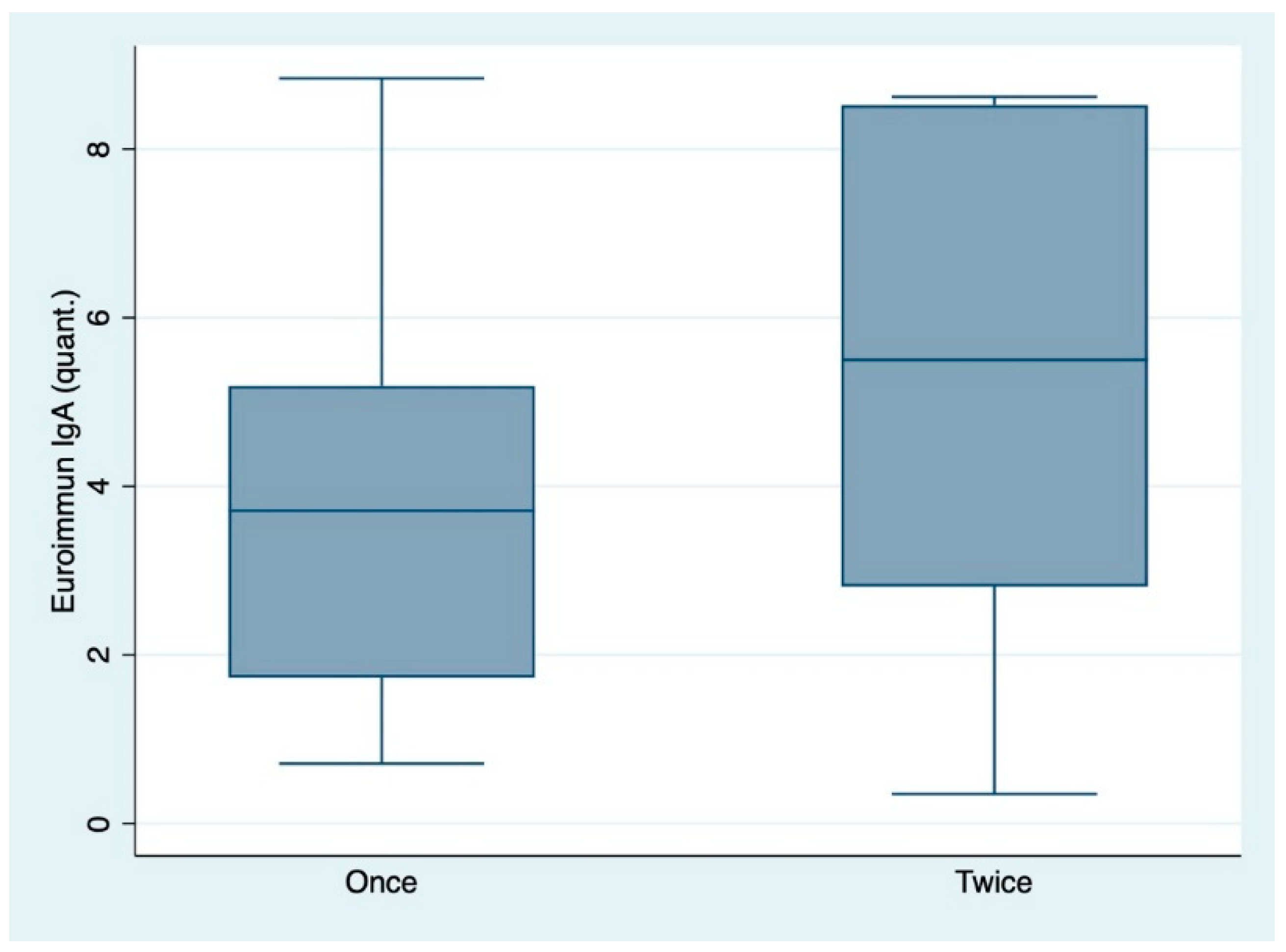
| IgA: All individuals | 27 | 3.87 (2.28) | 3.71 (1.73, 5.19) | 53 | 5.39 (2.82) | 5.50 (2.81, 8.52) | 0.0217 |
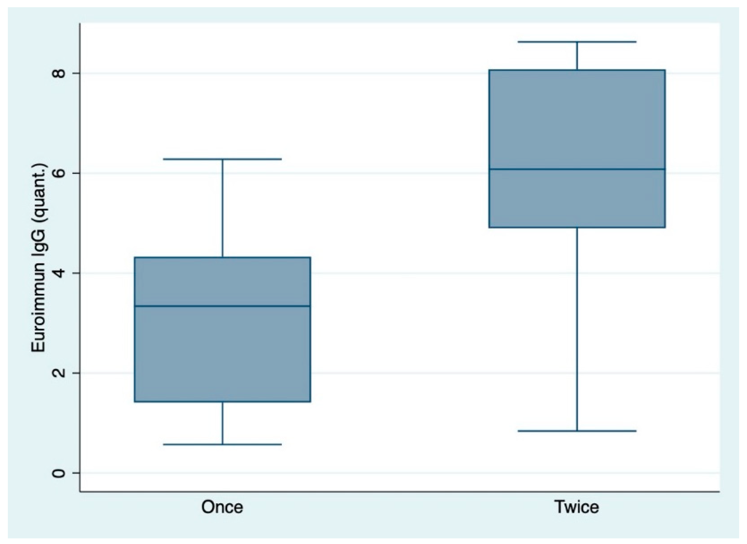
| IgG: All individuals | 27 | 2.96 (1.69) | 3.34 (1.41, 4.33) | 53 | 5.98 (2.18) | 6.08 (4.90, 8.08) | <0.0001 |
Publisher’s Note: MDPI stays neutral with regard to jurisdictional claims in published maps and institutional affiliations. |
© 2021 by the authors. Licensee MDPI, Basel, Switzerland. This article is an open access article distributed under the terms and conditions of the Creative Commons Attribution (CC BY) license (http://creativecommons.org/licenses/by/4.0/).
Share and Cite
Dörschug, A.; Frickmann, H.; Schwanbeck, J.; Yilmaz, E.; Mese, K.; Hahn, A.; Groß, U.; Zautner, A.E. Comparative Assessment of Sera from Individuals after S-Gene RNA-Based SARS-CoV-2 Vaccination with Spike-Protein-Based and Nucleocapsid-Based Serological Assays. Diagnostics 2021, 11, 426. https://doi.org/10.3390/diagnostics11030426
Dörschug A, Frickmann H, Schwanbeck J, Yilmaz E, Mese K, Hahn A, Groß U, Zautner AE. Comparative Assessment of Sera from Individuals after S-Gene RNA-Based SARS-CoV-2 Vaccination with Spike-Protein-Based and Nucleocapsid-Based Serological Assays. Diagnostics. 2021; 11(3):426. https://doi.org/10.3390/diagnostics11030426
Chicago/Turabian StyleDörschug, Anja, Hagen Frickmann, Julian Schwanbeck, Elif Yilmaz, Kemal Mese, Andreas Hahn, Uwe Groß, and Andreas E. Zautner. 2021. "Comparative Assessment of Sera from Individuals after S-Gene RNA-Based SARS-CoV-2 Vaccination with Spike-Protein-Based and Nucleocapsid-Based Serological Assays" Diagnostics 11, no. 3: 426. https://doi.org/10.3390/diagnostics11030426
APA StyleDörschug, A., Frickmann, H., Schwanbeck, J., Yilmaz, E., Mese, K., Hahn, A., Groß, U., & Zautner, A. E. (2021). Comparative Assessment of Sera from Individuals after S-Gene RNA-Based SARS-CoV-2 Vaccination with Spike-Protein-Based and Nucleocapsid-Based Serological Assays. Diagnostics, 11(3), 426. https://doi.org/10.3390/diagnostics11030426