Article

Quantification of Sodium Relaxation Times and Concentrations as Surrogates of Proteoglycan Content of Patellar CARTILAGE at 3T MRI

1
Department of Diagnostic and Interventional Radiology, Medical Faculty, University Dusseldorf, D-40225 Dusseldorf, Germany
2
Clinic of Nuclear Medicine, Medical Faculty, University Dusseldorf, D-40225 Dusseldorf, Germany
3
Institute of Radiology, University Hospital Erlangen, Friedrich-Alexander-Universität Erlangen-Nürnberg (FAU), D-91054 Erlangen, Germany
4
German Cancer Research Center (DKFZ), Division of Medical Physics in Radiology, D-69120 Heidelberg, Germany
5
Russell H. Morgan Department for Radiology and Radiological Science, The Johns Hopkins University School of Medicine, Baltimore, MD 21205-2196, USA
6
F. M. Kirby Research Center for Functional Brain Imaging, Kennedy Krieger Institute, Baltimore, MD 21205-2196, USA
*
Author to whom correspondence should be addressed.
Diagnostics 2021, 11(12), 2301; https://doi.org/10.3390/diagnostics11122301
Submission received: 5 November 2021 / Revised: 3 December 2021 / Accepted: 6 December 2021 / Published: 8 December 2021
(This article belongs to the Special Issue Advanced MRI Techniques for Musculoskeletal Imaging 2.0)

Abstract

:
Sodium MRI has the potential to depict cartilage health accurately, but synovial fluid can influence the estimation of sodium parameters of cartilage. Therefore, this study aimed to reduce the impact of synovial fluid to render the quantitative compositional analyses of cartilage tissue technically more robust. Two dedicated protocols were applied for determining sodium T 1 and T 2 * relaxation times. For each protocol, data were acquired from 10 healthy volunteers and one patient with patellar cartilage damage. Data recorded with multiple repetition times for T 1 measurement and multi-echo data acquired with an additional inversion recovery pulse for T 2 * measurement were analysed using biexponential models to differentiate longitudinal relaxation components of cartilage ( T 1 , car ) and synovial fluid ( T 1 , syn ), and short ( T 2 s * ) from long ( T 2 l * ) transversal relaxation components. Sodium relaxation times and concentration estimates in patellar cartilage were successfully determined: T 1 , car = 14.5 ± 0.7 ms; T 1 , syn = 37.9 ± 2.9 ms; c( T 1 -protocol) = 200 ± 48 mmol/L; T 2 s * = 0.4 ± 0.1 ms; T 2 l * = 12.6 ± 0.7 ms; c( T 2 * -protocol) = 215 ± 44 mmol/L for healthy volunteers. In conclusion, a robust determination of sodium relaxation times is possible at a clinical field strength of 3T to quantify sodium concentrations, which might be a valuable tool to determine cartilage health.

1. Introduction

Joint cartilage serves as a mechanical buffer and plays a pivotal role in the joint’s health and functionality. Therefore, cartilage loss is a hallmark change in the pathogenesis of several degenerative and inflammatory joint diseases such as osteoarthritis, rheumatoid arthritis and psoriatic arthritis and is associated with pronounced disease burden and functional disability [1,2,3,4]. While some easier to implement techniques like 1H T2 mapping have been shown to be a feasible tool for differentiating healthy and degraded cartilage, they detect mainly structural changes in the collagen matrix of cartilage [5]. However, prior to the manifestation of irreversible structural cartilage degradation, early and potentially reversible changes in cartilage composition occur and proteoglycans (PG) are lost. For the detection of compositional changes, techniques with high PG specificity such as delayed gadolinium-enhanced MRI, glycosaminoglycan (GAG) chemical exchange saturation transfer or sodium (23Na) MRI have emerged [4,6,7,8,9,10,11,12,13].
The GAG side chains of PGs consist of carboxyl and sulphate groups and create a fixed charge density (FCD) [14]. The negatively charged FCD attracts positively charged 23Na ions, which can be measured by 23Na MRI and, consequently, the intra-tissue 23Na concentration indicates the PG content [15]. Therefore, the 23Na signal can be used to assess the compositional make-up of cartilage and to detect PG depletion as a sign of early degeneration. Beyond 23Na concentrations, 23Na relaxation times of cartilage change with degeneration, as shown in enzymatically degraded cartilage samples [16].
Despite its high sensitivity for PGs, the signal-to-noise ratio (SNR) of 23Na MRI of cartilage is up to three orders of magnitude lower compared to conventional proton MRI [17]. This is due to lower abundance of 23Na as compared to hydrogen in humans and the generally lower nuclear magnetic resonance sensitivity [18]. Transverse relaxation of the 23Na signal in cartilage follows a biexponential decay with a short ( T 2 s * ) and a long component ( T 2 l * ). The already low SNR and fast decay of the 23Na signal with increasing echo times (TEs) requires the use of ultra-short echo-time (UTE) sequences [19]. 23Na MRI is usually performed with lower spatial resolution than conventional proton MRI to keep image acquisition periods manageable, yet this comes at the expense of more and more severe partial volume effects (PVEs). In articular cartilage, the 23Na signal from the synovial fluid that surrounds the cartilage spills into the cartilage signal, thereby leading to inaccurate quantification of 23Na concentration and relaxation times [20]. For 23Na MRI on clinical 3T MRI scanners (that incurs lower SNR and more PVEs as compared to ≥7T MRI scanners [21]), this aspect becomes very relevant as it challenges correct classification of pixels as cartilage or synovial fluid and artificially increases T 1 and T 2 * relaxation times of patellar cartilage. Therefore, methods to reduce the influence of synovial fluid on 23Na relaxation times and concentrations, especially in regions prone to PVEs, are highly desirable to advance the clinical applicability of 23Na MRI in cartilage assessment.
Human patellar cartilage 23Na T 1 ( T 1 , car = 21 ms) and T 2 * relaxation times ( T 2 s * = 0.8 ms; T 2 l * = 19.7 ms; biexponential model) have been shown to be shorter than those of synovial fluid ( T 1 , syn = 48 ms; T 2 * = 47 ms, monoexponential model) when imaged at 4.7T and a high in-plane resolution of 1.5 × 1.5 mm2 [20]. However, even at this superior resolution, clear differentiation of cartilage and synovial fluid was only possible in certain areas adjacent to the joint capsule and not possible for the patellar cartilage. A potential way to suppress the signal of synovial fluid is the application of an inversion pulse to null the synovial fluid signal, which was introduced previously and can easily be applied for T 2 * measurements [22,23,24]. However, the determination of T 1 with additional inversion pulses is challenging, as these directly interfere with cartilage T 1 relaxation.
In this study, we set out (1) to determine 23Na T 1 and T 2 * relaxation times of patellar cartilage in the presence of PVEs at 3T. We aimed to develop a robust method for T 1 measurements of cartilage and to evaluate T 2 * while using an inversion recovery (IR) method for fluid suppression. We then (2) aimed to apply the measured relaxation times to determine 23Na concentration maps. We hypothesized, in addition to successfully estimating the above 23Na parameters, to be able to observe a trend towards different relaxation times and concentrations between healthy controls and patients.

2. Materials and Methods

This study was conducted with two separate MRI protocols for measuring 23Na T 1 (protocol 1) and T 2 * (protocol 2). Although it would be preferable to measure both T 1 and T 2 * in a single MRI session, the approach with separate protocols ensures that more data points can be acquired and thus, more stable results should be obtained for the estimation of T 1 and T 2 * parameters.

2.1. Study Population

Two age-matched cohorts of healthy volunteers were examined, one for each study protocol ( T 1 protocol: 4 females, 6 males, mean age 23 ± 3 years, minimum/maximum 19/29 years; T 2 * protocol: 3 females, 7 males, mean age 23 ± 2 years, minimum/maximum 19/28 years). Participants were excluded from the healthy volunteer group if any degenerative joint disease of the knee or cartilage damage was known. Participants were also excluded if they reported a history of acute or chronic knee pain as well as previous surgery to the index knee.
Two patients with retropatellar chondropathy, one for each protocol, were studied using the same protocols as above. For the T 1 protocol, one 66 years old female patient with established osteochondrosis of the left knee was included. Following the MRI Osteoarthritis Knee Score (MOAKS) [25], which classifies cartilage lesions from 0 (no defects) to 3 (severe defects) according to the percentage extent of any defect and the percentage extent of full-thickness cartilage defects (Figure 1), their patellar cartilage lesions were classified as grade 2/1 (any/full-thickness) in the medial retropatellar region and 2/0 (any/full-thickness) in the lateral subregion. For the T 2 * protocol, a 30-year-old female patient was included with posttraumatic cartilage defects of the right knee, which were classified as grade 3/1 (any/full-thickness) in the medial and grade 1/1 (any/full-thickness) in the lateral subregion according to the MOAKS classification.
The study was approved by the local ethics committee (Ethics Committee, Medical Faculty of the Heinrich-Heine-University Düsseldorf, study number 4733R), and written informed consent was obtained from all volunteers and patients.

2.2. MRI

All images were acquired using a 3T MRI scanner (Siemens MAGNETOM Prisma, Siemens Healthineers, Erlangen, Germany) and a dual-tuned 23Na/1H surface coil (RAPID Biomedical GmbH, Rimpar, Germany) with a 11 cm circular 23Na resonator and a 18 cm × 24 cm rectangular 1H resonator. Once all participants were placed in the feet-first and supine position, the dual-tuned coil was placed on top of the right knee of healthy volunteers and on top of the affected knee of patients. For reference purposes, patients were also studied using a dedicated 1H 15-channel knee coil (Tx/Rx Knee 15 Flare Coil, Siemens Healthineers, Erlangen, Germany) in line with clinical standard routines. Consequently, the coil had to be replaced between the 23Na and 1H measurements.
For quantification of 23Na concentrations, three cylindrical phantoms (diameter: 1.5 cm, length: 10 cm) with different 23Na concentrations (50 mmol/L, 100 mmol/L and 200 mmol/L) and a fixed agarose content of 4% (ROTI®Garose, Carl ROTH GmbH & Co. KG, Karlsruhe, Germany) were manufactured by B.K. and attached medial to the examined knee [11,23,26]. All 23Na MRI was performed using a density-adapted 3D radial sequence (DA-3D-RAD), which was developed by Nagel et al. and has been shown to improve SNR compared to conventional 3D radial sequences through more efficient k-space sampling [27]. Furthermore, inversion recovery pulses and spoiler gradients are usable with this sequence.

2.2.1. Characterization of the 23Na Coil

The sensitivity of the 23Na coil was evaluated by measuring a homogenous water phantom and the sensitivity image was then used to correct the 23Na images of all participants on a pixel-by-pixel basis [28]. The phantom was of cylindrical shape (diameter: 18 cm; height: 7.5 cm) and filled with 100 mmol/L NaCl solution.
To assess B1 homogeneity of the surface coil for 23Na MRI, the field dependency was mapped with the same 100 mmol/L NaCl phantom as above using the double-angle method [29,30]. To this end, two 23Na images with varying FAs were acquired. The applied sequence parameters for these measurements are summarized in Table 1.

2.2.2. MRI Sequence Parameters

Two separate study protocols were designed for relaxation time measurements. For both protocols, a 1H localizer sequence and two sequences for the adjustment of the required reference voltage were acquired first, one for 1H MRI and one for 23Na MRI [31]. Manual B0 shimming was performed prior to DA-3D-RAD image acquisition. High-resolution 1H MRI was performed using the DA-3D-RAD sequence as the anatomic reference. The parameters for 23Na MRI with the DA-3D-RAD differed between protocol 1 and protocol 2 as summarized in Table 1.
For 23Na T 1 determination images were acquired with 17 different TR to obtain a high number of data points for the later explained fitting procedure. For 23Na T 2 * determination a multi-echo DA-3D-RAD was acquired three times with four different TEs each time, resulting in 12 data points. This interleaving acquisition pattern of different TEs with multiple sequences was chosen because the readout time would not allow shorter spacing between TE per single sequence. To minimize synovial fluid-induced PVEs and their bearing on T 2 * estimation, a rectangular inversion recovery pulse with an inversion time of TI = 24 ms and an inversion pulse duration of 1 ms was used to null the synovial fluid signal [11,24,32].
For clinical reference purposes, patients were further examined using a conventional knee coil with proton density (PD)-weighted fat-saturated and T 1 -weighted sequences. The parameters for the additional sequences to study the patients are detailed in Table 2.

2.3. Image Post-Processing

The images were reconstructed using a Hann Filter to increase SNR and reduce Gibbs ringing. The 23Na images were motion-corrected using the in-house developed software stroketool [33], which utilises a cross-correlation algorithm based on advanced normalization tools [34]. For further data evaluation and ROI definition, in-house developed MATLAB (MathWorks, Natick, MA, USA, R2018a) scripts were used. To correct displacement between the 1H and 23Na DA-3D-RAD images, regions-of-interest (ROIs) were drawn around the agarose phantoms in the 1H and one 23Na DA-3D-RAD image. The calculated shift between these ROIs was then used to overlay the 1H DA-3D-RAD image precisely with the 23Na image.
Relaxation curve fitting was performed using the mean values of the cartilage ROI for more stable results than would be obtained with pixelwise fitting. The data of protocol 1 was fitted with a biexponential two-pool model as indicated in Equation (1) to determine T 1 for both cartilage ( T 1 , car ) and synovial fluid ( T 1 , syn ) from the signal intensity S(t):
S ( t ) = S ( 0 ) · ( p car · ( 1 e t T 1 , car ) + ( 1 p car ) · ( 1 e t T 1 , syn ) ) + noise
In this approach, p car designates the fraction of the cartilage T 1 , car of the total T 1 signal relaxation, while ( 1 p car ) is the fraction of synovial fluid, assuming that signal only comes from these two pools. The values of p car follow the condition 0 < p car < 1 .
Data from protocol 2 were used to determine transverse relaxation times for the patellar cartilage using a validated biexponential model introduced by our group previously (Equation (2)), assuming a short and a long component T 2 s * and T 2 l * [31]:
S ( t ) = S ( 0 ) · ( p s · e t T 2 s * + ( 1 p s ) · e t T 2 l * ) + noise
Here, p s defines the fraction of the T 2 s * signal decrease in the T 2 * signal decrease as a whole, satisfying the condition 0 < p s < 1 .
Finally, the sodium images were used to determine the sodium concentration in the retropatellar cartilage. Agarose phantoms for the quantification of sodium concentrations were manufactured as described previously [31]. The relaxation times of the phantoms were: T 1 = 38.6 ms; T 2 s * = 4.5 ms; T 2 l * = 15.3 ms; p s = 66.8%. The relaxation times were used to correct the influence of the relaxation time difference between agarose phantoms and cartilage on their signal ratio. For the in-vivo cartilage measurements acquired in this study, the arithmetic means of the determined longitudinal relaxation times for the group of volunteers measured with protocol 1 and the transverse relaxation times determined for the group of volunteers measured with protocol 2 were applied for this correction.
The corrected signal from the agarose phantoms was fitted linearly to calculate the 23Na concentration of the cartilage. Agarose phantoms were excluded if positioned too far from the surface coil causing insufficient excitation flip angles, which had been determined previously by B1 mapping in the course of the characterization of the 23Na coil. To estimate 23Na concentrations in cartilage, the partial volume correction method of previous studies of our group [31] was applied. The spatial dimensions of the patellar cartilage are comparable to and sometimes smaller than the voxel size of the 23Na images. When the voxel of the 23Na image is not completely filled with cartilage, the 23Na concentration of the cartilage in this voxel is often averaged with background noise, resulting in the underestimation of 23Na concentration in that voxel depending on how much volume of the voxel is filled with cartilage. To reduce this effect, the higher resolution of the 1H images and the ROIs drawn on those images are used to calculate the volume fraction of cartilage in each 23Na voxel. The volume fraction is averaged over the whole ROI and the concentration values in the ROI are multiplied by the inverse of this volume fraction. Finally, the resulting 23Na concentration estimates were divided by 0.75, because ~25% of cartilage is made up by solids that do not contribute to the 23Na signal [24,35,36].
23Na concentration maps were determined for both protocols. Specifically, for protocol 1 we selected the image acquired at a TR of 70 ms to minimize T 1 weighting of the 23Na signal. For protocol 2, the shortest available TE (TE = 0.3 ms) was selected to ensure high SNR and to minimize T2 weighting. The ROIs of the retropatellar cartilage were independently drawn by two experienced radiologists on the 1H DA-3D-RAD images. Radiologist #1 (M.F.; 5 years of experience in musculoskeletal imaging) defined the ROIs twice (i.e., two weeks apart to allow for sufficient washout) to allow for intra-reader reliability assessment, while radiologist #2 (D.B.A.; 5 years of experience in musculoskeletal imaging) defined them once for inter-reader reliability assessment.
Segmentations were performed using in-house developed MATLAB scripts by manually delineating the outer contours of the patellar cartilage in the 1H DA-3D-RAD images. Synovial fluid was not intentionally included in the ROIs; however, for the biexponential T 1 fitting described in Equation (1), it is expected to affect the signal in the ROIs anyway, because of PVEs caused by the large voxel size of the 23Na images and the close proximity of the synovial fluid to the cartilage.

2.4. Statistical Analysis

Statistical analysis was performed with SPSS (IBM Corp. Released 2020. IBM SPSS Statistics for Windows, Version 27.0. Armonk, NY: IBM Corp.). For the healthy controls, descriptive statistics (mean, standard deviation, minimum, median, maximum) were calculated for each protocol. For the relaxation times of the patients, the standard deviation was calculated across the respective measurements of the radiologists; the standard deviation of the 23Na concentration of the patients was determined across the pixelwise deviation of 23Na concentrations in the cartilage ROI.
To test whether the different settings (TE, TR, inversion pulse) in protocol 1 and 2 resulted in significant differences in 23Na concentration estimates, the mean concentration values between the volunteer groups were compared using the Wilcoxon signed-rank test with a significance level of p < 0.05. For assessing intra- and inter-reader reliability, single intraclass correlation coefficients (sICC) and average intraclass correlation coefficients (aICC) were calculated for the biexponential model parameters T 1 , car , T 1 , syn , p car , T 2 s * , T 2 l * , p s and 23Na concentrations of protocol 1 and 2 in healthy volunteers. The resulting intraclass correlation coefficients were categorized according to Koo et al. [37].

3. Results

Relaxation time measurements were successfully performed on all participants. For the first measurement of radiologist #1, T 1 results were T 1 , car = 14.45 ± 0.74 ms; T 1 , syn = 37.91 ± 2.92 ms; p car = 77.30 ± 3.73% for healthy volunteers measured with protocol 1 (Figure 2a) and T 1 , car = 15.42 ± 0.08 ms; T 1 , syn = 39.78 ± 0.14 ms; p car = 71.23 ± 0.22% for the patient measured with protocol 1. Other associated T 1 results are summarized in Table 3. Intra- and inter-reader testing resulted in sICC( T 1 , car ) = 0.93; aICC( T 1 , car ) = 0.83; sICC( T 1 , syn ) = 0.94; aICC( T 1 , syn ) = 0.83; sICC( p car ) = 0.92 and aICC( p car ) = 0.69.
For T 2 * results, the first measurements of radiologist #1 were T 2 s * = 0.358 ± 0.147 ms; T 2 l * = 12.62 ± 0.73 ms; p s = 34.39 ± 4.78% for healthy volunteers measured with protocol 2 (exemplary fit in Figure 2b) and T 2 s * = 0.105 ± 0.001 ms; T 2 l * = 13.99 ± 0.01 ms; p s = 25.86 ± 0.06% for the patient measured with protocol 2. Other associated T 2 * results are summarized in Table 4. Intra- and inter-reader testing resulted in sICC( T 2 s * ) = 0.99; aICC( T 2 s * ) = 0.99; sICC( T 2 l * ) = 0.78; aICC( T 2 l * ) = 0.86; sICC( p s ) = 0.99 and aICC( p s ) = 0.98.
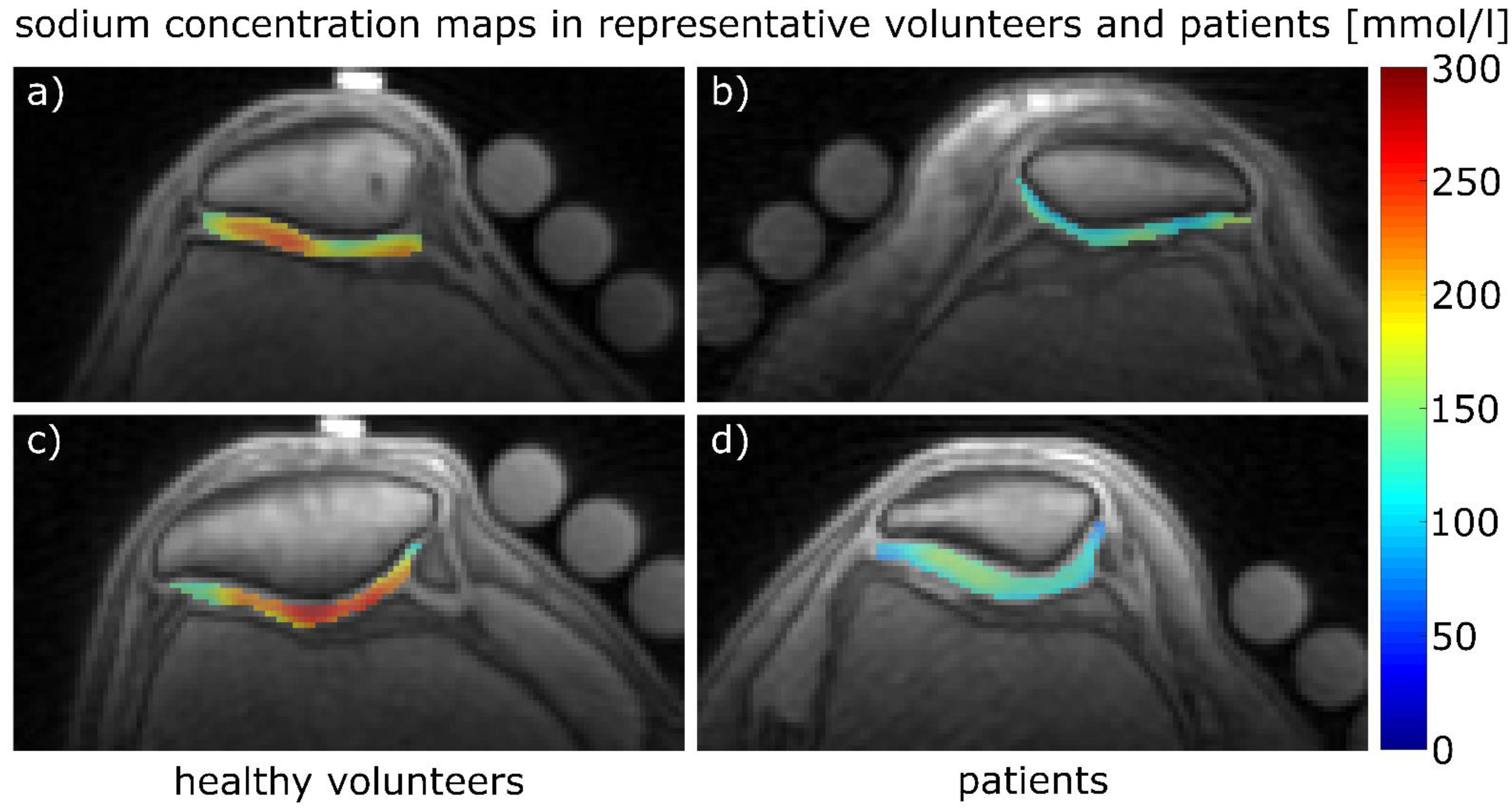
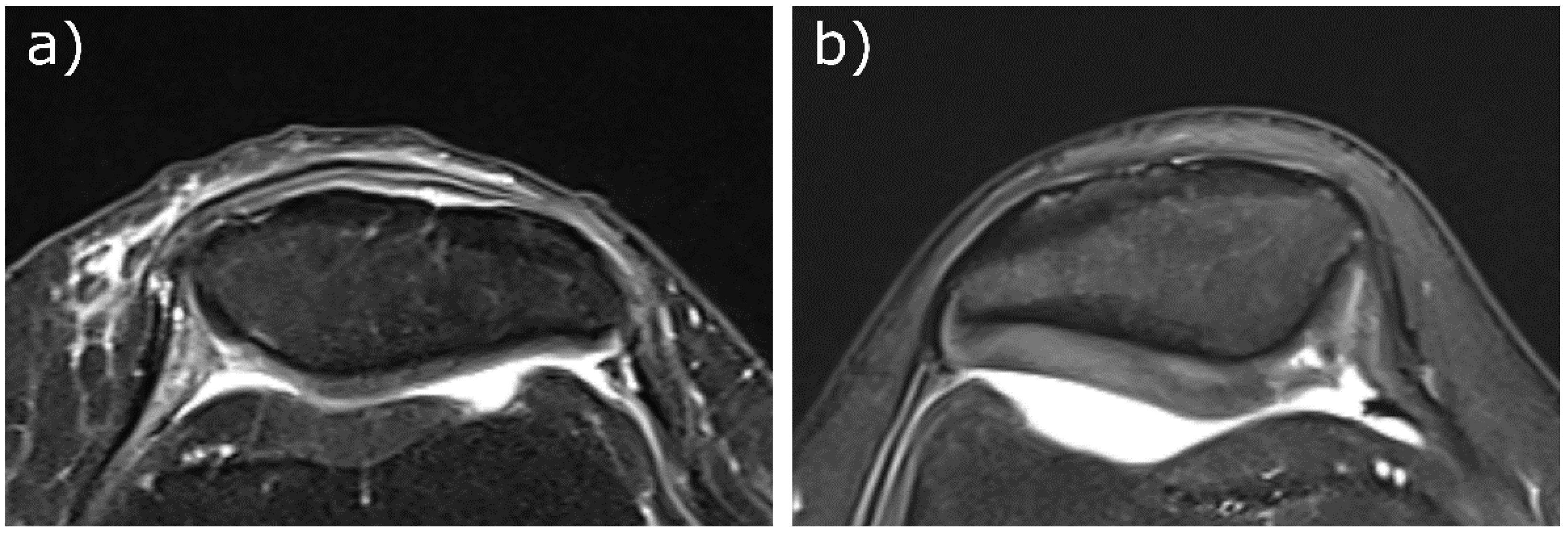
The 23Na concentrations estimated from the first measurement of radiologist #1 for protocol 1 were 200 ± 48 mmol/L for the healthy volunteers (n = 10) and 158 ± 30 mmol/L for the patient, while for protocol 2, the values were 215 ± 44 mmol/L for the healthy volunteers (n = 9) and 135 ± 29 mmol/L for the patient. Proton images overlaid with 23Na concentration maps for volunteers and patients are shown in Figure 3. Transversal Proton Density-weighted fat-saturated images of the two patients are shown in Figure 4.
The 23Na concentration estimation results from all remaining participants are summarized in Table 5. Intra- and inter-reader testing resulted in sICC(protocol 1) = 0.94; aICC(protocol 1) = 0.78; sICC(protocol 2) = 0.90 and aICC(protocol 2) = 0.87. The concentration estimates between the healthy volunteer groups acquired for protocol 1 or 2 did not differ significantly (first measurement of radiologist #1: p-value= 0.441, data of remaining radiologist measurements in Table 5).

4. Discussion

In this study, we successfully determined (1) 23Na relaxation times of the patellar cartilage at a clinical field strength of 3T while minimizing the effects of PVEs caused by coarse spatial resolution and (2) calculated 23Na concentrations based on the relaxation times. The results of the sICC and aICC indicated good-to-excellent (0.78–0.99) intra- and moderate-to-excellent (0.69–0.99) inter-reader reliability for these parameters. We observed decreased T 2 s * and increased T 2 l * and T 1 , car relaxation times in two patients with patellar cartilage degeneration as compared to healthy volunteers. However, further studies with more participants are necessary to draw more definitive conclusions.
In efforts to render such quantitative studies more reliable, the influence of synovial fluid on relaxation time quantification needs to be reduced. If not well controlled, the variable contribution of synovial fluid to the observed signal may confound 23Na quantification and further challenge the differentiation of healthy volunteers and patients with patellar cartilage degeneration or osteoarthritis. We successfully demonstrated the feasibility of using an inversion pulse for fluid suppression in T 2 * determination and biexponential modeling for T 1 determination.
To our knowledge, applying a biexponential two-pool model to the 23Na signal for T 1 determination has not been performed before in this context. To our mind, it has distinct advantages. First, it reduces the influence of synovial fluid on relaxation time measurements in cartilage by including T 1 , syn as fitting parameter in Equation (1), thus separating the longitudinal relaxation times of cartilage and synovial fluid. In widely used monoexponential one-pool models, these parameters would otherwise be merged in a singular parameter T 1 , hampering accurate depiction of T 1 in cartilage. Second, it allows T 1 , syn of synovial fluid and the signal fraction p car to be estimated as additional parameters. It remains to be seen whether these parameters are of diagnostic value. Furthermore, applying inversion pulses for fluid suppression also reduces the signal from cartilage, which may prove problematic at lower field strengths than 3T.
Determining the 23Na relaxation times of cartilage is not only useful for a more accurate estimation of 23Na concentration by correcting the difference in signal ratio between cartilage and reference phantoms caused by different relaxation behaviour, but the relaxation times themselves can be an indicator for cartilage health [16]. In the aforementioned study, relaxation times of bovine patellar cartilage specimens were measured at 2T before and after enzymatic degradation by trypsin exposure, causing defined PG depletion [38]. Lower PG content was associated with decreases in T 2 s * and increases in T 2 l * and T 1 , car , which is in good agreement with our findings. Because data regarding the 23Na relaxation times in degraded cartilage is scarce and we only measured one patient per protocol, the following discussion is centred around the measurements performed on our healthy volunteers. An excerpt of study results from different authors regarding 23Na relaxation times in patellar cartilage is shown in Table 6.
Madelin et al. carried out extensive testing of knee cartilage relaxation times at 7T [39]. Their T 2 * estimates are comparable to our results, although acquired at higher field strength. Theoretically, p s should be 60%, assuming a one compartment model, based on quadrupole interactions without magnetic field inhomogeneities, but under experimental conditions in the presence of field inhomogeneities and different tissue properties, p s has been shown to deviate from the theoretical value of 60% [40]. Madelin et al. found slightly higher T 1 , car estimates than we did, which might be due to them using a MRI system with higher field strength (7T) compared to us (3T) and T 1 rising with increasing field strength [41]. Another reason could be the differences in their used TR values for T 1 measurement (30 ms to 250 ms). We focused on lower TR values to increase the number of data points available for the biexponential model, particularly for the shorter T 1 , car .
Feldman et al. measured 23Na relaxation times of the patellar cartilage and synovial fluid at 4.7T, reporting slightly longer 23Na longitudinal and transversal relaxation times compared to ours [20]. This group elaborated on the difficulty of separating voxels containing pure cartilage signal and pure fluid signal, which might have led to slightly overestimated T 1 , car and T 2 l * relaxation times as compared to our estimates. In addition, the previously discussed reasons for higher T 1 , car values because of different MRI scanner field strengths and different TR intervals for fitting apply here as well. Staroswiecki et al. determined 23Na transversal relaxation times by monoexponential fitting [21]. However, due to the low resolution of 23Na images, these results are likely confounded by partial volume effects secondary to synovial fluid, a bias that we sought to address in this study.
Relaxation time measurements have also been performed with triple quantum filtered (TQF) 23Na MRI, which inherently suppresses synovial fluid [42]. Reported values at 3T were slightly lower ( T 2 s * = 0.84 ± 0.06 ms; T 2 l * = 9.59 ± 0.35 ms) compared to our estimates. However, TQF signal acquisition has its own challenges in even lower SNR [18] and additional imaging artefacts caused by off-resonance effects [43]. Against this background, UTE sequences are therefore generally preferred for in-vivo 23Na MRI [44].
Our calculated 23Na concentrations in patients with degenerative patellar cartilage were lower compared to the mean and median values across all healthy volunteers. However, only two patients were studied by us. Chang et al. showed that 23Na concentration correlates with PG content and lower PG content is a sign of early-to-moderate degeneration [15,18]. The reduced 23Na concentrations of our patients may therefore indicate ongoing cartilage degeneration, which—as an imaging finding—is not surprising, as both patients already had morphologic cartilage defects. Future studies should therefore focus on patients with cartilage at risk (secondary to patient -or joint level- factors such as obesity, malalignment or previous injury) to further evaluate our method in such patients.
Madelin et al. reported 23Na concentrations for healthy volunteers and patients with (volunteers: 220–270 mmol/L; patients: 170–200 mmol/L) and without (volunteers: 180–210 mmol/L; patients: 170–190 mmol/L) the use of an inversion pulse for synovial fluid suppression [24]. Chang et al. reported similar findings at 7T comparing cartilage tissue after surgical cartilage resurfacing with native cartilage tissue of another joint area, both with (surgery: 108.9 ± 29.8 mmol/L; native: 249.8 ± 44.6 mmol/L) and without (surgery: 177.8 ± 54.1 mmol/L; native: 172.2 ± 30.3 mmol/L) applying an inversion pulse [15]. Our 23Na concentrations in healthy volunteers and patients are in good agreement with these data.
When interpreting our results, some limitations must be taken into consideration. One limitation is the long acquisition time, currently precluding T 1 and T 2 * measurements within a single clinical MRI study. Total study time for T 1 and T 2 * determination were 60 and 45 min, respectively. For future studies with increased patient numbers, the total measurement time for each protocol should be reduced to less than 30 min, which could be achievable by measuring less TE/TR data points for fitting combined with advanced techniques for image reconstruction [45,46,47,48]. However, these methods and their capability of producing stable results for cartilage data will need to be evaluated carefully.
While the patellar cartilage of patients was examined with additional sequences for clinical reference, the patellar cartilage of healthy volunteers was assessed by asking for history of pain or surgery in the examined knee. To further reduce the possibility of falsely including participants with cartilage damage in the healthy volunteer group, clinical reference MRI could also be performed for this group.
Furthermore, our healthy controls were not age-matched to the patients. In the intervertebral disks, a correlation between increasing participant age and reduced GAG chemical exchange saturation transfer effect has been shown [49]. A similar dependency might be possible for PG in articular cartilage and in larger clinical studies patients and healthy controls should preferably be age-matched to reduce the possibility of it confounding results. Additionally, future studies investigating the relation between age and PG content in cartilage could be conducted for clarification of this problem.
Other limitations include the low resolution of 23Na MRI in general, which limits its applicability for detecting smaller cartilage lesions and may be difficult to implement in smaller joints such as the ankle. The 23Na surface coil allowed evaluation of the patellofemoral joint only, while the femorotibial joint is not assessable using the present setup. The size of the 23Na resonator limits the sensible detection of 23Na signal in more distant areas like the femorotibial joint. It is important to note that inhomogenities of the B1 field were only considered regarding the placement of the Agarose phantoms by B1 field mapping. Spatial fluctuation of the B1 field will also reduce the efficiency of the fluid suppression of the inversion pulse applied in protocol 2, which could be mitigated by using adiabatic inversion pulses [23].

5. Conclusions

Synovial fluid can confound the measurement of 23Na relaxation times and concentrations in cartilage due to partial volume effects, especially at clinical field strength (≤3T). Therefore, in this study, two different methods to reduce the influence of synovial fluid, the usage of an inversion pulse to determine 23Na T 2 * and 23Na concentrations and the appliance of a biexponential two-pool model for determining 23Na T 1 , were successfully applied in healthy volunteers and two pilot patients with patellar cartilage damage. While the clinical value is necessarily reliant on a larger clinical database and is still unclear, this study introduces more parameters to quantitatively assess the tissue’s proteoglycan content as the key surrogate marker of early cartilage degeneration.

Author Contributions

Conceptualization, B.K., J.M.H., G.O., S.N., H.-J.W. and A.M.-L.; methodology, B.K., J.M.H., H.-J.W. and A.M.-L.; software, B.K., J.M.H., A.M.N., L.V.G. and H.-J.W.; validation, B.K., M.F., J.M.H., G.O., D.B.A., L.M.W., H.-J.W. and A.M.-L.; formal analysis, B.K., J.M.H., M.F. and D.B.A.; investigation, B.K., J.M.H. and A.M.-L.; resources, A.M.N. and L.V.G.; data curation, B.K. and J.M.H.; writing—original draft preparation, B.K., M.F., H.-J.W. and A.M.-L.; writing—review and editing, B.K., M.F., J.M.H., D.B.A., A.M.N., L.V.G., G.O., L.M.W., S.N., G.A., H.-J.W. and A.M.-L.; visualization, B.K., J.M.H. and A.M.-L.; supervision, B.K., L.M.W., S.N., G.A., H.-J.W., A.M.-L.; project administration, B.K., J.M.H., G.A., A.M.-L. All authors have read and agreed to the published version of the manuscript.

Funding

M.F., D.B.A., L.M.W. and S.N. were supported by an internal research grant of the local Research Committee of the Medical Faculty of Heinrich-Heine-University Düsseldorf. S.N. was additionally supported by grants from the “Deutsche Forschungsgemeinschaft” (DFG) (NE 2136/3-1). G.O. is supported by a grant from the National Institutes of Health (National Institute on Aging) (R00AG062230).

Institutional Review Board Statement

The study was conducted according to the guidelines of the Declaration of Helsinki, and approved by the Ethics Committee of the Medical Faculty, University of Dusseldorf, Germany (protocol code study 4733R).

Informed Consent Statement

Informed consent was obtained from all subjects involved in the study.

Data Availability Statement

Data can be provided by the authors upon reasonable request.

Conflicts of Interest

The authors declare no conflict of interest.

References

  1. MacKay, J.W.; Low, S.B.L.; Smith, T.O.; Toms, A.P.; McCaskie, A.W.; Gilbert, F.J. Systematic Review and Meta-Analysis of the Reliability and Discriminative Validity of Cartilage Compositional MRI in Knee Osteoarthritis. Osteoarthr. Cartil. 2018, 26, 1140–1152. [Google Scholar] [CrossRef] [PubMed] [Green Version]
  2. Sewerin, P.; Müller-Lutz, A.; Odendahl, S.; Eichner, M.; Schneider, M.; Ostendorf, B.; Schleich, C. Prevention of the Progressive Biochemical Cartilage Destruction under Methotrexate Therapy in Early Rheumatoid Arthritis. Clin. Exp. Rheumatol. 2018, 37, 179–185. [Google Scholar] [CrossRef]
  3. Abrar, D.B.; Schleich, C.; Frenken, M.; Vordenbäumen, S.; Richter, J.; Schneider, M.; Ostendorf, B.; Nebelung, S.; Sewerin, P. DGEMRIC in the Assessment of Pre-Morphological Cartilage Degeneration in Rheumatic Disease: Rheumatoid Arthritis vs. Psoriatic Arthritis. Diagnostics 2021, 11, 147. [Google Scholar] [CrossRef]
  4. Abrar, D.B.; Schleich, C.; Nebelung, S.; Frenken, M.; Ullrich, T.; Radke, K.L.; Antoch, G.; Vordenbäumen, S.; Brinks, R.; Schneider, M.; et al. Proteoglycan Loss in the Articular Cartilage Is Associated with Severity of Joint Inflammation in Psoriatic Arthritis—a Compositional Magnetic Resonance Imaging Study. Arthritis Res. Ther. 2020, 22, 124. [Google Scholar] [CrossRef] [PubMed]
  5. Matzat, S.J.; van Tiel, J.; Gold, G.E.; Oei, E.H.G. Quantitative MRI Techniques of Cartilage Composition. Quant. Imaging Med. Surg. 2013, 3, 162–174. [Google Scholar] [CrossRef]
  6. Bittersohl, B.; Kircher, J.; Miese, F.R.; Dekkers, C.; Habermeyer, P.; Fröbel, J.; Antoch, G.; Krauspe, R.; Zilkens, C. T2* Mapping and Delayed Gadolinium-Enhanced Magnetic Resonance Imaging in Cartilage (DGEMRIC) of Humeral Articular Cartilage-a Histologically Controlled Study. J. Shoulder Elb. Surg. 2015, 24, 1644–1652. [Google Scholar] [CrossRef]
  7. Besselink, N.J.; Vincken, K.L.; Bartels, L.W.; van Heerwaarden, R.J.; Concepcion, A.N.; Marijnissen, A.C.A.; Spruijt, S.; Custers, R.J.H.; van der Woude, J.-T.A.D.; Wiegant, K.; et al. Cartilage Quality (DGEMRIC Index) Following Knee Joint Distraction or High Tibial Osteotomy. Cartilage 2020, 11, 19–31. [Google Scholar] [CrossRef] [PubMed] [Green Version]
  8. Schleich, C.; Bittersohl, B.; Miese, F.; Schmitt, B.; Müller-Lutz, A.; Sondern, M.; Antoch, G.; Krauspe, R.; Zilkens, C. Glycosaminoglycan Chemical Exchange Saturation Transfer at 3T MRI in Asymptomatic Knee Joints. Acta Radiol. 2016, 57, 627–632. [Google Scholar] [CrossRef] [PubMed]
  9. Abrar, D.B.; Schleich, C.; Radke, K.L.; Frenken, M.; Stabinska, J.; Ljimani, A.; Wittsack, H.-J.; Antoch, G.; Bittersohl, B.; Hesper, T.; et al. Detection of Early Cartilage Degeneration in the Tibiotalar Joint Using 3 T GagCEST Imaging: A Feasibility Study. Magn. Reson. Mater. Phys. Biol. Med. 2021, 34, 249–260. [Google Scholar] [CrossRef]
  10. Krishnamoorthy, G.; Nanga, R.P.R.; Bagga, P.; Hariharan, H.; Reddy, R. High Quality Three-dimensional GagCEST Imaging of in Vivo Human Knee Cartilage at 7 Tesla. Magn. Reson. Med. 2017, 77, 1866–1873. [Google Scholar] [CrossRef]
  11. Madelin, G.; Xia, D.; Brown, R.; Babb, J.; Chang, G.; Krasnokutsky, S.; Regatte, R.R. Longitudinal Study of Sodium MRI of Articular Cartilage in Patients with Knee Osteoarthritis: Initial Experience with 16-Month Follow-Up. Eur. Radiol. 2018, 28, 133–142. [Google Scholar] [CrossRef]
  12. Zbýň, Š.; Schreiner, M.; Juras, V.; Mlynarik, V.; Szomolanyi, P.; Laurent, D.; Scotti, C.; Haber, H.; Deligianni, X.; Bieri, O.; et al. Assessment of Low-Grade Focal Cartilage Lesions in the Knee With Sodium MRI at 7 T. Investig. Radiol. 2020, 55, 430–437. [Google Scholar] [CrossRef] [PubMed]
  13. Zbýň, Š.; Brix, M.O.; Juras, V.; Domayer, S.E.; Walzer, S.M.; Mlynarik, V.; Apprich, S.; Buckenmaier, K.; Windhager, R.; Trattnig, S. Sodium Magnetic Resonance Imaging of Ankle Joint in Cadaver Specimens, Volunteers, and Patients After Different Cartilage Repair Techniques at 7 T. Investig. Radiol. 2015, 50, 246–254. [Google Scholar] [CrossRef] [PubMed] [Green Version]
  14. Borthakur, A.; Mellon, E.; Niyogi, S.; Witschey, W.; Kneeland, J.B.; Reddy, R. Sodium AndT1ρ MRI for Molecular and Diagnostic Imaging of Articular Cartilage. NMR Biomed. 2006, 19, 781–821. [Google Scholar] [CrossRef] [PubMed]
  15. Chang, G.; Madelin, G.; Sherman, O.H.; Strauss, E.J.; Xia, D.; Recht, M.P.; Jerschow, A.; Regatte, R.R. Improved Assessment of Cartilage Repair Tissue Using Fluid-Suppressed 23Na Inversion Recovery MRI at 7 Tesla: Preliminary Results. Eur. Radiol. 2012, 22, 1341–1349. [Google Scholar] [CrossRef]
  16. Insko, E.K.; Kaufman, J.H.; Leigh, J.S.; Reddy, R. Sodium NMR Evaluation of Articular Cartilage Degradation. Magn. Reson. Med. 1999, 41, 30–34. [Google Scholar] [CrossRef]
  17. Ladd, M.E.; Bachert, P.; Meyerspeer, M.; Moser, E.; Nagel, A.M.; Norris, D.G.; Schmitter, S.; Speck, O.; Straub, S.; Zaiss, M. Pros and Cons of Ultra-High-Field MRI/MRS for Human Application. Prog. Nucl. Magn. Reson. Spectrosc. 2018, 109, 1–50. [Google Scholar] [CrossRef]
  18. Madelin, G.; Lee, J.-S.; Regatte, R.R.; Jerschow, A. Sodium MRI: Methods and Applications. Prog. Nucl. Magn. Reson. Spectrosc. 2014, 79, 14–47. [Google Scholar] [CrossRef] [Green Version]
  19. Madelin, G.; Regatte, R.R. Biomedical Applications of Sodium MRI in Vivo. J. Magn. Reson. Imaging 2013, 38, 511–529. [Google Scholar] [CrossRef] [Green Version]
  20. Feldman, R.E.; Stobbe, R.; Watts, A.; Beaulieu, C. Sodium Imaging of the Human Knee Using Soft Inversion Recovery Fluid Attenuation. J. Magn. Reson. 2013, 234, 197–206. [Google Scholar] [CrossRef]
  21. Staroswiecki, E.; Bangerter, N.K.; Gurney, P.T.; Grafendorfer, T.; Gold, G.E.; Hargreaves, B.A. In Vivo Sodium Imaging of Human Patellar Cartilage with a 3D Cones Sequence at 3 T and 7 T. J. Magn. Reson. Imaging 2010, 32, 446–451. [Google Scholar] [CrossRef] [Green Version]
  22. Rong, P.; Regatte, R.R.; Jerschow, A. Clean Demarcation of Cartilage Tissue 23Na by Inversion Recovery. J. Magn. Reson. 2008, 193, 207–209. [Google Scholar] [CrossRef] [Green Version]
  23. Lee, J.-S.; Xia, D.; Madelin, G.; Regatte, R.R. Sodium Inversion Recovery MRI on the Knee Joint at 7 T with an Optimal Control Pulse. J. Magn. Reson. 2016, 262, 33–41. [Google Scholar] [CrossRef] [PubMed] [Green Version]
  24. Madelin, G.; Babb, J.; Xia, D.; Chang, G.; Krasnokutsky, S.; Abramson, S.B.; Jerschow, A.; Regatte, R.R. Articular Cartilage: Evaluation with Fluid-Suppressed 7.0-T Sodium MR Imaging in Subjects with and Subjects without Osteoarthritis. Radiology 2013, 268, 481–491. [Google Scholar] [CrossRef] [Green Version]
  25. Hunter, D.J.; Guermazi, A.; Lo, G.H.; Grainger, A.J.; Conaghan, P.G.; Boudreau, R.M.; Roemer, F.W. Evolution of Semi-Quantitative Whole Joint Assessment of Knee OA: MOAKS (MRI Osteoarthritis Knee Score). Osteoarthr. Cartil. 2011, 19, 990–1002. [Google Scholar] [CrossRef] [PubMed] [Green Version]
  26. Madelin, G.; Poidevin, F.; Makrymallis, A.; Regatte, R.R. Classification of Sodium MRI Data of Cartilage Using Machine Learning. Magn. Reson. Med. 2015, 74, 1435–1448. [Google Scholar] [CrossRef] [Green Version]
  27. Nagel, A.M.; Laun, F.B.; Weber, M.A.; Matthies, C.; Semmler, W.; Schad, L.R. Sodium MRI Using a Density-Adapted 3D Radial Acquisition Technique. Magn. Reson. Med. 2009, 62, 1565–1573. [Google Scholar] [CrossRef]
  28. Haneder, S.; Konstandin, S.; Morelli, J.N.; Nagel, A.M.; Zoellner, F.G.; Schad, L.R.; Schoenberg, S.O.; Michaely, H.J. Quantitative and Qualitative 23 Na MR Imaging of the Human Kidneys at 3 T: Before and after a Water Load. Radiology 2011, 260, 857–865. [Google Scholar] [CrossRef] [Green Version]
  29. Cunningham, C.H.; Pauly, J.M.; Nayak, K.S. Saturated Double-Angle Method for Rapid B1+ Mapping. Magn. Reson. Med. 2006, 55, 1326–1333. [Google Scholar] [CrossRef] [PubMed]
  30. Lommen, J.; Konstandin, S.; Krämer, P.; Schad, L.R. Enhancing the Quantification of Tissue Sodium Content by MRI: Time-Efficient Sodium B 1 Mapping at Clinical Field Strengths. NMR Biomed. 2016, 29, 129–136. [Google Scholar] [CrossRef]
  31. Müller-Lutz, A.; Kamp, B.; Nagel, A.M.; Ljimani, A.; Abrar, D.; Schleich, C.; Wollschläger, L.; Nebelung, S.; Wittsack, H.-J. Sodium MRI of Human Articular Cartilage of the Wrist: A Feasibility Study on a Clinical 3T MRI Scanner. Magn. Reson. Mater. Phys. Biol. Med. 2021, 34, 241–248. [Google Scholar] [CrossRef] [PubMed]
  32. Madelin, G.; Lee, J.-S.; Inati, S.; Jerschow, A.; Regatte, R.R. Sodium Inversion Recovery MRI of the Knee Joint in Vivo at 7T. J. Magn. Reson. 2010, 207, 42–52. [Google Scholar] [CrossRef] [PubMed] [Green Version]
  33. Wittsack, H.; Lanzman, R.S.; Mathys, C.; Janssen, H.; Mödder, U.; Blondin, D. Statistical Evaluation of Diffusion-weighted Imaging of the Human Kidney. Magn. Reson. Med. 2010, 64, 616–622. [Google Scholar] [CrossRef]
  34. Avants, B.B.; Tustison, N.J.; Song, G.; Cook, P.A.; Klein, A.; Gee, J.C. A Reproducible Evaluation of ANTs Similarity Metric Performance in Brain Image Registration. Neuroimage 2011, 54, 2033–2044. [Google Scholar] [CrossRef] [Green Version]
  35. Wang, M.; Tsang, A.; Tam, V.; Chan, D.; Cao, P.; Wu, E.X. Multiparametric MR Investigation of Proteoglycan Diffusivity, T2 Relaxation, and Concentration in an Ex Vivo Model of Intervertebral Disc Degeneration. J. Magn. Reson. Imaging 2020, 51, 1390–1400. [Google Scholar] [CrossRef] [PubMed]
  36. Borthakur, A.; Shapiro, E.M.; Akella, S.V.S.; Gougoutas, A.; Kneeland, J.B.; Reddy, R. Quantifying Sodium in the Human Wrist in Vivo by Using MR Imaging. Radiology 2002, 224, 598–602. [Google Scholar] [CrossRef]
  37. Koo, T.K.; Li, M.Y. A Guideline of Selecting and Reporting Intraclass Correlation Coefficients for Reliability Research. J. Chiropr. Med. 2016, 15, 155–163. [Google Scholar] [CrossRef] [Green Version]
  38. Hafner, T.; Schock, J.; Post, M.; Abrar, D.B.; Sewerin, P.; Linka, K.; Knobe, M.; Kuhl, C.; Truhn, D.; Nebelung, S. A Serial Multiparametric Quantitative Magnetic Resonance Imaging Study to Assess Proteoglycan Depletion of Human Articular Cartilage and Its Effects on Functionality. Sci. Rep. 2020, 10, 15106. [Google Scholar] [CrossRef]
  39. Madelin, G.; Jerschow, A.; Regatte, R.R. Sodium Relaxation Times in the Knee Joint in Vivo at 7T. NMR Biomed. 2012, 25, 530–537. [Google Scholar] [CrossRef] [Green Version]
  40. Burstein, D.; Springer, C.S. Sodium MRI Revisited. Magn. Reson. Med. 2019, 82, 521–524. [Google Scholar] [CrossRef] [Green Version]
  41. Nagel, A.M.; Umathum, R.; Rösler, M.B.; Ladd, M.E.; Litvak, I.; Gor’kov, P.L.; Brey, W.W.; Schepkin, V.D. 39 K and 23 Na Relaxation Times and MRI of Rat Head at 21.1 T. NMR Biomed. 2016, 29, 759–766. [Google Scholar] [CrossRef]
  42. Borthakur, A.; Hancu, I.; Boada, F.E.; Shen, G.X.; Shapiro, E.M.; Reddy, R. In Vivo Triple Quantum Filtered Twisted Projection Sodium MRI of Human Articular Cartilage. J. Magn. Reson. 1999, 141, 286–290. [Google Scholar] [CrossRef] [PubMed]
  43. Zaric, O.; Juras, V.; Szomolanyi, P.; Schreiner, M.; Raudner, M.; Giraudo, C.; Trattnig, S. Frontiers of Sodium MRI Revisited: From Cartilage to Brain Imaging. J. Magn. Reson. Imaging 2021, 54, 58–75. [Google Scholar] [CrossRef] [PubMed]
  44. Bangerter, N.K.; Kaggie, J.D.; Taylor, M.D.; Hadley, J.R. Sodium MRI Radiofrequency Coils for Body Imaging. NMR Biomed. 2016, 29, 107–118. [Google Scholar] [CrossRef] [PubMed]
  45. Lachner, S.; Utzschneider, M.; Zaric, O.; Minarikova, L.; Ruck, L.; Zbýň, Š.; Hensel, B.; Trattnig, S.; Uder, M.; Nagel, A.M. Compressed Sensing and the Use of Phased Array Coils in 23Na MRI: A Comparison of a SENSE-Based and an Individually Combined Multi-Channel Reconstruction. Z. Med. Phys. 2021, 31, 48–57. [Google Scholar] [CrossRef] [PubMed]
  46. Madelin, G.; Chang, G.; Otazo, R.; Jerschow, A.; Regatte, R.R. Compressed Sensing Sodium MRI of Cartilage at 7T: Preliminary Study. J. Magn. Reson. 2012, 214, 360–365. [Google Scholar] [CrossRef] [Green Version]
  47. Kratzer, F.J.; Flassbeck, S.; Schmitter, S.; Wilferth, T.; Magill, A.W.; Knowles, B.R.; Platt, T.; Bachert, P.; Ladd, M.E.; Nagel, A.M. 3D Sodium (23 Na) Magnetic Resonance Fingerprinting for Time-efficient Relaxometric Mapping. Magn. Reson. Med. 2021, 86, 2412–2425. [Google Scholar] [CrossRef] [PubMed]
  48. Khajehim, M.; Christen, T.; Tam, F.; Graham, S.J. Streamlined Magnetic Resonance Fingerprinting: Fast Whole-Brain Coverage with Deep-Learning Based Parameter Estimation. Neuroimage 2021, 238, 118237. [Google Scholar] [CrossRef] [PubMed]
  49. Müller-Lutz, A.; Schleich, C.; Pentang, G.; Schmitt, B.; Lanzman, R.S.; Matuschke, F.; Wittsack, H.-J.; Miese, F. Age-Dependency of Glycosaminoglycan Content in Lumbar Discs: A 3t GagcEST Study. J. Magn. Reson. Imaging 2015, 42, 1517–1523. [Google Scholar] [CrossRef]
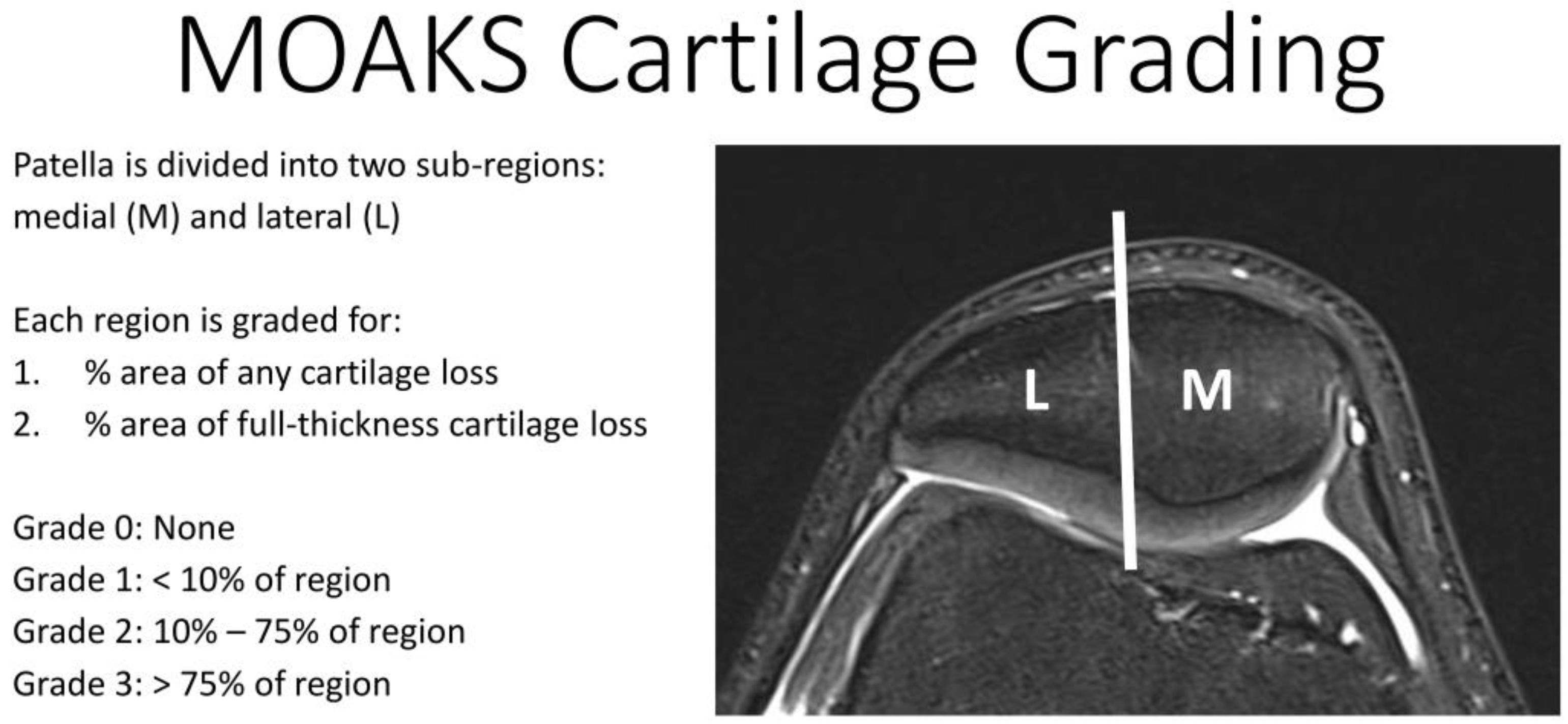
Figure 1. MRI Osteoarthritis Knee Score (MOAKS). This system divides cartilage surfaces within the retropatellar knee into the lateral (L) and the medial (M) subregions. Cartilage lesions in each subregion are then analyzed using scores based on the amount of any cartilage loss as a percentage of the subregion and amount of full-thickness cartilage loss as percentage of subregion.
Diagnostics 11 02301 g001
Figure 2. (a) Data points and biexponential fitting results to determine longitudinal 23Na relaxation times of the patellar cartilage of a representative healthy volunteer measured with protocol 1. The signal was normalized to the mean signal value at TR = 70 ms. The corresponding fitting results were: T 1 , car = 14.1 ms; T 1 , syn = 35.1 ms; p car = 79.6%; R 2 = 0.9957. (b) Data points and biexponential fitting results to determine transverse 23Na relaxation times of the patellar cartilage of a representative healthy volunteer measured with protocol 2. The signal was normalized to the mean signal value at TE = 0.3 ms. The corresponding fitting results were T 2 s * = 0.5 ms; T 2 l * = 12.5 ms; p s = 41.0%; R 2 = 0.9906.
Diagnostics 11 02301 g002
Figure 3. Color-coded 23Na concentration maps of the patellar cartilage (overlaid onto the corresponding 1H images). (a) Healthy volunteer (male, 22 years, right knee) measured with protocol 1. (b) Patient (female, 66 years, left knee) with moderate cartilage defects, some of which are severe on the medial side (MRI Osteoarthritis Knee Score (MOAKS): (2/1) medial, (2/0) lateral) measured with protocol 1. (c) Healthy volunteer (male, 25 years, right knee) measured with protocol 2. (d) Patient (female, 30 years, right knee) with extensive cartilage defects on the medial side and singular full-thickness cartilage defects on both sides (MOAKS: (3/1) medial, (1/1) lateral) measured with protocol 2. The corresponding mean 23Na concentration estimates were (a) 216 ± 42 mmol/L, (b) 158 ± 30 mmol/L, (c) 226 ± 64 mmol/L and (d) 135 ± 29 mmol/L. Partially visualized are agarose phantom tubes that were used for signal normalization. The scale on the right indicates 23Na concentration (mmol/L).
Diagnostics 11 02301 g003
Figure 4. Transversal Proton Density-weighted fat-saturated images of the two patients. (a) Patient (female, 66 years, left knee, protocol 1) with moderate long-distance cartilage loss medial and lateral (10–75% of the subregion) and medial short-distance (<10% of the subregion) full-thickness defect (MRI Osteoarthritis Knee Score (MOAKS): (2/1) medial, (2/0) lateral). (b) Patient (female, 30 years, right knee, protocol 2) with very long-distance cartilage loss medial (>75% of the subregion) with short-stretch full-thickness defect (<10% of the subregion) and short-stretch, full-thickness lateral defect (<10% of the subregion), the latter is not visible in the depicted slice (MOAKS: (3/1) medial, (1/1) lateral).
Diagnostics 11 02301 g004
Table 1. Sequence parameters for 23Na coil characterisation (coil sensitivity and B1 mapping) and for participant measurements (protocol 1, protocol 2 and 1H imaging).
23Na Coil
Sensitivity
B1 MappingProtocol 1
( T 1   Protocol )
Protocol 2
( T 2 *   Protocol )
1H Imaging
Sequence typeDA-3D-RADDA-3D-RADDA-3D-RADDA-3D-RADDA-3D-RAD
Nucleus23Na23Na23Na23Na1H
Orientationtratratratratra
Repetition time (ms)603008/9/10/11/12/13/14/15/16/18/20/23/26/30/40/50/708430
Echo time (ms)0.30.30.3(0.30/6.45/12.60/18.80) (1.50/7.65/13.80/20.00) (3.00/9.15/15.30/21.50)0.8
Inversion time (ms)---24-
Inversion pulse
Duration (ms)
---1-
Field of View (mm)180 × 180 × 180180 × 180 × 180180 × 180 × 180180 × 180 × 180180 × 180 × 180
Projections50,00050,000900090009000
Pixel size (mm/px)3 × 3 × 33 × 3 × 33 × 3 × 33 × 3 × 31 × 1 × 1
Flip angle (°)9040/80909010
Pulse duration (ms)0.50.50.50.50.2
Readout time (ms)55551
Averages122111
Total examination time (h:min:s)10:00:0016:40:0000:57:4500:37:4800:04:30
Abbreviations: tra: transversal, DA-3D-RAD: density-adapted 3D radial sequence.
Table 2. Additional sequences and their acquisition parameters applied for patients.
PD-Weighted fs T 1 - Weighted
Sequence typeTSETSE
Turbo Factor38109
GRAPPA22
Orientationcor/tra/sagsag
Repetition time (ms)4980864
Echo time (ms)4213
Field of View (mm)160 × 160160 × 160
Image matrix (px)512 × 512512 × 512
Pixel size (mm/px)0.3 × 0.30.3 × 0.3
Flip angle (°)180180
Slices3535
Slice thickness (mm)33
Total examination time (min:s)09:5703:10
Abbreviations: cor: coronal, tra: transversal, sag: sagittal, PD: proton density, fs: fat saturated, TSE: turbospin echo, GRAPPA: generalized autocalibrating partial parallel acquisition.
Table 3. Longitudinal 23Na relaxation times of the patellar cartilage of 10 healthy volunteers (4 females, 6 males, mean age 23 ± 3 years) and one patient (female, age 66 years) measured with protocol 1.
Radiologist/ Volunteers Patient
MeasurementMeanStdMinMedianMax
1/114.450.7413.2414.4715.3315.42
T 1 , car (ms)1/214.580.7413.2414.7815.3615.60
2/114.610.6813.2614.8015.2915.60
1/137.912.9235.0737.7944.2439.78
T 1 , syn (ms)1/238.322.8935.0239.2444.5039.65
2/138.882.8635.0739.2443.8139.99
1/177.303.7372.8078.5382.6471.23
p car (%)1/276.543.7372.7175.3482.6071.03
2/175.104.1568.1073.5882.5570.69
1/10.99170.00450.98080.99260.99590.9829
R 2 1/20.99180.00400.98310.99320.99620.9907
2/10.99180.00450.98060.99300.99710.9903
Abbreviations: std: standard deviation, min: minimum, max: maximum, p car : fraction of T 1 , car of the total T 1 relaxation, R 2 : coefficient of determination for T 1 fit.
Table 4. Transverse 23Na relaxation times of the patellar cartilage in 10 healthy volunteers (3 females, 7 males, mean age 23 ± 2 years) and one patient (female, age 30 years) measured with protocol 2.
Radiologist/ Volunteers Patient
MeasurementMeanStdMinMedianMax
1/10.3580.1470.1030.3530.6630.105
T 2 s * (ms)1/20.3650.1550.1040.3700.6770.107
2/10.3650.1740.1050.3570.7510.107
1/112.620.7311.3012.5513.7413.99
T 2 l * (ms)1/212.770.7011.3512.7813.7414.00
2/112.790.7211.4912.7113.9814.00
1/134.394.7825.3534.4841.4425.86
p s (%)1/234.114.9225.3734.2841.7226.00
2/133.815.0925.2834.6641.7925.99
1/10.98560.00880.96430.98790.99310.9623
R21/20.98700.00970.96310.99100.99600.9657
2/10.98610.00930.96400.98880.99490.9650
Abbreviations: std: standard deviation, min: minimum, max: maximum, p s : fraction of T 2 s * of the total T 2 * relaxation, R 2 : coefficient of determination for T 2 * fit.
Table 5. 23Na concentration estimates of the patellar cartilage of 10 healthy volunteers (4 females, 6 males, mean age 23 ± 3 years) and one patient (female, age 66 years) measured with protocol 1 (TE = 0.3 ms and TR = 70 ms) and of nine healthy volunteers (3 females, 7 males, mean age 23 ± 2 years) and one patient (female, age 30 years) measured with protocol 2 (TE = 0.3 ms and TR = 84 ms). The listed p-values indicate the results of Wilcoxon signed-rank tests comparing the mean values of the healthy volunteers of protocol 1 and 2. Note that one healthy volunteer from protocol 2 was excluded from quantification of 23Na concentration because two agarose phantoms had to be excluded, leading to an insufficient number of data points for the fitting of the phantoms.
Radiologist/ Volunteers Patient
MeasurementMeanStdMinMedianMaxp-Value
Protocol 11/1215441662032910.441135
23Na-Conc.1/2204401691872760.859129
(mmol/L)2/1218521512032970.374136
Protocol 21/120048130199267-158
23Na-Conc.1/219445134188261-152
(mmol/L)2/120439134201291-136
Abbreviations: std: standard deviation, min: minimum, max: maximum, conc.: concentration.
Table 6. Summary of results for 23Na relaxation times in patellar cartilage and synovial fluid from different authors.
Magnetic Field Strength of MRI Scanner (T)23Na Relaxation Time Results for Patellar Cartilage and Synovial Fluid
T 1 , c a r   ( ms ) T 1 , s y n   ( ms ) T 2 s *   ( ms ) T 2 l *   ( ms ) p s   ( % )
Madelin et al. [39]7.017.7 ± 2.6 -0.5 ± 0.1 11.4 ± 1.8 39 ± 4
Feldman et al. [20]4.721 ± 148 ± 3 0.8 ± 0.2 19.7 ± 0.565 ± 12
Staroswiecki et al. [21]7.0---13.2 ± 1.5 -
3.0---15.5 ± 1.3 -
Abbreviations: p s –fraction of T 2 s * of the total T 2 * relaxation.
Publisher’s Note: MDPI stays neutral with regard to jurisdictional claims in published maps and institutional affiliations.