
Association between Endometrial Polyps and Chronic Endometritis: Is It Time for a Paradigm Shift in the Pathophysiology of Endometrial Polyps in Pre-Menopausal Women? Results of a Systematic Review and Meta-Analysis

 ,
,  ,
,  ,
, 
Abstract
1. Introduction
2. Materials and Methods
2.1. Study Design
2.2. Search Strategy
2.3. Inclusion Criteria
2.4. Study Selection and Data Extraction
2.5. Study Outcomes
2.6. Data Synthesis and Analysis
3. Results
3.1. Study Selection
3.2. Included Studies
3.2.1. Patients
3.2.2. Diagnosis of CE
3.3. Synthesis of Results
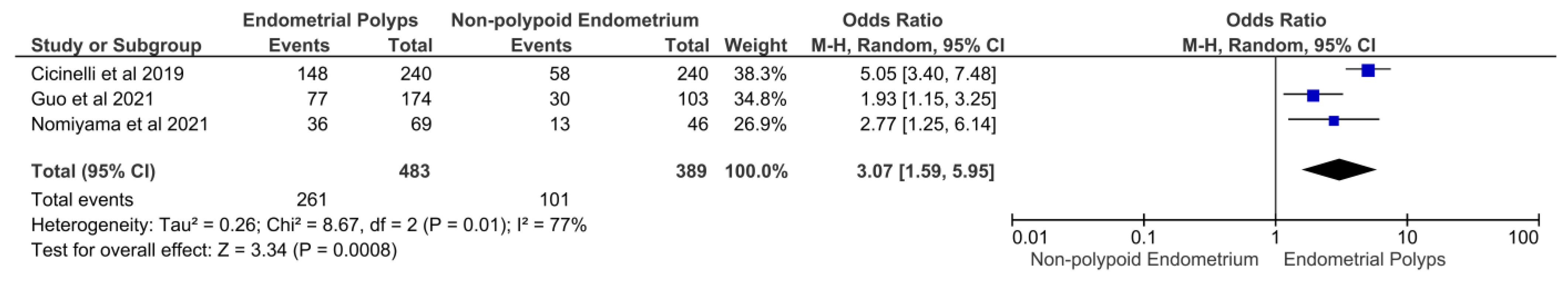
3.3.1. Primary Outcome: Prevalence of CE in Women with Eps
3.3.2. Secondary Outcome: Prevalence of CD-138-Positive EPs among EPs
3.3.3. Tertiary Outcomes
3.3.4. Prevalence of CE in Women with a Single EP Versus Women with Multiple EPs
3.3.5. Investigation of Sources of Heterogeneity across Studies
4. Discussion
4.1. Main Results and Implications
4.2. Strengths and Limitations
5. Conclusions
Author Contributions
Funding
Institutional Review Board Statement
Informed Consent Statement
Conflicts of Interest
Appendix A

| Study ID | Study Design | Country | Patients (Number) | Women Characteristics | CE Definition | Study Outcomes |
|---|---|---|---|---|---|---|
| Mollo et al. 2011 [32] | Prospective controlled | Italy | 42 | -Infertile women Study group: n = 21 women affected by EPs Control group: n = 21 women without EPs | na | To assess levels of interferon-gamma (IFN-g) both in serum and in endometrial biopsy samples from infertile patients with EPs as compared to women without EPs |
| Kitaya et al. 2012 [4] | Cross-sectional | Japan | 75 | -Infertile women with 3 or more IVF failures Macropolypoid endometrium group: n = 23 Micropolypoid endometrium group: n = 25 Nonpolypoid endometrium group: n = 27 | ≥5 stromal CD138 + plasmacytes in 20 HPF | To characterize the local mononuclear cell subsets in infertile patients with endometrial macropolyps versus micropolyps |
| Carvalho et al. 2013 [33] | Prospective cohort | Brazil | 435 | Infertile patients | High vascular density with endothelial proliferation and swelling, hyaline thickening of the vessel wall with luminal occlusion, fibrinoid degeneration of the vessel wall and small vessel thrombosis | To describe morphological vascular changes in endometrial samples from asymptomatic infertile patients and their association with CE and EPs |
| Fang et al. 2016 [9] | Prospective controlled | China | 30 | Infertile women Group A (EPs)+: n = 10 Group B (EPs+CE)+: n = 10 Control group: n = 10 | ≥5 CD138-positive cells in 10 HPF | To characterize the intrauterine microbial communities in patients suffering from endometrial polyps combined with or without chronic endometritis and the intrauterine population difference compared to healthy donors |
| Kosei et al. 2017 [30] | na | Georgia | 130 | Group A (EPs): n = 34 Group B (micropolyps): n = 30 Group C (EPs + micropolyps): n = 36 Control Group: n = 30 healthy women | na | To study the relationship between the morphofunctional characteristics of the endometrium, hormonal homeostasis and microbiocenosis of the reproductive system in patients with endometrial polyps |
| Tolani et al. 2020 [34] | Retrospective cohort study | USA | 201 | Women with infertility or recurrent miscarriage Group A: Normal uterine cavity n = 167 Group B: Abnormal uterine cavity n = 34 | ≥1 plasma cells per 10 HPF | To compare plasma cell infiltrate in patients with normal and abnormal cavity evaluations |
| Study ID | Main Findings |
|---|---|
| Mollo et al. 2011 [32] | Higher concentrations of IFN-ℽ were detected in the serum and the endometrium of infertile patients with EPs. The possible role of an inflammatory factor in a proliferative pathology represents a novel insight into the understanding of EPs and their relationship with infertility. |
| Kitaya et al. 2012 [4] | Compared with the non-polypoid endometrium, macropolypoid endometrium contained a lower density of pan-leukocytes, pan-T cells, and NK cells, whereas micropolypoid endometrium had a higher density of pan-leukocytes and B cells, along with a lower density of NK cells. |
| Carvalho et al. 2013 [33] | Endometrial samples from infertile patients present a broad spectrum of vascular changes, most of them associated with CE. This association is also identified in EPs. It is possible that the vessel axis of functional polyps may originate from the evolution of the vascular changes associated with CE. This would place EPs among the spectrum of inflammatory endometrial diseases. |
| Fang et al. 2016 [9] | Uterine microbiomes between patients with EP and the healthy are significantly different, and all the potentially important variations of uterine microbes may cause EP, but not definitively related to CE. |
| Kosei et al. 2017 [30] | Progesterone deficiency and local immune imbalance with severe hypofunctional NK cells against viral and fungal infestations result in excessive endometrial cell proliferation and development of an isolated polyp. |
| Tolani et al. 2020 [34] | Intracavitary pathology is associated with a significantly higher frequency and density of endometrial plasma cells (71% vs. 41%). |
References
- Resta, L.; Palumbo, M.; Rossi, R.; Piscitelli, D.; Grazia Fiore, M.; Cicinelli, E. Histology of micro polyps in chronic endometritis. Histopathology 2012, 60, 670–674. [Google Scholar] [CrossRef]
- Kitaya, K.; Takeuchi, T.; Mizuta, S.; Matsubayashi, H.; Ishikawa, T. Endometritis: New time, new concepts. Fertil. Steril. 2018, 110, 344–350. [Google Scholar] [CrossRef] [PubMed]
- Li, Y.; Xu, S.; Yu, S.; Huang, C.; Lin, S.; Chen, W.; Mo, M.; Lian, R.; Diao, L.; Ding, L.; et al. Diagnosis of chronic endometritis: How many CD138+ cells/HPF in endometrial stroma affect pregnancy outcome of infertile women? Am. J. Reprod. Immunol. 2021, 85, e13369. [Google Scholar] [CrossRef] [PubMed]
- Kitaya, K.; Tada, Y.; Taguchi, S.; Funabiki, M.; Hayashi, T.; Nakamura, Y. Local mononuclear cell infiltrates in infertile patients with endometrial macropolyps versus micropolyps. Hum. Reprod. 2012, 27, 3474–3480. [Google Scholar] [CrossRef] [PubMed]
- Bouet, P.E.; El Hachem, H.; Monceau, E.; Gariépy, G.; Kadoch, I.J.; Sylvestre, C. Chronic endometritis in women with recurrent pregnancy loss and recurrent implantation failure: Prevalence and role of office hysteroscopy and immunohistochemistry in diagnosis. Fertil. Steril. 2016, 105, 106–110. [Google Scholar] [CrossRef]
- Kitaya, K.; Yasuo, T. Immunohistochemistrical and clinicopathological characterization of chronic endometritis. Am. J. Reprod. Immunol. 2011, 66, 410–415. [Google Scholar] [CrossRef]
- Cicinelli, E.; De Ziegler, D.; Nicoletti, R.; Colafiglio, G.; Saliani, N.; Resta, L.; Rizzi, D.; De Vito, D. Chronic endometritis: Correlation among hysteroscopic, histologic, and bacteriologic findings in a prospective trial with 2190 consecutive office hysteroscopies. Fertil. Steril. 2008, 89, 677–684. [Google Scholar] [CrossRef]
- Molina, N.M.; Sola-Leyva, A.; Jose Saez-Lara, M.; Plaza-Diaz, J.; Tubic-Pavlovic, A.; Romero, B.; Clavero, A.; Mozas-Moreno, J.; Fontes, J.; Altmäe, S. New Opportunities for Endometrial Health by Modifying Uterine Microbial Composition: Present or Future? Biomolecules 2020, 10, 593. [Google Scholar] [CrossRef]
- Fang, R.L.; Chen, L.X.; Shu, W.S.; Yao, S.Z.; Wang, S.W.; Chen, Y.Q. Barcoded sequencing reveals diverse intrauterine microbiomes in patients suffering with endometrial polyps. Am. J. Transl. Res. 2016, 8, 1581–1592. [Google Scholar]
- Kitaya, K.; Nagai, Y.; Arai, W.; Sakuraba, Y.; Ishikawa, T. Characterization of Microbiota in Endometrial Fluid and Vaginal Secretions in Infertile Women with Repeated Implantation Failure. Mediators Inflamm. 2019, 2019, 4893437. [Google Scholar] [CrossRef] [PubMed]
- Kitaya, K.; Matsubayashi, H.; Takaya, Y.; Nishiyama, R.; Yamaguchi, K.; Takeuchi, T.; Ishikawa, T. Live birth rate following oral antibiotic treatment for chronic endometritis in infertile women with repeated implantation failure. Am. J. Reprod. Immunol. 2017, 78, e12719. [Google Scholar] [CrossRef] [PubMed]
- Baker, J.M.; Chase, D.M.; Herbst-Kralovetz, M.M. Uterine Microbiota: Residents, Tourists, or Invaders? Front. Immunol. 2018, 9, 208. [Google Scholar] [CrossRef] [PubMed]
- Zargar, M.; Ghafourian, M.; Nikbakht, R.; Mir Hosseini, V.; Moradi Choghakabodi, P. Evaluating Chronic Endometritis in Women with Recurrent Implantation Failure and Recurrent Pregnancy Loss by Hysteroscopy and Immunohistochemistry. J. Minim. Invasive Gynecol. 2020, 27, 116–121. [Google Scholar] [CrossRef] [PubMed]
- Cicinelli, E.; Matteo, M.; Tinelli, R.; Lepera, A.; Alfonso, R.; Indraccolo, U.; Marrocchella, S.; Greco, P.; Resta, L. Prevalence of chronic endometritis in repeated unexplained implantation failure and the IVF success rate after antibiotic therapy. Hum. Reprod. 2015, 30, 323–330. [Google Scholar] [CrossRef]
- Zolghadri, J.; Momtahan, M.; Aminian, K.; Ghaffarpasand, F.; Tavana, Z. The value of hysteroscopy in diagnosis of chronic endometritis in patients with unexplained recurrent spontaneous abortion. Eur. J. Obstet. Gynecol. Reprod. Biol. 2011, 155, 217–220. [Google Scholar] [CrossRef] [PubMed]
- Vitagliano, A.; Saccardi, C.; Noventa, M.; Di Spiezio Sardo, A.; Saccone, G.; Cicinelli, E.; Pizzi, S.; Andrisani, A.; Litta, P.S. Effects of chronic endometritis therapy on in vitro fertilization outcome in women with repeated implantation failure: A systematic review and meta-analysis. Fertil. Steril. 2018, 110, 103–112. [Google Scholar] [CrossRef]
- Buzzaccarini, G.; Vitagliano, A.; Andrisani, A.; Santarsiero, C.M.; Cicinelli, R.; Nardelli, C.; Ambrosini, G.; Cicinelli, E. Chronic endometritis and altered embryo implantation: A unified pathophysiological theory from a literature systematic review. J. Assist. Reprod. Genet. 2020, 37, 2897–2911. [Google Scholar] [CrossRef]
- Kitaya, K.; Matsubayashi, H.; Yamaguchi, K.; Nishiyama, R.; Takaya, Y.; Ishikawa, T.; Yasuo, T.; Yamada, H. Chronic Endometritis: Potential Cause of Infertility and Obstetric and Neonatal Complications. Am. J. Reprod. Immunol. 2016, 75, 13–22. [Google Scholar] [CrossRef]
- Kitaya, K. Prevalence of chronic endometritis in recurrent miscarriages. Fertil. Steril. 2011, 95, 1156–1158. [Google Scholar] [CrossRef]
- Cicinelli, E.; Matteo, M.; Tinelli, R.; Pinto, V.; Marinaccio, M.; Indraccolo, U.; De Ziegler, D.; Resta, L. Chronic endometritis due to common bacteria is prevalent in women with recurrent miscarriage as confirmed by improved pregnancy outcome after antibiotic treatment. Reprod. Sci. 2014, 21, 640–647. [Google Scholar] [CrossRef]
- Cicinelli, E.; Matteo, M.; Trojano, G.; Mitola, P.C.; Tinelli, R.; Vitagliano, A.; Crupano, F.M.; Lepera, A.; Miragliotta, G.; Resta, L. Chronic endometritis in patients with unexplained infertility: Prevalence and effects of antibiotic treatment on spontaneous conception. Am. J. Reprod. Immunol. 2018, 79, e12782. [Google Scholar] [CrossRef] [PubMed]
- Cicinelli, E.; Resta, L.; Nicoletti, R.; Zappimbulso, V.; Tartagni, M.; Saliani, N. Endometrial micropolyps at fluid hysteroscopy suggest the existence of chronic endometritis. Hum. Reprod. 2005, 20, 1386–1389. [Google Scholar] [CrossRef] [PubMed]
- Cicinelli, E.; Resta, L.; Nicoletti, R.; Tartagni, M.; Marinaccio, M.; Bulletti, C.; Colafiglio, G. Detection of chronic endometritis at fluid hysteroscopy. J. Minim. Invasive Gynecol. 2005, 12, 514–518. [Google Scholar] [CrossRef] [PubMed]
- Cicinelli, E.; Vitagliano, A.; Kumar, A.; Lasmar, R.B.; Bettocchi, S.; Haimovich, S.; Kitaya, K.; de Ziegler, D.; Simon, C.; Moreno, I.; et al. Unified diagnostic criteria for chronic endometritis at fluid hysteroscopy: Proposal and reliability evaluation through an international randomized-controlled observer study. Fertil. Steril. 2019, 112, 162–173. [Google Scholar] [CrossRef]
- Guo, G.L.; Chen, S.Y.; Zhang, W.; Zhang, C.; He, L. Diagnosis value of hysteroscopy for chronic endometritis. Clin. Exp. Obstet. Gynecol. 2013, 40, 250–252. [Google Scholar]
- Song, D.; Li, T.C.; Zhang, Y.; Feng, X.; Xia, E.; Huang, X.; Xiao, Y. Correlation between hysteroscopy findings and chronic endometritis. Fertil. Steril. 2019, 111, 772–779. [Google Scholar] [CrossRef]
- Fagioli, R.; Vitagliano, A.; Carugno, J.; Castellano, G.; De Angelis, M.C.; Di Spiezio Sardo, A. Hysteroscopy in postmenopause: From diagnosis to the management of intrauterine pathologies. Climacteric 2020, 23, 360–368. [Google Scholar] [CrossRef]
- Luerti, M.; Vitagliano, A.; Di Spiezio Sardo, A.; Angioni, S.; Garuti, G.; De Angelis, C. Italian School of Minimally Invasive Gynecological Surgery Hysteroscopists Group. Effectiveness of Hysteroscopic Techniques for Endometrial Polyp Removal: The Italian Multicenter Trial. J. Minim. Invasive Gynecol. 2019, 26, 1169–1176. [Google Scholar] [CrossRef]
- Kuroda, K.; Takamizawa, S.; Motoyama, H.; Tsutsumi, R.; Sugiyama, R.; Nakagawa, K.; Sugiyama, R.; Kuribayashi, Y. Analysis of the therapeutic effects of hysteroscopic polypectomy with and without doxycycline treatment on chronic endometritis with endometrial polyps. Am. J. Reprod. Immunol. 2021, 85, e13392. [Google Scholar] [CrossRef]
- Kosei, N.; Zakharenko, N.; Herman, D. Endometrial Polyps in Women of Reproductive Age: Clinical and Pathogene-Tic Variations. Georgian Med. News 2017, 273, 16–22. [Google Scholar]
- Moher, D.; Liberati, A.; Tetzlaff, J.; Altman, D.G.; PRISMA Group. Preferred reporting items for systematic reviews and meta-analyses: The PRISMA statement. PLoS Med. 2009, 6, e1000097. [Google Scholar] [CrossRef]
- Mollo, A.; Stile, A.; Alviggi, C.; Granata, M.; De Placido, G.; Perrella, A.; d’Antonio, A.; Cicinelli, E. Endometrial polyps in infertile patients: Do high concentrations of interferon-gamma play a role? Fertil. Steril. 2011, 96, 1209–1212. [Google Scholar] [CrossRef] [PubMed]
- Carvalho, F.M.; Aguiar, F.N.; Tomioka, R.; de Oliveira, R.M.; Frantz, N.; Ueno, J. Functional endometrial polyps in infertile asymptomatic patients: A possible evolution of vascular changes secondary to endometritis. Eur. J. Obstet. Gynecol. Reprod. Biol. 2013, 170, 152–156. [Google Scholar] [CrossRef] [PubMed]
- Tolani, A.; Ryan, E.E.; Folkins, A.K.; Lathi, R.B. Does the Timing of Endometrial Biopsy Impact the Detection of Endometrial Plasma Cells? Fertil. Steril. 2020, 114, e192. [Google Scholar] [CrossRef]
- Sklyarova, V.; Kyshakevych, I.; Volosovsky, P.; Sklyarov, P.; Kupchak, I. Epidemiological Features of Chronic Endometritis in Reproductive Age Women with Disorders of Reproductive Health. Georgian Med. News 2020, 304–305, 27–32. [Google Scholar]
- Inaba, K.; Wada-Hiraike, O.; Harada, M.; Hirota, Y.; Koga, K.; Fujii, T.; Osuga, Y. Dienogest suppresses cellular proliferation status of endometrial polyps and acts differently depending on the morphological type. Women’s Health 2020, 16, 1745506520952003. [Google Scholar] [CrossRef]
- Song, D.; Feng, X.; Zhang, Q.; Xia, E.; Xiao, Y.; Xie, W.; Li, T.C. Prevalence and confounders of chronic endometritis in premenopausal women with abnormal bleeding or reproductive failure. Reprod. Biomed. Online 2018, 36, 78–83. [Google Scholar] [CrossRef]
- Nomiyama, M.; Yamasaki, F.; Tokunaga, M.; Ohbuchi, Y.; Sago, N.; Arima, K.; Nishiyama, W.; Hashiguchi, M.; Kojima, K. Endometrial polyps with increased plasma cells are associated with chronic endometritis in infertility patients: Hysteroscopic findings and post-polypectomy pregnancy rates. Reprod. Med. Biol. 2021, 20, 494–504. [Google Scholar] [CrossRef]
- Guo, L.; Gu, F.; Tan, J.; Luo, L.; Gao, J.; Zhou, C. Multiple endometrial polyps is associated with higher risk of chronic endometritis in reproductive-aged women. J. Obstet. Gynaecol. Res. 2021, 47, 389–396. [Google Scholar] [CrossRef]
- Cicinelli, E.; Bettocchi, S.; de Ziegler, D.; Loizzi, V.; Cormio, G.; Marinaccio, M.; Trojano, G.; Crupano, F.M.; Francescato, R.; Vitagliano, A.; et al. Chronic Endometritis, a Common Disease Hidden Behind Endometrial Polyps in Premenopausal Women: First Evidence From a Case-Control Study. J. Minim. Invasive Gynecol. 2019, 26, 1346–1350. [Google Scholar] [CrossRef]
- Volodarsky-Perel, A.; Badeghiesh, A.; Shrem, G.; Steiner, N.; Tulandi, T. Chronic Endometritis in Fertile and Infertile Women Who Underwent Hysteroscopic Polypectomy. J. Minim. Invasive Gynecol. 2020, 27, 1112–1118. [Google Scholar] [CrossRef] [PubMed]
- Indraccolo, U.; Di Iorio, R.; Matteo, M.; Corona, G.; Greco, P.; Indraccolo, S.R. The pathogenesis of endometrial polyps: A systematic semi-quantitative review. Eur. J. Gynaecol. Oncol. 2013, 34, 5–22. [Google Scholar] [PubMed]
- Cicinelli, E.; Cicinelli, R.; Vitagliano, A. Antibiotic therapy for chronic endometritis and its reproductive implications: A step forward, with some uncertainties. Fertil. Steril. 2021, 115, 1445–1446. [Google Scholar] [CrossRef] [PubMed]
- Cicinelli, E.; Cicinelli, R.; Vitagliano, A. Consistent evidence on the detrimental role of severe chronic endometritis on in vitro fertilization outcome and the reproductive improvement after antibiotic therapy: On the other hand, mild chronic endometritis appears a more intricate matter. Fertil. Steril. 2021, 116, 345–346. [Google Scholar] [CrossRef]
- Vitagliano, A.; Saccardi, C.; Litta, P.S.; Noventa, M. Chronic endometritis: Really so relevant in repeated IVF failure? Am. J. Reprod. Immunol. 2017, 78. [Google Scholar] [CrossRef]
- Vitagliano, A.; Noventa, M.; Gizzo, S. Autoimmunity, systemic inflammation, and their correlation with repeated implantation failure and recurrent miscarriage: Is chronic endometritis the missing piece of the jigsaw? Am. J. Reprod. Immunol. 2016, 77, 672–677. [Google Scholar] [CrossRef]
- Gelardi, M.; Netti, G.S.; Giancaspro, R.; Spadaccino, F.; Pennella, A.; Fiore, V.; La Gatta, E.; Grilli, G.M.; Cassano, M.; Ranieri, E. Chronic rhinosinusitis with nasal polyposis (CRSwNP): The correlation between expression of Galectin-10 and Clinical-Cytological Grading (CCG). Am. J. Rhinol. Allergy 2021, 14. [Google Scholar] [CrossRef]
- Ashktorab, H.; Brim, H.; Hassan, S.; Nouraie, M.; Gebreselassie, A.; Laiyemo, A.O.; Kibreab, A.; Aduli, F.; Latella, G.; Brant, S.R.; et al. Inflammatory polyps occur more frequently in inflammatory bowel disease than other colitis patients. BMC Gastroenterol. 2020, 20, 170. [Google Scholar] [CrossRef]
- Humphrey, P.A. Polypoid/Papillary cystitis. J. Urol. 2013, 189, 1091–1092. [Google Scholar] [CrossRef]
- Cicinelli, E.; Vitagliano, A.; Loizzi, V.; De Ziegler, D.; Fanelli, M.; Bettocchi, S.; Nardelli, C.; Trojano, G.; Cicinelli, R.; Minervini, C.F.; et al. Altered Gene Expression Encoding Cytochines, Grow Factors and Cell Cycle Regulators in the Endometrium of Women with Chronic Endometritis. Diagnostics 2021, 11, 471. [Google Scholar] [CrossRef]
- Terzić, J.; Grivennikov, S.; Karin, E.; Karin, M. Inflammation and colon cancer. Gastroenterology 2010, 138, 2101–2114.e5. [Google Scholar] [CrossRef] [PubMed]
- Sun, Y.; Zhang, J.; Bai, W. Higher Prevalence of Endometrial Polyps in Patients with Fallopian Tube Obstruction: A Case-control Study. J. Minim. Invasive Gynecol. 2019, 26, 935–940. [Google Scholar] [CrossRef] [PubMed]
- Holzer, I.; Ott, J.; Kurz, C.; Hofstetter, G.; Hager, M.; Kuessel, L.; Parry, J.P. Is Chronic Endometritis Associated with Tubal Infertility? A Prospective Cohort Study. J. Minim. Invasive Gynecol. 2021, 28, 1876–1881. [Google Scholar] [CrossRef] [PubMed]
- Chen, P.; Chen, P.; Guo, Y.; Fang, C.; Li, T. Interaction Between Chronic Endometritis Caused Endometrial Microbiota Disorder and Endometrial Immune Environment Change in Recurrent Implantation Failure. Front. Immunol. 2021, 12, 748447. [Google Scholar] [CrossRef]
- Chen, W.; Wei, K.; He, X.; Wei, J.; Yang, L.; Li, L.; Chen, T.; Tan, B. Identification of Uterine Microbiota in Infertile Women Receiving in vitro Fertilization With and Without Chronic Endometritis. Front. Cell Dev. Biol. 2021, 9, 693267. [Google Scholar] [CrossRef] [PubMed]
- Moreno, I.; Cicinelli, E.; Garcia-Grau, I.; Gonzalez-Monfort, M.; Bau, D.; Vilella, F.; Ziegler, D.D.; Resta, L.; Valbuena, D.; Simon, C. The diagnosis of chronic endometritis in infertile asymptomatic women: A comparative study of histology, microbial cultures, hysteroscopy, and molecular microbiology. Am. J. Obstet. Gynecol. 2018, 218, 602.e1–602.e16. [Google Scholar] [CrossRef] [PubMed]
- Pirtea, P.; Cicinelli, E.; De Nola, R.; de Ziegler, D.; Ayoubi, J.M. Endometrial causes of recurrent pregnancy losses: Endometriosis, adenomyosis, and chronic endometritis. Fertil. Steril. 2021, 115, 546–560. [Google Scholar] [CrossRef]
- Vitale, S.G.; Capriglione, S.; Peterlunger, I.; La Rosa, V.L.; Vitagliano, A.; Noventa, M.; Valenti, G.; Sapia, F.; Angioli, R.; Lopez, S.; et al. The Role of Oxidative Stress and Membrane Transport Systems during Endometriosis: A Fresh Look at a Busy Corner. Oxid. Med. Cell. Longev. 2018, 2018, 7924021. [Google Scholar] [CrossRef]
- Freitag, N.; Pour, S.J.; Fehm, T.N.; Toth, B.; Markert, U.R.; Weber, M.; Togawa, R.; Kruessel, J.-S.; Baston-Buest, D.M.; Bielfeld, A.P. Are uterine natural killer and plasma cells in infertility patients associated with endometriosis, repeated implantation failure, or recurrent pregnancy loss? Arch. Gynecol. Obstet. 2020, 302, 1487–1494. [Google Scholar] [CrossRef]
- Noventa, M.; Scioscia, M.; Schincariol, M.; Cavallin, F.; Pontrelli, G.; Virgilio, B.; Vitale, S.G.; Laganà, A.S.; Dessole, F.; Cosmi, E.; et al. Imaging Modalities for Diagnosis of Deep Pelvic Endometriosis: Comparison between Trans-Vaginal Sonography, Rectal Endoscopy Sonography and Magnetic Resonance Imaging. A Head-to-Head Meta-Analysis. Diagnostics 2019, 9, 225. [Google Scholar] [CrossRef]
- Takebayashi, A.; Kimura, F.; Kishi, Y.; Ishida, M.; Takahashi, A.; Yamanaka, A.; Takahashi, K.; Suginami, H.; Murakami, T. The association between endometriosis and chronic endometritis. PLoS ONE 2014, 9, e88354. [Google Scholar]
- Cicinelli, E.; Trojano, G.; Mastromauro, M.; Vimercati, A.; Marinaccio, M.; Mitola, P.C.; Resta, L.; de Ziegler, D. Higher prevalence of chronic endometritis in women with endometriosis: A possible etiopathogenetic link. Fertil. Steril. 2017, 108, 289–295.e1. [Google Scholar] [CrossRef] [PubMed]
- Kim, M.R.; Kim, Y.A.; Jo, M.Y.; Hwang, K.J.; Ryu, H.S. High frequency of endometrial polyps in endometriosis. J. Am. Assoc. Gynecol. Laparosc. 2003, 10, 46–48. [Google Scholar] [CrossRef]
- Shen, L.; Wang, Q.; Huang, W.; Wang, Q.; Yuan, Q.; Huang, Y.; Lei, H. High prevalence of endometrial polyps in endometriosis-associated infertility. Fertil. Steril. 2011, 95, 2722–2724.e1. [Google Scholar] [CrossRef] [PubMed]
- García-Peñarrubia, P.; Ruiz-Alcaraz, A.J.; Martínez-Esparza, M.; Marín, P.; Machado-Linde, F. Hypothetical roadmap towards endometriosis: Prenatal endocrine-disrupting chemical pollutant exposure, anogenital distance, gut-genital microbiota and subclinical infections. Hum. Reprod Update 2020, 26, 214–246. [Google Scholar] [CrossRef]
- Khan, K.N.; Kitajima, M.; Yamaguchi, N.; Fujishita, A.; Nakashima, M.; Ishimaru, T.; Masuzaki, H. Role of prostaglandin E2 in bacterial growth in women with endometriosis. Hum. Reprod. 2012, 27, 3417–3424. [Google Scholar] [CrossRef][Green Version]
- Kobayashi, H.; Higashiura, Y.; Shigetomi, H.; Kajihara, H. Pathogenesis of endometriosis: The role of initial infection and subsequent sterile inflammation (review). Mol. Med. Rep. 2014, 9, 9–15. [Google Scholar] [CrossRef]
- Chadchan, S.B.; Cheng, M.; Parnell, L.A.; Yin, Y.; Schriefer, A.; Mysorekar, I.U.; Kommagani, R. Antibiotic therapy with metronidazole reduces endometriosis disease progression in mice: A potential role for gut microbiota. Hum. Reprod. 2019, 34, 1106–1116. [Google Scholar] [CrossRef]
- Vitagliano, A.; Sardo, A.D.S.; Saccone, G.; Valenti, G.; Sapia, F.; Kamath, M.S.; Blaganje, M.; Andrisani, A.; Ambrosini, G. Endometrial scratch injury for women with one or more previous failed embryo transfers: A systematic review and meta-analysis of randomized controlled trials. Fertil. Steril. 2018, 110, 687–702.e2. [Google Scholar] [CrossRef]
- Seval, M.M.; Şükür, Y.E.; Özmen, B.; Kan, Ö.; Sönmezer, M.; Berker, B.; Atabekoğlu, C. Does adding endometrial scratching to diagnostic hysteroscopy improve pregnancy rates in women with recurrent in-vitro fertilization failure? Gynecol. Endocrinol. 2016, 32, 957–960. [Google Scholar] [CrossRef]
- Vitagliano, A.; Andrisani, A.; Alviggi, C.; Vitale, S.G.; Valenti, G.; Sapia, F.; Favilli, A.; Martins, W.P.; Raine-Ferring, N.; Polanski, L.; et al. Endometrial scratching for infertile women undergoing a first embryo transfer: A systematic review and meta-analysis of published and unpublished data from randomized controlled trials. Fertil. Steril. 2019, 111, 734–746.e2. [Google Scholar] [CrossRef] [PubMed]


| Study ID | Study Design | Country | Patients (Number) | Women Characteristics | CE Definition | Study Outcomes |
|---|---|---|---|---|---|---|
| Song et al. 2018 [37] | Retrospective cohort study | China | 1551 | Premenopausal women with abnormal uterine bleeding or reproductive failure. | ≥1 CD-138 positive plasma cell per 10 HPF | To examine the prevalence of chronic endometritis in a consecutive series of endometrial biopsies and to identify confounding variables that may affect the prevalence of chronic endometritis |
| Cicinelli et al. 2019 [40] | Retrospective case-control study | Italy | 480 | Premenopausal women with AUB. Group A: n = 240 women with EPs (diagnosed at hysteroscopy and histology) Group B: included 240 patients without evidence of EPs at hysteroscopy. | >1 CD-138 positive plasma cell per 10 HPF | To investigate the correlation between endometrial polyps (EPs) and chronic endometritis (CE) |
| Volodarsky-Perel et al. 2019 [41] | Retrospective cohort | Canada | 277 | Patients undergoing hysteroscopic polipectomy Group A: Infertile (n = 137) Group B: Fertile (n = 140) | ≥1 plasma cells per 10 HPF | (1) To evaluate the prevalence of CE in infertile women with EPs compared with infertile women with EPs (2) To investigate the prevalence of CE in women with primary infertility compared with those with secondary infertility |
| Inaba et al. 2020 [36] | Retrospective case-control study | Japan | 40 | 4 groups of 10 patients each by the shape of the polyp (sessile type or pedunculated type) and Dienogest treatment prior to the operation | >5 CD138-positive cells per 10 HPF | To investigate the effects of Dienogest on the proliferation and inflammation of endometrial polyps |
| Sklyarova et al. 2020 [35] | na | Ukraine | 133 | Reproductive age women with reproductive health disorders Group I: 30 patients with recurrent pregnancy loss Group II: 47 women with primary infertility Group III: 36 women who had a polyp or endometrial polyps detected during routine ultrasound. Control group: 20 women | na | To analyze the incidence of chronic endometritis in women of reproductive age with reproductive health disorders |
| Guo et al. 2021 [39] | Cross-sectional study | China | 277 | Premenopausal patients who have undergone hysteroscopic inspection with gynecologic conditions for different reasons Group A: single EP: n = 82 Group B: ≥6 EPs: n = 92 Control group: n = 103 | ≥5 CD138-positive cells in 10 HPF | To determine whether single endometrial polyp (EP) or multiple EPs (polyp number ≥ 6) are associated with CE |
| Kuroda et al. 2021 [29] | Cross-sectional study | Japan | 222 | Infertile patients undergoing hysteroscopic polipectomy -Group A: women with CE who received doxicicline after polypectomy: n = 62 -Group B: women with CE who did not receive doxicicline after polypectomy: n = 160. | ≥5 CD138-positive cells in 10 HPF | To compare the therapeutic effects of hysteroscopic polypectomy with and without doxycycline treatment on CE |
| Nomiyama et al. 2021 [38] | Retrospective cohort study | Japan | 245 | Women with a suspicion of EPs undergone diagnostic hysteroscopy Group 1: 38 patients with CD138 + EPs Group 2: 31 patients with CD138 − EPs Group 3: no EPs | ≥10 CD138-positive cells in 20 HPF | To determine the prevalence of CE in groups 1, 2 and 3 |
| Study ID | Main Findings |
|---|---|
| Song et al. 2018 [37] |
|
| Cicinelli et al. 2019 [40] |
|
| Volodarsky-Perel et al. 2019 [41] |
|
| Inaba et al. 2020 [36] |
|
| Sklyarova et al. 2020 [35] |
|
| Guo et al. 2021 [39] | Multiple EPs were positively associated with CE among reproductive-aged women (58.7%) compared to single EP (28%) and controls (29.1%), suggesting a possible hidden etiopathogenetic link between chronic inflammation and multiple EPs. © |
| Kuroda et al. 2021 [29] | CE was present in 92.6% of women with EPs. Most CE patients with endometrial polyps had been cured by polypectomy without doxycycline (88.8% vs. 58.1%). Clinical pregnancy rate within 6 months was higher in women who did not receive antibiotics (63.2% vs. 43.8%). © |
| Nomiyama et al. 2021 [38] | Infertile patients with EPs have higher prevalence of CE compared to those without EPs. Women with CD-138-positive EPs have higher rate of CE compared to those with CD-138-negative EPs and those without EPs (68.4% vs. 32.2% vs. 28.3%). © |
Publisher’s Note: MDPI stays neutral with regard to jurisdictional claims in published maps and institutional affiliations. |
© 2021 by the authors. Licensee MDPI, Basel, Switzerland. This article is an open access article distributed under the terms and conditions of the Creative Commons Attribution (CC BY) license (https://creativecommons.org/licenses/by/4.0/).
Share and Cite
Vitagliano, A.; Cialdella, M.; Cicinelli, R.; Santarsiero, C.M.; Greco, P.; Buzzaccarini, G.; Noventa, M.; Cicinelli, E. Association between Endometrial Polyps and Chronic Endometritis: Is It Time for a Paradigm Shift in the Pathophysiology of Endometrial Polyps in Pre-Menopausal Women? Results of a Systematic Review and Meta-Analysis. Diagnostics 2021, 11, 2182. https://doi.org/10.3390/diagnostics11122182
Vitagliano A, Cialdella M, Cicinelli R, Santarsiero CM, Greco P, Buzzaccarini G, Noventa M, Cicinelli E. Association between Endometrial Polyps and Chronic Endometritis: Is It Time for a Paradigm Shift in the Pathophysiology of Endometrial Polyps in Pre-Menopausal Women? Results of a Systematic Review and Meta-Analysis. Diagnostics. 2021; 11(12):2182. https://doi.org/10.3390/diagnostics11122182
Chicago/Turabian StyleVitagliano, Amerigo, Mariangela Cialdella, Rossana Cicinelli, Carla Mariaflavia Santarsiero, Pantaleo Greco, Giovanni Buzzaccarini, Marco Noventa, and Ettore Cicinelli. 2021. "Association between Endometrial Polyps and Chronic Endometritis: Is It Time for a Paradigm Shift in the Pathophysiology of Endometrial Polyps in Pre-Menopausal Women? Results of a Systematic Review and Meta-Analysis" Diagnostics 11, no. 12: 2182. https://doi.org/10.3390/diagnostics11122182
APA StyleVitagliano, A., Cialdella, M., Cicinelli, R., Santarsiero, C. M., Greco, P., Buzzaccarini, G., Noventa, M., & Cicinelli, E. (2021). Association between Endometrial Polyps and Chronic Endometritis: Is It Time for a Paradigm Shift in the Pathophysiology of Endometrial Polyps in Pre-Menopausal Women? Results of a Systematic Review and Meta-Analysis. Diagnostics, 11(12), 2182. https://doi.org/10.3390/diagnostics11122182