
Breast Carcinomatous Lymphangitis as an Unusual Presentation of Ovarian Cancer


1
Oncology Institute of Southern Switzerland, Ente Ospedaliero Cantonale, 6501 Bellinzona, Switzerland
2
Imaging Institute of Southern Switzerland, Ente Ospedaliero Cantonale, 6501 Bellinzona, Switzerland
3
Faculty of Biomedical Sciences, Università della Svizzera italiana, 6900 Lugano, Switzerland
4
Faculty of Biology and Medicine, University of Lausanne, 1011 Lausanne, Switzerland
5
Cantonal Institute of Pathology, Ente Ospedaliero Cantonale, 6600 Locarno, Switzerland
*
Author to whom correspondence should be addressed.
Diagnostics 2021, 11(11), 2106; https://doi.org/10.3390/diagnostics11112106
Submission received: 14 September 2021
/
Revised: 3 November 2021
/
Accepted: 11 November 2021
/
Published: 14 November 2021
(This article belongs to the Collection Interesting Images)
Abstract
:We describe the case of a 45-year-old woman with an unusual presentation of metastatic ovarian cancer. The patient presented to the oncological clinic with a three-week history of skin rash on the right breast. She underwent a chest and abdomen CT scan, which showed skin thickening of the right breast, right pleural effusion and bilateral cystic ovarian masses. Biopsy of a left ovarian lesion by diagnostic laparoscopy revealed the presence of ovarian serous carcinoma. Biopsy of the breast skin lesion revealed the presence of carcinomatous lymphangitis and immunohistochemistry documented the ovarian origin.
Figure 1.
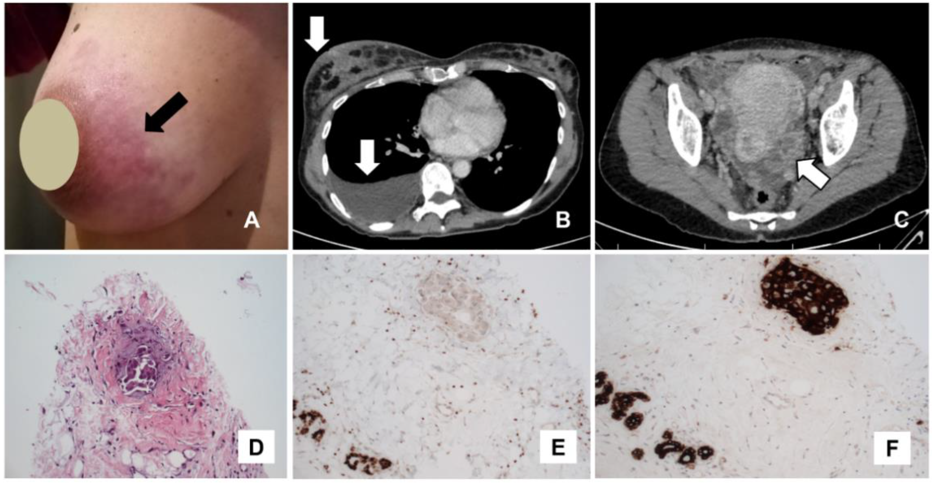
A 45-year-old woman presented to the oncological clinic with a three-week history of skin rash on the right breast ((A); arrow). The patient underwent a chest and abdomen CT scan (B,C), which showed skin thickening of the right breast ((B); upper arrow), right pleural effusion ((B); lower arrow) and bilateral cystic ovarian masses ((C); arrow). Biopsy of a left ovarian lesion by diagnostic laparoscopy revealed the presence of ovarian serous carcinoma. The patient performed other staging exams (breast ultrasound and mammography) which were suspicious for carcinomatous lymphangitis. Biopsy of the breast skin lesion revealed the presence of carcinomatous lymphangitis (D), whereas immunohistochemistry showed negativity for CK20, GATA3 (E) and positivity for CK7 (F), PAX8 and WT1. The immunochemistry pattern demonstrated the ovarian origin of breast lesions. After the diagnosis of metastatic disease, the patient underwent chemotherapy with carboplatin and paclitaxel with partial radiological response after three cycles. Due to inoperable disease, the patient continued chemotherapy with the addition of bevacizumab, obtaining partial treatment response at last follow-up (about one year after the diagnosis of carcinomatous lymphangitis). Carcinomatous lymphangitis may be a metastatic manifestation of different tumors; the most common primary sites are breast, lung and stomach, whereas in rare cases it can be due to ovarian cancer [1,2,3,4,5,6,7,8]. In the described case, an integrated diagnostic approach was very useful to detect breast carcinomatous lymphangitis as an uncommon presentation of metastatic ovarian cancer.
Figure 1.
A 45-year-old woman presented to the oncological clinic with a three-week history of skin rash on the right breast ((A); arrow). The patient underwent a chest and abdomen CT scan (B,C), which showed skin thickening of the right breast ((B); upper arrow), right pleural effusion ((B); lower arrow) and bilateral cystic ovarian masses ((C); arrow). Biopsy of a left ovarian lesion by diagnostic laparoscopy revealed the presence of ovarian serous carcinoma. The patient performed other staging exams (breast ultrasound and mammography) which were suspicious for carcinomatous lymphangitis. Biopsy of the breast skin lesion revealed the presence of carcinomatous lymphangitis (D), whereas immunohistochemistry showed negativity for CK20, GATA3 (E) and positivity for CK7 (F), PAX8 and WT1. The immunochemistry pattern demonstrated the ovarian origin of breast lesions. After the diagnosis of metastatic disease, the patient underwent chemotherapy with carboplatin and paclitaxel with partial radiological response after three cycles. Due to inoperable disease, the patient continued chemotherapy with the addition of bevacizumab, obtaining partial treatment response at last follow-up (about one year after the diagnosis of carcinomatous lymphangitis). Carcinomatous lymphangitis may be a metastatic manifestation of different tumors; the most common primary sites are breast, lung and stomach, whereas in rare cases it can be due to ovarian cancer [1,2,3,4,5,6,7,8]. In the described case, an integrated diagnostic approach was very useful to detect breast carcinomatous lymphangitis as an uncommon presentation of metastatic ovarian cancer.

Author Contributions
Conceptualization, B.M. and M.D.G.; investigation, B.M., P.M. and M.D.G.; data curation, B.M., P.M. and M.D.G.; writing—original draft preparation, B.M. and G.T.; writing—review and editing, M.D.G. and G.T. All authors have read and agreed to the published version of the manuscript.
Funding
This research received no external funding.
Institutional Review Board Statement
Not applicable.
Informed Consent Statement
Written informed consent was obtained from the patient to publish this paper.
Data Availability Statement
Original data supporting the reported results are available contacting the authors.
Conflicts of Interest
The authors declare no conflict of interest.
References
- Cruz, R.P.; Rodini, G.P.; da Rosa, M.D.; Cabral, V.D.; Cambruzzi, E.; Ferrandina, G.; Ribeiro, R. Intestinal lymphangitis carcinomatosa related to ovarian cancer: Case report and review of the literature. Gynecol. Oncol. Rep. 2020, 33, 100606. [Google Scholar] [CrossRef] [PubMed]
- Martynychen, M.G.; Rabelo, L.M.; Silva, R.L.; Escuissato, D.L. Carcinomatous lymphangitis as the initial manifestation of ovarian adenocarcinoma. J. Bras. Pneumol. 2007, 33, 609–611. [Google Scholar] [CrossRef] [PubMed]
- Hominal, S.; Falchero, L.; Perol, M.; Guérin, J.C. Lymphangite carcinomateuse [Carcinomatous lymphangitis]. Presse Med. 1999, 28, 979–984. [Google Scholar] [PubMed]
- Burns, Z.; Omar, L.; Compton, L.; Hayes, J.; Sahoo, S.; Merchant, K. Lymphangitic spread of invasive lobular carcinoma to the contralateral breast. Clin. Imaging 2019, 58, 187–190. [Google Scholar] [CrossRef] [PubMed]
- Burbano, J.; Salazar-González, A.; Echeverri, C.; Rendón, G.; Gaviria, M.; Pareja, R. Cutaneous lymphangitic carcinomatosis: A rare metastasis from cervical cancer. Gynecol. Oncol. Rep. 2018, 26, 1–3. [Google Scholar] [CrossRef] [PubMed]
- Lebeck Lee, C.; Zwerner, J.; O’Brian, B.; Eng, C. Cutaneous Lymphangitic Carcinomatosis as the First Sign of Recurrent Malignancy in a Patient With a History of Rectal Adenocarcinoma. Clin. Colorectal. Cancer 2021. [Google Scholar] [CrossRef] [PubMed]
- Biswas, A.; Sriram, P.S. Getting the whole picture: Lymphangitic carcinomatosis. Am. J. Med. 2015, 128, 837–840. [Google Scholar] [CrossRef] [PubMed] [Green Version]
- Massollo, M.; Fiz, F.; Bottoni, G.; Ugolini, M.; Paparo, F.; Puppo, C.; Provinciali, N.; Iacozzi, M.; Altrinetti, V.; Cistaro, A.; et al. To Enhance or Not to Enhance? The Role of Contrast Medium 18F-FDG PET/CT in Recurrent Ovarian Carcinomas. Medicina 2021, 57, 561. [Google Scholar] [CrossRef] [PubMed]
Publisher’s Note: MDPI stays neutral with regard to jurisdictional claims in published maps and institutional affiliations. |
© 2021 by the authors. Licensee MDPI, Basel, Switzerland. This article is an open access article distributed under the terms and conditions of the Creative Commons Attribution (CC BY) license (https://creativecommons.org/licenses/by/4.0/).
Share and Cite
MDPI and ACS Style
Muoio, B.; Treglia, G.; Migliora, P.; Del Grande, M. Breast Carcinomatous Lymphangitis as an Unusual Presentation of Ovarian Cancer. Diagnostics 2021, 11, 2106. https://doi.org/10.3390/diagnostics11112106
AMA Style
Muoio B, Treglia G, Migliora P, Del Grande M. Breast Carcinomatous Lymphangitis as an Unusual Presentation of Ovarian Cancer. Diagnostics. 2021; 11(11):2106. https://doi.org/10.3390/diagnostics11112106
Chicago/Turabian StyleMuoio, Barbara, Giorgio Treglia, Paola Migliora, and Maria Del Grande. 2021. "Breast Carcinomatous Lymphangitis as an Unusual Presentation of Ovarian Cancer" Diagnostics 11, no. 11: 2106. https://doi.org/10.3390/diagnostics11112106
Note that from the first issue of 2016, this journal uses article numbers instead of page numbers. See further details here.