
Main Diagnostic Pitfalls in Reading the Sacroiliac Joints on MRI

 , , and
Abstract
:1. Introduction
2. Mechanical Changes and Osteoarthritis
3. Osteitis Condensans Ilii and Post-Partum Changes
3.1. Osteitis Condensans Ilii
3.2. Post-Partum
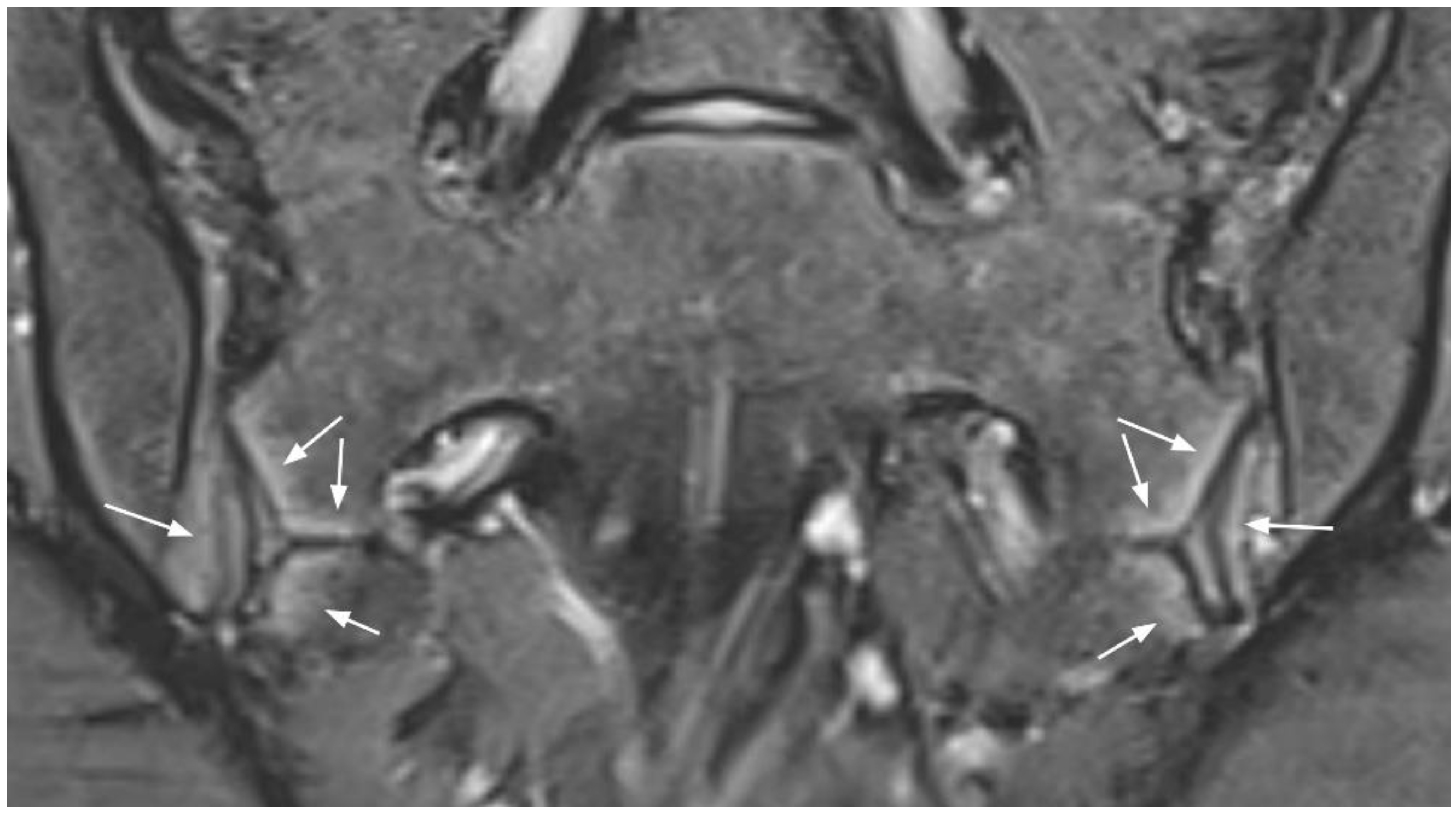
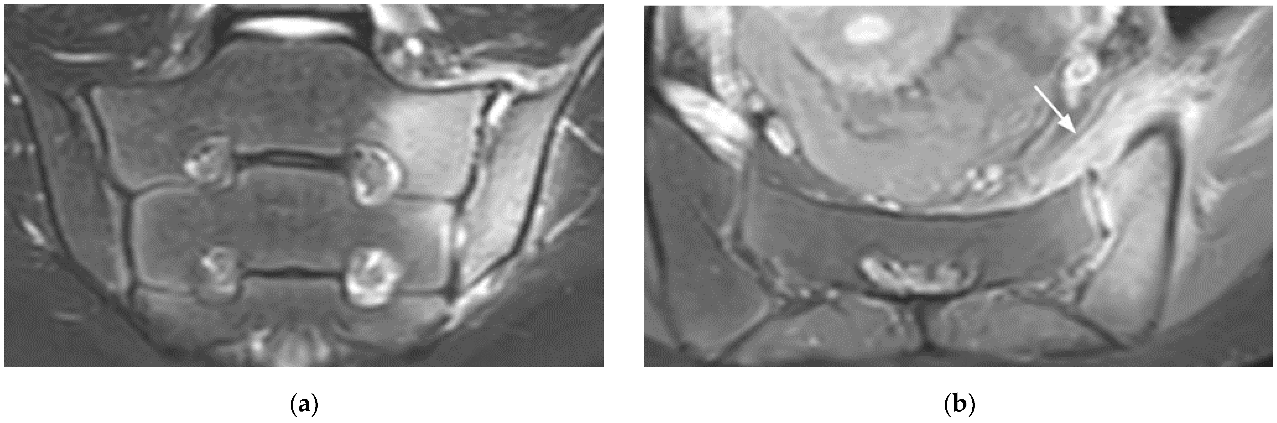
4. Anatomical Variants
4.1. Dysmorphic SIJ Aspect
4.2. Accessory SIJ
4.3. Isolated Synostosis
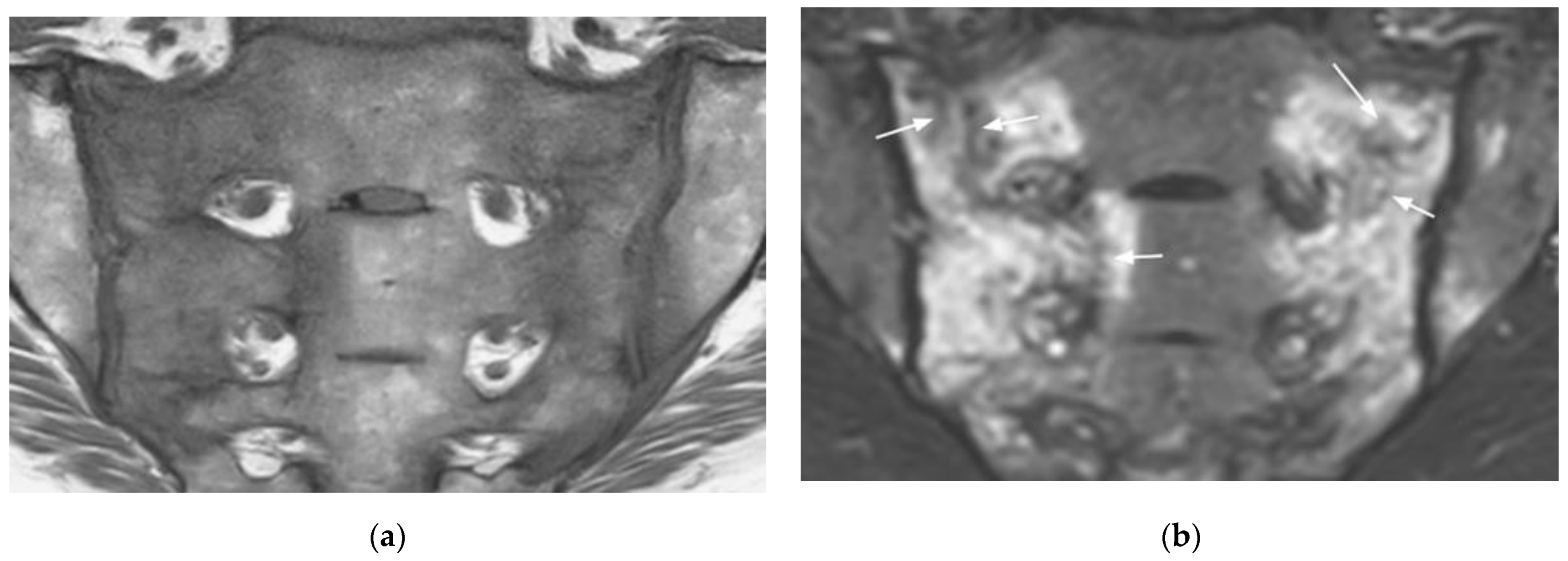
5. Vessels
6. Pediatric SIJ
7. Bone Insufficiency Fractures
8. Infectious Sacroiliitis
9. Hyperparathyroidism
10. Malignancies
11. Conclusions and Perspectives
Funding
Institutional Review Board Statement
Informed Consent Statement
Conflicts of Interest
References
- Rudwaleit, M.; Van Der Heijde, D.; Landewé, R.; Listing, J.; Akkoç, N.; Brandt, J.; Braun, J.; Chou, C.T.; Estévez, E.C.; Dougados, M.; et al. The development of Assessment of SpondyloArthritis international Society classification criteria for axial spondyloarthritis (part II): Validation and final selection. Ann. Rheum. Dis. 2009, 68, 777–783. [Google Scholar] [CrossRef] [Green Version]
- Lambert, R.; Bakker, P.A.C.; Van Der Heijde, D.; Weber, U.; Rudwaleit, M.; Hermann, K.-G.; Sieper, J.; Baraliakos, X.; Bennett, A.; Braun, J.; et al. Defining active sacroiliitis on MRI for classification of axial spondyloarthritis: Update by the ASAS MRI working group. Ann. Rheum. Dis. 2016, 75, 1958–1963. [Google Scholar] [CrossRef]
- Maksymowych, W.P.; Lambert, R.G.; Østergaard, M.; Pedersen, S.J.; Machado, P.M.; Weber, U.; Bennett, A.N.; Braun, J.; Burgos-Vargas, R.; De Hooge, M.; et al. MRI lesions in the sacroiliac joints of patients with spondyloarthritis: An update of definitions and validation by the ASAS MRI working group. Ann. Rheum. Dis. 2019, 78, 1550–1558. [Google Scholar] [CrossRef]
- Barnsley, L.; Paiva, J.; Barnsley, L. Frequency of pertinent MRI abnormalities of the sacroiliac joints of patients without spondyloarthropathies: A systematic review of the literature. Skelet. Radiol. 2021, 1–8. [Google Scholar] [CrossRef]
- Jans, L.; Coeman, L.; Van Praet, L.; Carron, P.; Elewaut, D.; Bosch, F.V.D.; Jaremko, J.L.; Huysse, W.; Verstraete, K.L. How sensitive and specific are MRI features of sacroiliitis for diagnosis of spondyloarthritis. J. Belg. Soc. Radiol. 2014, 97, 202–205. [Google Scholar] [CrossRef] [Green Version]
- Campos-Correia, D.; Sudoł-Szopińska, I.; Afonso, P.D. Are We Overcalling Sacroillitis on MRI? Differential diagnosis that every rheumatologist should know—Part II. Acta Reumatol. Port. 2019, 44, 42–56. [Google Scholar]
- Weber, U.; Jurik, A.G.; Zejden, A.; Larsen, E.; Jørgensen, S.H.; Rufibach, K.; Schioldan, C.; Schmidt-Olsen, S. Frequency and Anatomic Distribution of Magnetic Resonance Imaging Features in the Sacroiliac Joints of Young Athletes. Arthritis Rheumatol. 2018, 70, 736–745. [Google Scholar] [CrossRef]
- Weber, U.; Jurik, A.G.; Zejden, A.; Larsen, E.; Jørgensen, S.H.; Rufibach, K.; Schioldan, C.; Schmidt-Olsen, S. MRI of the sacroiliac joints in athletes: Recognition of non-specific bone marrow oedema by semi-axial added to standard semi-coronal scans. Rheumatology 2019, 59, 1381–1390. [Google Scholar] [CrossRef]
- Jans, L.; Van Praet, L.; Elewaut, D.; Bosch, F.V.D.; Carron, P.; Jaremko, J.; Behaeghe, M.; Denis, A.; Huysse, W.; Lambrecht, V.; et al. MRI of the SI joints commonly shows non-inflammatory disease in patients clinically suspected of sacroiliitis. Eur. J. Radiol. 2014, 83, 179–184. [Google Scholar] [CrossRef]
- Eshed, I.; Miloh-Raz, H.; Dulitzki, M.; Lidar, Z.; Aharoni, D.; Liberman, B.; Lidar, M. Peripartum changes of the sacroiliac joints on MRI: Increasing mechanical load correlating with signs of edema and inflammation kindling spondyloarthropathy in the genetically prone. Clin. Rheumatol. 2015, 34, 1419–1426. [Google Scholar] [CrossRef]
- Ma, L.; Gao, Z.; Zhong, Y.; Meng, Q. Osteitis condensans ilii may demonstrate bone marrow edema on sacroiliac joint magnetic resonance imaging. Int. J. Rheum. Dis. 2017, 21, 299–307. [Google Scholar] [CrossRef] [Green Version]
- Poddubnyy, D.; Weineck, H.; Diekhoff, T.; Redeker, I.; Gobejishvili, N.; Llop, M.; Rodriguez, V.R.; Proft, F.; Protopopov, M.; Haibel, H.; et al. Clinical and imaging characteristics of osteitis condensans ilii as compared with axial spondyloarthritis. Rheumatology 2020, 59, 3798–3806. [Google Scholar] [CrossRef]
- Agten, C.A.; Zubler, V.; Zanetti, M.; Binkert, C.A.; Kolokythas, O.; Prentl, E.; Buck, F.M.; Pfirrmann, C.W.A. Postpartum Bone Marrow Edema at the Sacroiliac Joints May Mimic Sacroiliitis of Axial Spondyloarthritis on MRI. Am. J. Roentgenol. 2018, 211, 1306–1312. [Google Scholar] [CrossRef]
- Hermann, K.-G.; Halle, H.; Reißhauer, A.; Schink, T.; Vsianska, L.; Mühler, M.R.; Lembcke, A.; Hamm, B.; Bollow, M. Peripartale Veränderungen des Beckenringes: Wie sinnvoll ist die Magnetresonanztomografie? RöFo—Fortschr. Auf Dem Geb. Der R 2007, 179, 1243–1250. [Google Scholar] [CrossRef]
- Renson, T.; Depicker, A.; De Craemer, A.-S.; Deroo, L.; Varkas, G.; de Hooge, M.; Carron, P.; Jans, L.; Herregods, N.; Dehaene, I.; et al. High prevalence of spondyloarthritis-like MRI lesions in postpartum women: A prospective analysis in relation to ma-ternal, child and birth characteristics. Ann. Rheum. Dis. 2020, 7, 929–934. [Google Scholar] [CrossRef]
- Hoballah, A.; Lukas, C.; Leplat, C.; Taourel, P.; Pialat, J.-B.; Sans, N.; Ramos-Pascual, S.; Cyteval, C. MRI of sacroiliac joints for the diagnosis of axial SpA: Prevalence of inflammatory and structural lesions in nulliparous, early postpartum and late postpartum women. Ann. Rheum. Dis. 2020, 79, 1063–1069. [Google Scholar] [CrossRef]
- Seven, S.; Østergaard, M.; Morsel-Carlsen, L.; Sørensen, I.J.; Bonde, B.; Thamsborg, G.; Lykkegaard, J.J.; Hendricks, O.; Jørgensen, N.R.; Pedersen, S.J. Magnetic Resonance Imaging of Lesions in the Sacroiliac Joints for Differentiation of Patients with Axial Spondyloarthritis from Control Subjects with or without Pelvic or Buttock Pain: A Prospective, Cross-Sectional Study of 204 Participants. Arthritis Rheumatol. 2019, 71, 2034–2046. [Google Scholar] [CrossRef]
- Depicker, A.; Renson, T.; Craemer, A.D.; Hooge, M.D.; Deroo, L.; Varkas, G.; Herregods, N.; Jans, L.B.; Roelens, K.; Dehaene, I.; et al. SAT0526 the course of aberrant findings on MRI of the sacroiliac joints over 6 months in postpartum women. Ann. Rheum Dis. 2019, 78, 1354–1355. [Google Scholar] [CrossRef]
- Germann, C.; Kroismayr, D.; Brunner, F.; Pfirrmann, C.W.A.; Sutter, R.; Zubler, V. Influence of pregnancy/childbirth on long-term bone marrow edema and subchondral sclerosis of sacroiliac joints. Skelet. Radiol. 2021, 50, 1617–1628. [Google Scholar] [CrossRef]
- El Rafei, M.; Badr, S.; Lefebvre, G.; Machuron, F.; Capon, B.; Flipo, R.-M.; Cotten, A. Sacroiliac joints: Anatomical variations on MR images. Eur. Radiol. 2018, 28, 5328–5337. [Google Scholar] [CrossRef]
- Kiil, R.M.; Jurik, A.G.; Zejden, A. Anatomical variation at the sacroiliac joints in young adults: Estimated prevalence by CT and concomitant diagnostics by MRI. Skelet. Radiol. 2021, 1–11. [Google Scholar] [CrossRef]
- Ziegeler, K.; Hermann, K.G.A.; Diekhoff, T. Anatomical Joint Form Variation in Sacroiliac Joint Disease: Current Concepts and New Perspectives. Curr. Rheumatol. Rep. 2021, 23, 1–6. [Google Scholar] [CrossRef] [PubMed]
- Benz, R.M.; Daikeler, T.; Mameghani, A.T.; Tamborrini, G.; Studler, U. Synostosis of the Sacroiliac Joint as a Developmental Variant, or Ankylosis Due to Sacroiliitis? Arthritis Rheumatol. 2014, 66, 2367. [Google Scholar] [CrossRef] [Green Version]
- Weiss, P.F.; Brandon, T.G.; Bohnsack, J.; Heshin-Bekenstein, M.; Francavilla, M.L.; Jaremko, J.L.; Liao, L.; McHugh, A.; Oberle, E.J.; Rumsey, D.; et al. Variability in Interpretation of Magnetic Resonance Imaging of the Pediatric Sacroiliac Joint. Arthritis Rheum. 2020, 73, 841–848. [Google Scholar] [CrossRef]
- Chauvin, N.A.; Xiao, R.; Brandon, T.G.; Biko, D.M.; Francavilla, M.; Khrichenko, D.; Weiss, P.F. MRI of the Sacroiliac Joint in Healthy Children. Am. J. Roentgenol. 2019, 212, 1–7. [Google Scholar] [CrossRef]
- Herregods, N.; Jans, L.B.O.; Chen, M.; Paschke, J.; De Buyser, S.L.; Renson, T.; Dehoorne, J.; Joos, R.; Lambert, R.G.W.; Jaremko, J.L. Normal subchondral high T2 signal on MRI mimicking sacroiliitis in children: Frequency, age distribution, and relationship to skeletal maturity. Eur. Radiol. 2020, 31, 3498–3507. [Google Scholar] [CrossRef]
- Herregods, N.; Maksymowych, W.; Jans, L.; Otobo, T.; Sudoł-Szopińska, I.; Meyers, A.; Van Rossum, M.; Kirkhus, E.; Panwar, J.; Appenzeller, S.; et al. Atlas of MRI findings of sacroiliitis in pediatric sacroiliac joints to accompany the updated preliminary OMERACT pediatric JAMRIS (Juvenile Idiopathic Arthritis MRI Score) scoring system: Part I: Active lesions. Semin. Arthritis Rheum. 2021, 51, 1089–1098. [Google Scholar] [CrossRef]
- Herregods, N.; Lambert, R.G.; Schiettecatte, E.; Dehoorne, J.; Renson, T.; Laloo, F.; Berghe, T.V.D.; Jans, L.B.; Jaremko, J.L. Blurring and irregularity of the subchondral cortex in pediatric sacroiliac joints on T1 images: Incidence of normal findings that can mimic erosions. Arthritis Rheum. 2021. [Google Scholar] [CrossRef] [PubMed]
- Lyders, E.; Whitlow, C.; Baker, M.; Morris, P. Imaging and Treatment of Sacral Insufficiency Fractures. Am. J. Neuroradiol. 2009, 31, 201–210. [Google Scholar] [CrossRef] [Green Version]
- Hermet, M.; Minichiello, E.; Flipo, R.M.; Dubost, J.J.; Allanore, Y.; Ziza, J.M.; Gaudin, P.; Thomas, T.; Dernis, E.; Glace, B.; et al. Infectious sacroiliitis: A retrospective, multicentre study of 39 adults. BMC Infect. Dis. 2012, 12, 305. [Google Scholar] [CrossRef] [Green Version]
- Kang, Y.; Hong, S.H.; Kim, J.Y.; Yoo, H.J.; Choi, J.-Y.; Yi, M.; Kang, H.S. Unilateral Sacroiliitis: Differential Diagnosis Between Infectious Sacroiliitis and Spondyloarthritis Based on MRI Findings. Am. J. Roentgenol. 2015, 205, 1048–1055. [Google Scholar] [CrossRef] [PubMed]
- Montandon, C.; Costa, M.A.B.; Carvalho, T.N.; Montadon, J.M.E.T.; Santos, K.I.S. Sacroiliitis: Imaging evaluation. Radiolog. Brasil. 2007, 40, 53–60. [Google Scholar] [CrossRef] [Green Version]
- Stürzenbecher, A.; Braun, J.; Paris, S.; Biedermann, T.; Hamm, B.; Bollow, M. MR imaging of septic sacroiliitis. Skelet. Radiol. 2000, 29, 439–446. [Google Scholar] [CrossRef]
- Wang, D.; Yin, H.; Liu, W.; Li, Z.; Ren, J.; Wang, K.; Han, D. Comparative analysis of the diagnostic values of T2 mapping and diffusion-weighted imaging for sacroiliitis in ankylosing spondylitis. Skelet. Radiol. 2020, 49, 1597–1606. [Google Scholar] [CrossRef] [PubMed]
- Bray, T.J.P.; Sakai, N.; Dudek, A.; Fisher, C.; Rajesparan, K.; Lopes, A.; Ciurtin, C.; Sen, D.; Bainbridge, A.; Hall-Craggs, M.A. Histographic analysis of oedema and fat in inflamed bone marrow based on quantitative MRI. Eur. Radiol. 2020, 30, 5099–5109. [Google Scholar] [CrossRef] [PubMed] [Green Version]
- Lefebvre, G.; Bergère, A.; El Rafei, M.; Duhamel, A.; Teixeira, P.; Cotten, A. T2 Mapping of the Sacroiliac Joints With 3-T MRI: A Preliminary Study. Am. J. Roentgenol. 2017, 209, 389–394. [Google Scholar] [CrossRef] [PubMed]
- Jans, L.B.O.; Chen, M.; Elewaut, D.; Bosch, F.V.D.; Carron, P.; Jacques, P.; Wittoek, R.; Jaremko, J.L.; Herregods, N. MRI-based Synthetic CT in the Detection of Structural Lesions in Patients with Suspected Sacroiliitis: Comparison with MRI. Radiology 2021, 298, 343–349. [Google Scholar] [CrossRef]
- Kepp, F.H.; Huber, F.A.; Wurnig, M.C.; Mannil, M.; Kaniewska, M.; Guglielmi, R.; Del Grande, F.; Guggenberger, R. Differentiation of inflammatory from degenerative changes in the sacroiliac joints by machine learning supported texture analysis. Eur. J. Radiol. 2021, 140, 109755. [Google Scholar] [CrossRef] [PubMed]


Publisher’s Note: MDPI stays neutral with regard to jurisdictional claims in published maps and institutional affiliations. |
© 2021 by the authors. Licensee MDPI, Basel, Switzerland. This article is an open access article distributed under the terms and conditions of the Creative Commons Attribution (CC BY) license (https://creativecommons.org/licenses/by/4.0/).
Share and Cite
Badr, S.; Jacques, T.; Lefebvre, G.; Boulil, Y.; Abou Diwan, R.; Cotten, A. Main Diagnostic Pitfalls in Reading the Sacroiliac Joints on MRI. Diagnostics 2021, 11, 2001. https://doi.org/10.3390/diagnostics11112001
Badr S, Jacques T, Lefebvre G, Boulil Y, Abou Diwan R, Cotten A. Main Diagnostic Pitfalls in Reading the Sacroiliac Joints on MRI. Diagnostics. 2021; 11(11):2001. https://doi.org/10.3390/diagnostics11112001
Chicago/Turabian StyleBadr, Sammy, Thibaut Jacques, Guillaume Lefebvre, Youssef Boulil, Ralph Abou Diwan, and Anne Cotten. 2021. "Main Diagnostic Pitfalls in Reading the Sacroiliac Joints on MRI" Diagnostics 11, no. 11: 2001. https://doi.org/10.3390/diagnostics11112001
APA StyleBadr, S., Jacques, T., Lefebvre, G., Boulil, Y., Abou Diwan, R., & Cotten, A. (2021). Main Diagnostic Pitfalls in Reading the Sacroiliac Joints on MRI. Diagnostics, 11(11), 2001. https://doi.org/10.3390/diagnostics11112001