Article

Retrospective Definition of Clostridioides difficile PCR Ribotypes on the Basis of Whole Genome Polymorphisms: A Proof of Principle Study

1
BioMérieux, Open Innovation and Partnerships, 3 Route du Port Michaud, 38390 La Balme Les Grottes, France
2
BioMérieux, Applied Maths NV, 9830 Sint-Martens-Latem, Belgium
3
BioMérieux, Industry, 69290 Craponne, France
4
BioMérieux, 69280 Marcy l’Etoile, France
5
Department of Medical Microbiology and Immunology, Creighton University School of Medicine, 2500 California Plaza, Omaha, NE 68178, USA
*
Author to whom correspondence should be addressed.
Diagnostics 2020, 10(12), 1078; https://doi.org/10.3390/diagnostics10121078
Submission received: 17 November 2020 / Revised: 8 December 2020 / Accepted: 10 December 2020 / Published: 12 December 2020
(This article belongs to the Section Diagnostic Microbiology and Infectious Disease)

Abstract

:
Clostridioides difficile is a cause of health care-associated infections. The epidemiological study of C. difficile infection (CDI) traditionally involves PCR ribotyping. However, ribotyping will be increasingly replaced by whole genome sequencing (WGS). This implies that WGS types need correlation with classical ribotypes (RTs) in order to perform retrospective clinical studies. Here, we selected genomes of hyper-virulent C. difficile strains of RT001, RT017, RT027, RT078, and RT106 to try and identify new discriminatory markers using in silico ribotyping PCR and De Bruijn graph-based Genome Wide Association Studies (DBGWAS). First, in silico ribotyping PCR was performed using reference primer sequences and 30 C. difficile genomes of the five different RTs identified above. Second, discriminatory genomic markers were sought with DBGWAS using a set of 160 independent C. difficile genomes (14 ribotypes). RT-specific genetic polymorphisms were annotated and validated for their specificity and sensitivity against a larger dataset of 2425 C. difficile genomes covering 132 different RTs. In silico PCR ribotyping was unsuccessful due to non-specific or missing theoretical RT PCR fragments. More successfully, DBGWAS discovered a total of 47 new markers (13 in RT017, 12 in RT078, 9 in RT106, 7 in RT027, and 6 in RT001) with minimum q-values of 0 to 7.40 × 10⁻⁵, indicating excellent marker selectivity. The specificity and sensitivity of individual markers ranged between 0.92 and 1.0 but increased to 1 by combining two markers, hence providing undisputed RT identification based on a single genome sequence. Markers were scattered throughout the C. difficile genome in intra- and intergenic regions. We propose here a set of new genomic polymorphisms that efficiently identify five hyper-virulent RTs utilizing WGS data only. Further studies need to show whether this initial proof-of-principle observation can be extended to all 600 existing RTs.

1. Introduction

Clostridioides difficile (C. difficile), formerly known as Clostridium difficile, is an anaerobic, spore-forming Gram-positive bacterial species that can survive in harsh environments. It can withstand high temperatures, exposure to ultraviolet light, toxic chemicals, and exposure to antibiotics. Colonization by C. difficile is asymptomatic. The development of disease is mostly driven by host factors and disruption of the gut microbiome by frequent consumption of antibiotics [1,2,3]. Toxigenic strains of C. difficile can be a lethal cause of C. difficile infection (CDI), which is commonly associated with post antibiotic diarrhea [4,5]. C. difficile is present in the environment and can be transmitted to patients or healthcare workers through contact with contaminated surfaces. Inter-human spread mainly occurs through the fecal–oral route. C. difficile spores are intrinsically resistant to antibiotics and remain viable during antibiotic treatment. Clindamycin, cephalosporins, and fluoroquinolones are considered as major antibiotics associated with CDI [6]. Food or water contamination, gastric acid-suppression, and asymptomatic carriage in the community are the potential risk factors of community acquired CDI [7]. One-third of the total CDI burden occurring in the USA in 2011 was community-associated [8]. CDI caused half a million hospital-acquired infections and 29,000 deaths in 2012 in the United States [8] and approximately 40,000 cases of CDI in Europe [9]. Only a limited number of studies reported emerging CDI in Asia [10]. The increasing incidence of CDI and rapid evolution of antibiotic resistance in C. difficile has become a global threat to public health [11,12,13].
CDI diagnosis allows early pathogen isolation and treatment of infection, thereby reducing the potential of CDI transmission. Various diagnostic procedures for CDI are available, including toxigenic culture, cell cytotoxic neutralization assay, glutamate dehydrogenase assay, the detection of toxins by enzyme immunoassays, nucleic acid amplification-based molecular tests, etc. [14,15,16,17,18,19]. Still, epidemiological C. difficile strain typing is necessary to identify outbreaks within a hospital or the wider community and facilitates understanding of the dissemination of infections. Ribotyping is a classical technique for C. difficile typing initially based on hybridization patterns of conserved ribosomal RNA probe sequences [20,21]. Ribotype (RT) analysis has also been extremely important in the long-term surveillance of CDI [22]. While traditional typing methods such as restriction endonuclease analysis (REA) and pulsed-field gel electrophoresis (PFGE) were widely used in the past, PCR-based ribotyping is the current method of choice for C. difficile typing [23,24]. PCR ribotyping is dependent on the amplification of the intergenic spacer region (ISR) between 16S and 23S rRNA genes [25,26,27,28]. Since most bacterial species encode multiple ribosomal alleles in their genomes, multiple fragments of different lengths are amplified when different species but also different strains are considered [25,26]. There are still considerable constraints on PCR ribotyping including elevated costs, a higher probability of false-positive results, and a lack of interlaboratory portability [10,29,30].
Bacterial whole genome sequencing (WGS) has the potential to provide more detailed epidemiological information than classical PCR ribotyping [23,31]. To further explore a WGS-based approach to C. difficile typing, backward compatibility with PCR ribotyping is essential [32]. Previous studies reported the association of RT001, RT017, and RT027 with lethal CDI and considered those isolates as hyper-virulent [33,34]. A survey conducted in the North East of England concluded that RT001, RT027, and RT106 were among the most prevalent and dangerous clones [35]. In the United States and Europe, RT001, RT014, RT020, RT027, and RT078 have been identified as predominant [36,37]. RT017 is a globally emerging toxigenic RT and can be found on almost every continent [38,39]. Thus, here, we tested both in silico PCR ribotyping and the De Bruijn graph-based Genome Wide Association Study (DBGWAS) [40] for their capacity to perform retrospective PCR ribotyping for C. difficile RT001, RT017, RT027, RT078, and RT106. These strains were chosen as a test set representing global, long-term circulating and clinically relevant epidemic strains. The primary study goal was to develop a proof-of-principle procedure for sequence-based C. difficile strain typing with retrospective compatibility to established PCR RTs.

2. Materials and Methods

2.1. In Silico PCR-Based Ribotyping

We performed in silico PCR using canonical ribotyping PCR primers. Based on the reference sequences 16S-USA and 23S-USA (Table 1), in silico PCR was performed using the subsequence search tool in BioNumerics v7.6 software (Applied Maths NV, Sint Martens-Latem, Belgium). Besides 7 genomes obtained from Creighton University, 23 C. difficile genomes of five selected RTs were downloaded from NCBI to verify in silico amplification of the ISR region (Supplementary Table S1).

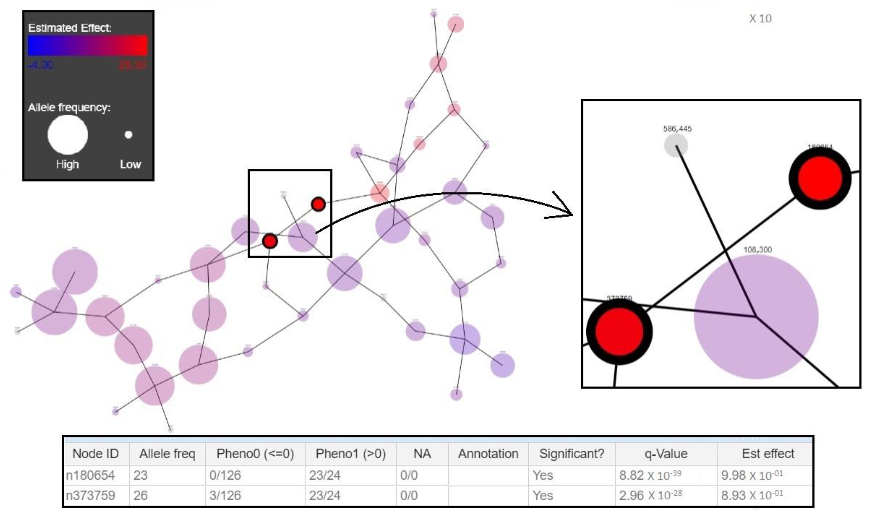
2.2. DBGWAS-Mediated Discovery New RT-Specific Markers

A total number of 160 C. difficile genome assemblies (training set) of 14 different RTs including hyper-virulent RT001, RT017, RT027, RT078, and RT106 were used for the discovery of unique RT genomic markers (Table 2). This small training set allowed for the development of discriminatory markers to characterize the five major RTs among the 14 different RTs. Genomes were collected from the National Center for Biotechnology and Information (NCBI; www.ncbi.nlm.nih.gov), Creighton University, and the Enterobase databases (https://enterobase.warwick.ac.uk). Metadata of these genomes are summarized in Supplementary Table S2.
To identify associations between variant genetic loci and PCR RT, a hypothesis-free DBGWAS method was used. DBGWAS defines genetic variants linked to phenotypic traits via single nucleotide polymorphism (SNP), insertions, deletions, and consequences of recombination [41,42]. We used an open source tool (https://gitlab.com/leoisl/dbgwas) [40]. The tool is able to cover variants in coding as well as non-coding regions of bacterial genomes. DBGWAS was performed keeping the tool parameters in the default setting for different C. difficile ribotypes (RT001, RT017, RT027, RT078, and RT106) and their RT-specific genetic variants observed in the training set. Each C. difficile RT was considered independently in this study. DBGWAS identified short signature sequences called (overlapping) k-mers, yielding a compact summary of all variations across a set of genomes [40]. Overlapping k-mers are called unitigs and were selected on the basis of their specific and unique presence in a particular RT. Q-values define test sensitivity and specificity and are Benjamini–Hochberg-transformed p-values for controlling the false-positive results in case of multiple testing [40,43]. R scripting was used to deal with large matrices defining the presence (1) or absence (0) of extracted unitigs in the training set of C. difficile genomes.

2.3. Validation of Markers

Validation of novel unitig markers was performed by means of BLAST searches against the test set of genomes (Table 3). A wide range of 2425 genomes covering 132 different C. difficile RTs was downloaded [44] and processed using a Linux shell script. These sequences represented PCR ribotyped strains from different countries and clinical and environmental specimens for which phylogenetic analyses were already performed by Frentrup et al. [44] (Supplementary Table S3). A database of this test set was created to perform local command line BLAST searches against the set of significant unitigs identified above. The specificity of all the unitigs was tested using strict parametric filters of 100% coverage and identity.

2.4. Statistically Reliable Ribotype Prediction

To evaluate the potential typing significance of the unitigs as compared to the classical ribotyping of C. difficile strains, sensitivity and specificity (selectivity) were computed for all the unitigs [45]. The efficiency of GWAS can be measured by assessing the false discovery rate (FDR) [46]. To increase the potential typing significance of our new method, combination statistics were performed. Sensitivity and specificity were also computed for certain combinations of two or more selected unitig sequences. The parameters defined were, next to the FDR, TP (true-positives, correctly predicting positive values, e.g., number of true RT017 predicted as RT017), FP (false-positives, missed negative values, e.g., number of non-RT017 genomes still predicted as RT017), FN (false-negatives, missed positive values), and TN (true-negatives, correctly rejected values).

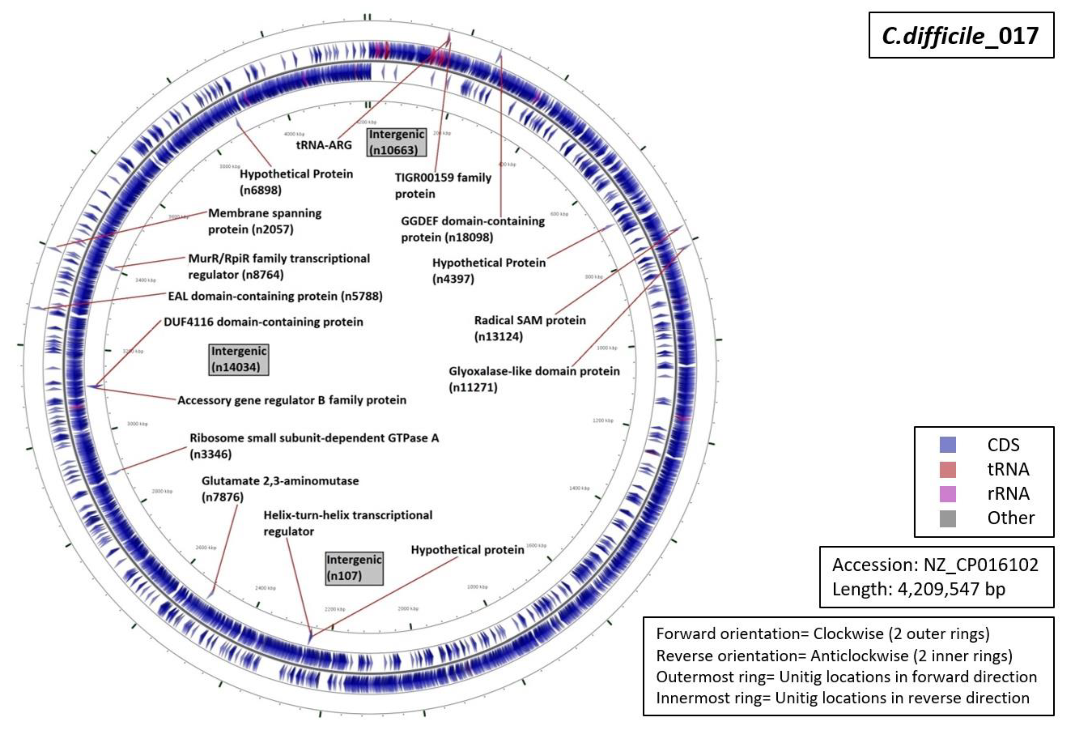
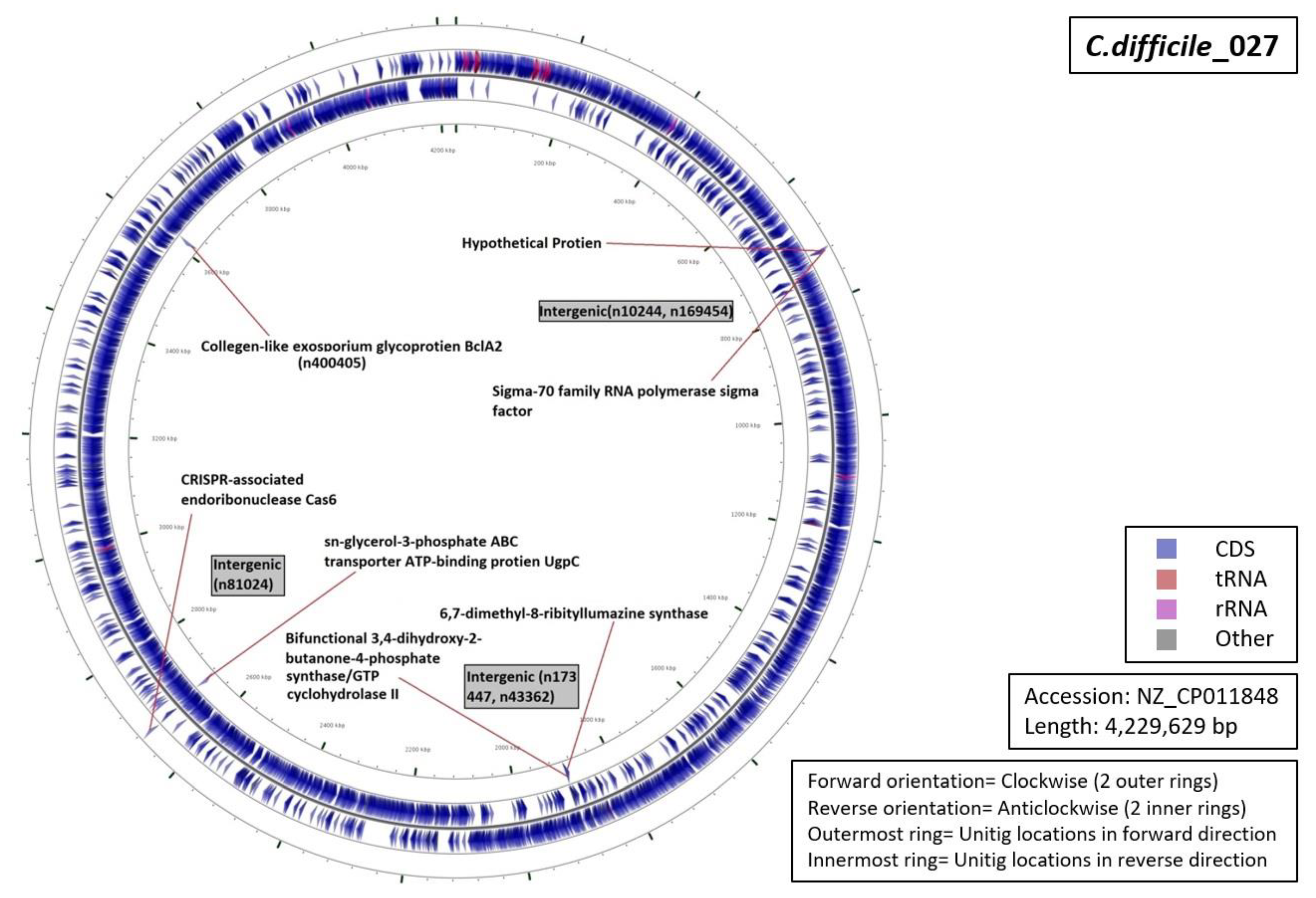
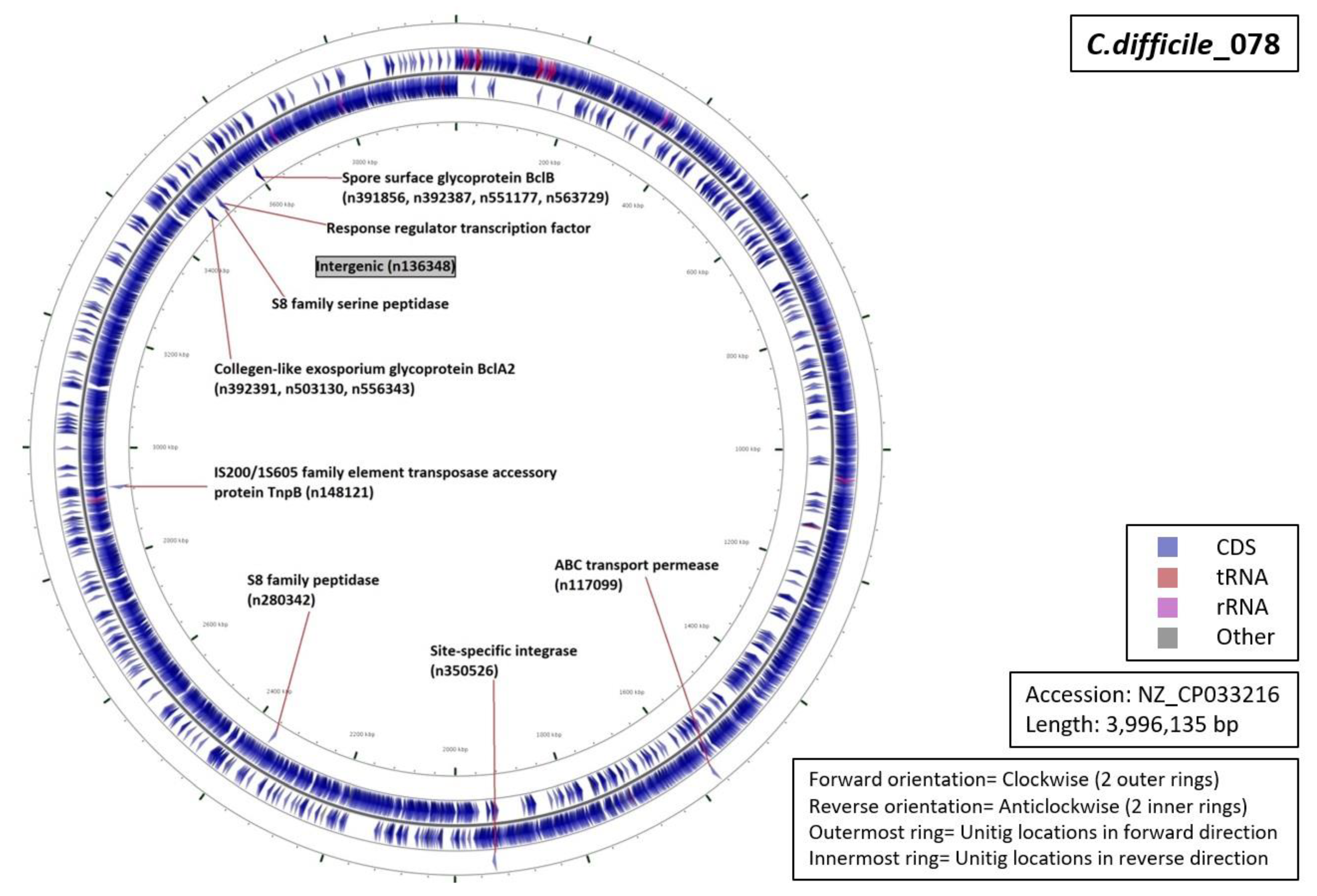
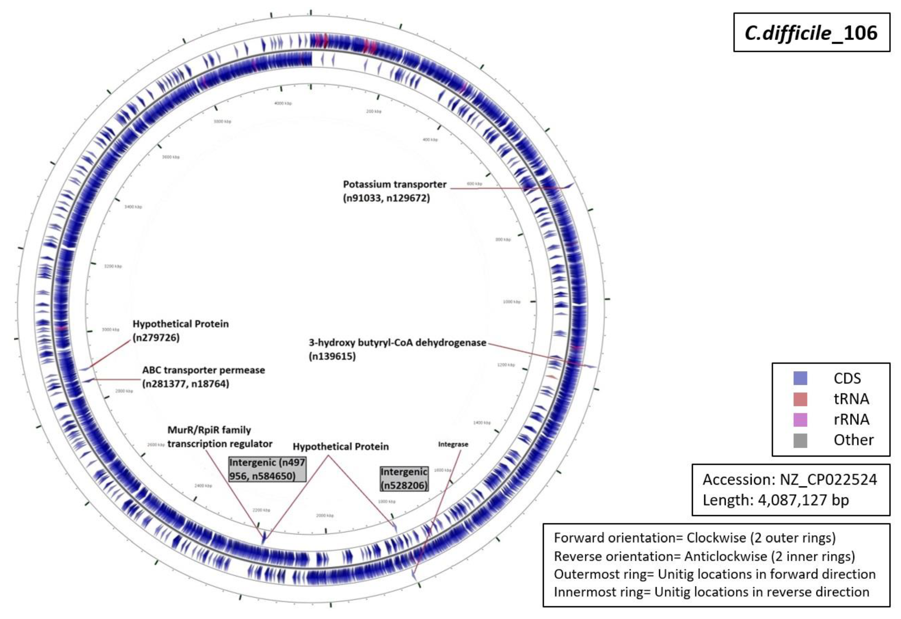
2.5. Functional Annotation of Unitigs

Selected unitigs were annotated using BLASTn alignment. Well-characterized C. difficile genomes were used as a reference to locate these new markers. Specific annotation for each marker was filtered out using minimum E-value, 100% identity, 100% coverage, and 0 gap score.

3. Results and Discussion

3.1. In Silico PCR

The visualization of amplified DNA sequences from the intergenic region between 16S and 23S ribosomal genes is the current Gold Standard for C. difficile typing [25,47]. In our study, in silico PCR for 30 randomly selected, well-characterized C. difficile genome sequences was essentially unsuccessful (Figure 1 and Supplementary Table S1). Genome sequences included generated insufficient numbers of differently sized fragments. The fragment sizes that were calculated were verified with the online tool available at http://insilico.ehu.es/PCR [10]. On the other hand, more recently sequenced C. difficile genomes were showing only one or even none of the expected amplified fragments (Supplementary Table S1). There is a substantial possibility that the PCR ribotyping fragments observed upon laboratory experimentation for these strains may not derive from ISR variants but rather from random amplification. Thus, in silico PCR failed to generate reliable RTs which prompted us to explore the feasibility of DBGWAS-based typing. Of note, we presume here that NGS-based methods are very likely to be more reliable than any of the many other molecular typing methods.

3.2. New Genotyping Markers

A total number of 47 RT-specific unitigs (13 for RT017, 12 for RT078, 9 for RT106, 7 for RT027, and 6 for RT001) were identified. The unitigs shared an average length of 56 base pairs (Table 4). DBGWAS generated compacted De Bruijn graphs (cDBG) containing the specific unitigs as nodes defining a genotypic association between a particular RT and the C. difficile genomes included (Figure 2). Unitigs that were specific for a particular RT were color-coded according to their association to the RT (red for positive association, blue for negative association) and minimum q-values were provided by subgraphs. Q-values for selected unitigs ranged from 0 to 7.40 × 10⁻⁵. Significantly associated unitigs were extracted as FASTA formatted sequences (Supplementary Table S4).

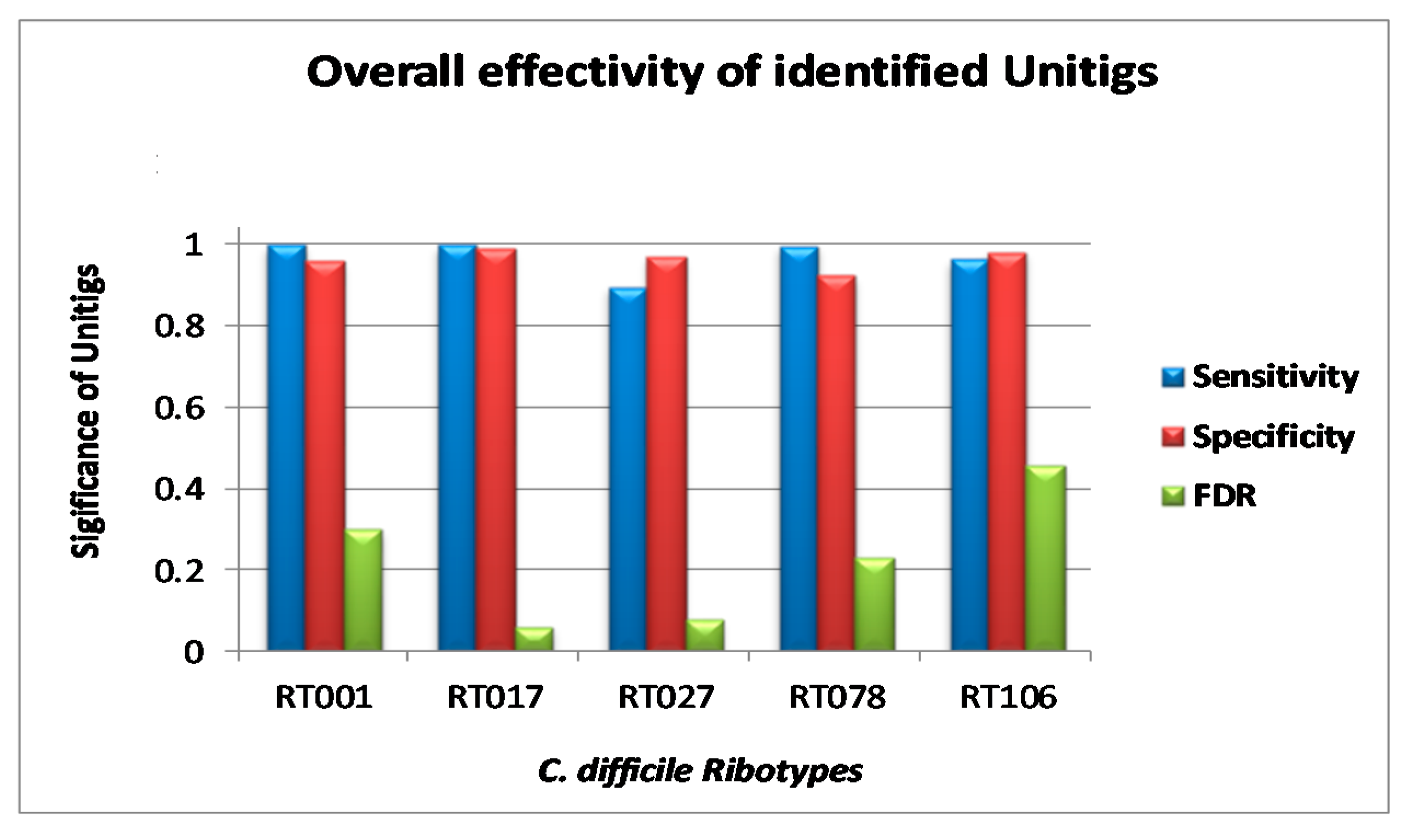
3.3. Validation of Markers

Unitigs showing 100% identity in all genomes belonging to a single RT in the validation set demonstrated the efficiency of these unique patterns to carry out in silico ribotyping. Although the individual unitig-based characterization of C. difficile strains was not absolute, it allowed RT determination with approximate sensitivity and specificity of between 0.90 and 1.0 (Figure 3). FDR for all the unitigs for RT017 was the lowest (0.06) followed by RT027 (0.08), RT078 (0.23), RT001 (0.30), and RT106 (0.46) (Figure 3).
Some of the unitigs were shared by closely related RTs. Unitigs for RT001 were able to identify the genetically closely related RT087, RT241, and RT012, which altogether form a clonal complex (CC) 141 [44]. One of the markers identified for RT017 showed no false-positives or false negatives. Other markers for RT017 initially generated a small number of false-positives, but 100% true-positives in the validation dataset. Markers for RT078 identified 78 out of 79 isolates of RT126 and all of the RT413 strains from the test dataset, likely due to the close genetic relatedness of these RTs (CC 1) [44,48,49]. Unitig sequences for RT106 were also able to identify C. difficile RT500 along with RT106 from the test set. Phylogenetic grouping of C. difficile genomes [44] showed that C. difficile core genome multi locus sequence typing (cgMLST) of RT106 and RT500 (CC 22) generated completely indistinguishable groupings. Considering closely related strains as true-positives based on their respective RTs, the FDRs for each unitig subset were found to be smaller, underscoring the biological consistency of the results. Adding genomes of RT413, RT126, and RT500 to the training set resulted in a decreased FDR rate. The continuously increasing number of publicly available C. difficile genome sequences will provide substantial opportunities for improvement of our new characterization technique.

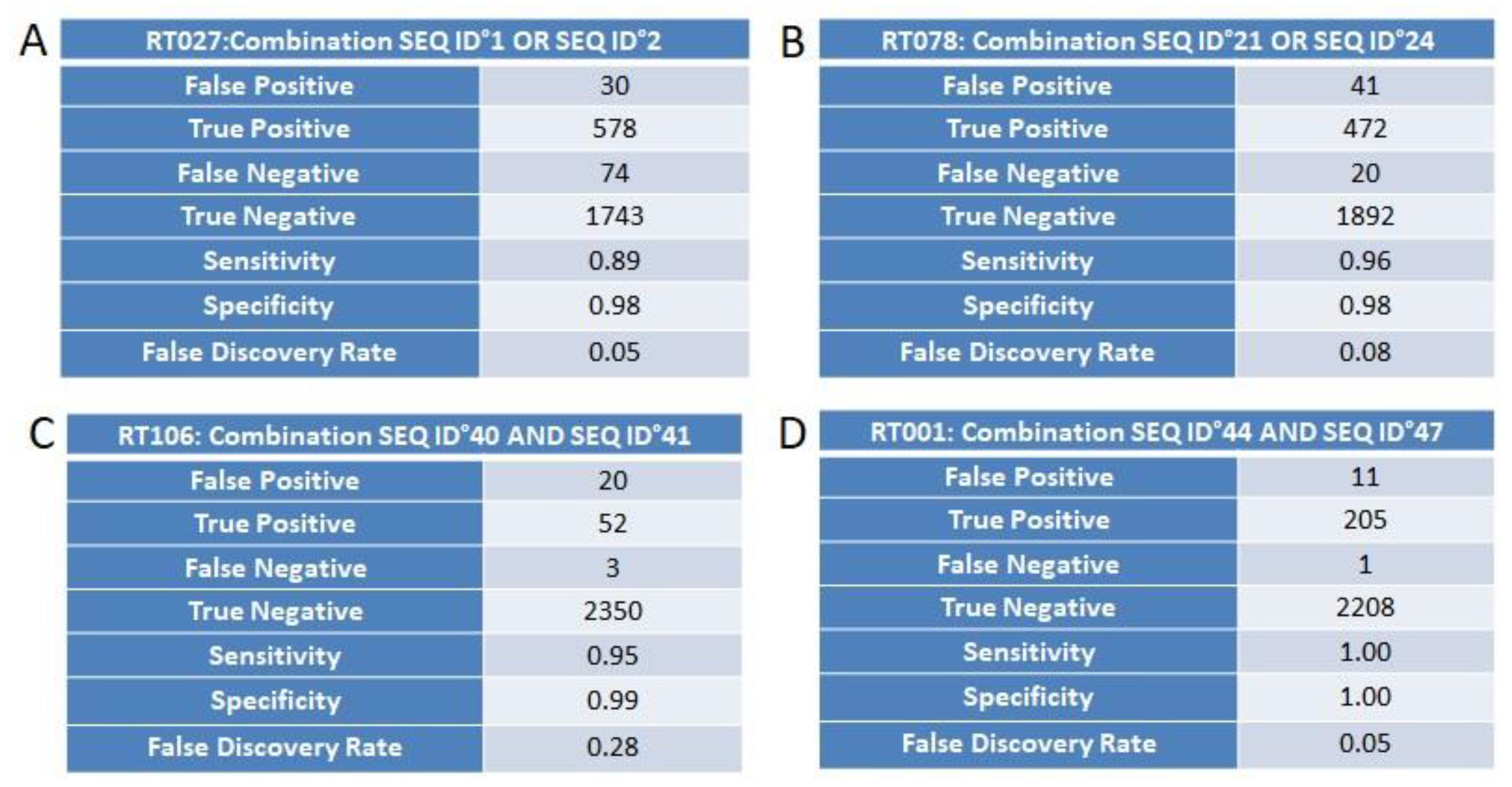
3.4. Marker Combination Study

For the ribotypes RT027, RT078, RT106, and RT001, every possible combination of RT-specific unitigs was created and tested for statistical significance. A combination of two unitigs was shown to increase sensitivity and specificity up to 1 and to reduce the FDR to 0.05 (Figure 4A–D). Each combination was defined on the basis of logical operators “AND/OR”. The AND operator symbolizes that both the markers in a combination need to be present with 100% identity, whereas the OR operator means that any one of the two markers in a combination need to be present at one time, again with 100% sequence identity. There is no combination required in the case of RT017. Conclusively, as clearly exemplified in Figure 4A–D, in certain cases, the combination of markers improves RT testing by suppression of the false discovery rate. Marker’s SEQ ID numbers and their sequences are given in Supplementary Table S4.

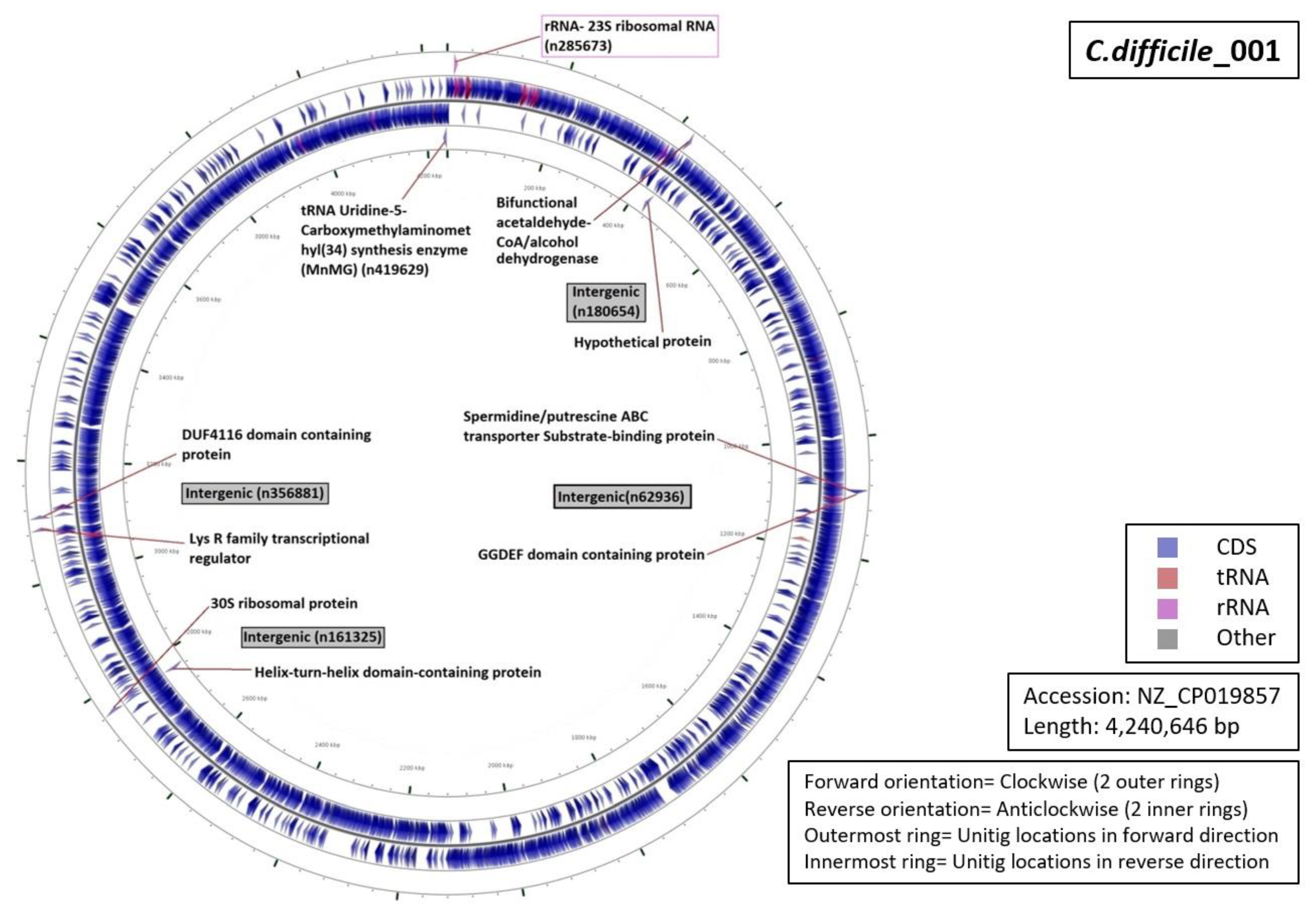
3.5. Functional Annotation of Markers

Functional characterization of the regions from which our unitigs originated demonstrated that 34% of the unitigs were localized in intergenic regions (five for RT027, four for RT001, three for of RT017 and RT106 each, and one for RT078 (Figure 5, Figure 6, Figure 7, Figure 8 and Figure 9). Six percent of all markers were left unannotated in RT001, RT027, and RT078 (one marker for each) (Table 4). Only RT001 was identified with a unitig marker residing within the rRNA-23S ribosomal gene showing at least some correspondence with ribotyping (Figure 5). This marker did not show sufficient diagnostic power and was thus not selected in the final set of markers. All other markers were observed to be scattered throughout the C. difficile genome. In RT078, one of these markers was identified in a mobile genetic element (Figure 8). Mostly, genes and intergenic regions, apart from the conserved ribosomal ISR, were observed to play a potential role in the unitig-mediated C. difficile typing.

4. Conclusions

Strain typing has a proven value in monitoring the persistence and spread of bacterial pathogens in human populations. For C. difficile, PCR ribotyping is the current first choice but may be challenged now that genome sequencing is an option. No single-step test or algorithm is available so far for correlating C. difficile RTs with WGS data. This implies that there may be an issue with the correlation between WGS-based epidemiological analysis and PCR ribotyping for C. difficile. Here, we show that DBGWAS identified unique genomic markers that would suit that specific purpose. A combination of two unitigs led to 100% sensitive and specific discrimination between five important RTs. We believe that this approach is highly promising, providing a clear opportunity to define backward compatibility between classical RTs and WGS data.

Supplementary Materials

The following are available online at https://www.mdpi.com/2075-4418/10/12/1078/s1. Table S1 describes a collection of genome sequences that were used for the in silico search of ribotypes based on amplification of tentative ISRs. Table S2 contains a training set of C. difficile genomes used for the initial DBGWAS. Table S3 contains the C. difficile genomes used for DBGWAS validation. Table S4 shows a review of all unitigs that are statistically significantly associated with specific ribotypes.

Author Contributions

Conceptualization, A.v.B., M.G., L.H., M.J., K.D.B.and R.V.G.; Formal analysis, M.G.; Investigation, M.G., A.v.B., L.H., M.J. and R.V.G.; Methodology, M.G. and K.D.B.; Software, M.J.; Supervision, A.v.B.; Writing—original draft, M.G.; Writing—review & editing, A.v.B., L.H., H.P., M.J., K.D.B.and R.G. All authors have read and agreed to the published version of the manuscript.

Funding

This research was supported and funded by bioMérieux, France; Creighton University School of Medicine, NE, USA; and the European Union’s Horizon 2020 research and innovation program entitled as Viral and Bacterial Adhesin Network Training (ViBrANT) under Marie Skłodowska-Curie Grant Agreement No. 765042.

Acknowledgments

During this study, M.G., L.H., H.P., M.J., K.D.B. and A.v.B. were employees of bioMérieux, a company designing, developing, and marketing tests in the domain of infectious diseases. The company was not involved in the design of the current study and the opinions expressed are those of the authors and may be different from formal company opinions and policies. We thank our colleagues from bioMérieux and Creighton University who provided expertise and insight that greatly assisted the research.

Conflicts of Interest

This research was conducted in association with RG from Creighton University School of Medicine, NE, USA which could be constructed as a potential conflict of interest.

References

  1. Walk, S.T.; Micic, D.; Jain, R.; Lo, E.S.; Trivedi, I.; Liu, E.W.; Almassalha, L.M.; Ewing, S.A.; Ring, C.; Galecki, A.T.; et al. Clostridium difficile Ribotype Does Not Predict Severe Infection. Clin. Infect. Dis. 2012, 55, 1661–1668. [Google Scholar] [CrossRef] [PubMed]
  2. Walker, A.S.; Eyre, D.W.; Wyllie, D.H.; Dingle, K.E.; Griffiths, D.; Shine, B.; Walker, A.S.; O’Connor, L.; Finney, J.; Vaughan, A.; et al. Relationship Between Bacterial Strain Type, Host Biomarkers, and Mortality in Clostridium difficile Infection. Clin. Infect. Dis. 2013, 56, 1589–1600. [Google Scholar] [CrossRef] [PubMed] [Green Version]
  3. McFarland, L.V. Renewed interest in a difficult disease: Clostridium difficile infections—Epidemiology and current treatment strategies. Curr. Opin. Gastroenterol. 2009, 25, 24–35. [Google Scholar] [CrossRef] [PubMed]
  4. Lessa, F.C.; Gould, C.V.; McDonald, L.C. Current Status of Clostridium difficile Infection Epidemiology. Clin. Infect. Dis. 2012, 55, S65–S70. [Google Scholar] [CrossRef] [PubMed] [Green Version]
  5. Wiegand, P.N.; Nathwani, D.; Wilcox, M.H.; Stephens, J.; Shelbaya, A.; Haider, S. Clinical and economic burden of Clostridium difficile infection in Europe: A systematic review of healthcare-facility-acquired infection. J. Hosp. Infect. 2012, 81, 1–14. [Google Scholar] [CrossRef] [PubMed]
  6. Deshpande, A.; Pasupuleti, V.; Thota, P.; Pant, C.; Rolston, D.D.K.; Sferra, T.J.; Hernandez, A.V.; Donskey, C.J. Community-associated Clostridium difficile infection and antibiotics: A meta-analysis. J. Antimicrob. Chemother. 2013, 68, 1951–1961. [Google Scholar] [CrossRef] [PubMed] [Green Version]
  7. Namiki, H.; Kobayashi, T. Long-term, low-dose of clarithromycin as a cause of community-acquired Clostridium difficile infection in a 5-year-old boy. Oxf. Med. Case Rep. 2018, 2018, omx106. [Google Scholar] [CrossRef] [Green Version]
  8. Lessa, F.C.; Mu, Y.; Bamberg, W.M.; Beldavs, Z.G.; Dumyati, G.K.; Dunn, J.R.; Farley, M.M.; Holzbauer, S.M.; Meek, J.I.; Phipps, E.C.; et al. Burden of Clostridium difficile infection in the United States. N. Engl. J. Med. 2015, 372, 825–834. [Google Scholar] [CrossRef] [Green Version]
  9. Davies, K.G.; Longshaw, C.M.; Davis, G.L.; Bouza, E.; Barbut, F.; Barna, Z.; Delmée, M.; Fitzpatrick, F.; Ivanova, K.; Kuijper, E.; et al. Underdiagnosis of Clostridium difficile across Europe: The European, multicentre, prospective, biannual, point-prevalence study of Clostridium difficile infection in hospitalised patients with diarrhoea (EUCLID). Lancet Infect. Dis. 2014, 14, 1208–1219. [Google Scholar] [CrossRef]
  10. Borren, N.Z.; Ghadermarzi, S.; Hutfless, S.; Ananthakrishnan, A.N. The emergence of Clostridium difficile infection in Asia: A systematic review and meta-analysis of incidence and impact. PLoS ONE 2017, 12, e0176797. [Google Scholar] [CrossRef] [Green Version]
  11. Balsells, E.; Shi, T.; Leese, C.; Lyell, I.; Burrows, J.; Wiuff, C.; Campbell, H.; Kyaw, M.H.; Nair, H. Global burden of Clostridium difficile infections: A systematic review and meta-analysis. J. Glob. Health 2018, 9, 010407. [Google Scholar] [CrossRef] [PubMed]
  12. Mills, J.P.; Rao, K.; Young, V.B. Probiotics for prevention of Clostridium difficile infection. Curr. Opin. Gastroenterol. 2018, 34, 3–10. [Google Scholar] [CrossRef] [PubMed]
  13. CDC. Biggest Threats Antibiotic/Antimicrobial Resistance; CDC: Atlanta, GA, USA, 2017. [Google Scholar]
  14. Burnham, C.-A.D.; Carroll, K.C. Diagnosis of Clostridium difficile Infection: An Ongoing Conundrum for Clinicians and for Clinical Laboratories. Clin. Microbiol. Rev. 2013, 26, 604–630. [Google Scholar] [CrossRef] [PubMed] [Green Version]
  15. Planche, T.; Wilcox, M. Reference assays for Clostridium difficile infection: One or two gold standards? J. Clin. Pathol. 2011, 64, 1–5. [Google Scholar] [CrossRef] [Green Version]
  16. She, R.C.; Durrant, R.J.; Petti, C.A. Evaluation of Enzyme Immunoassays to Detect Clostridium difficile Toxin from Anaerobic Stool Culture. Am. J. Clin. Pathol. 2009, 131, 81–84. [Google Scholar] [CrossRef]
  17. Shetty, N.; Wren, M.; Coen, P. The role of glutamate dehydrogenase for the detection of Clostridium difficile in faecal samples: A meta-analysis. J. Hosp. Infect. 2011, 77, 1–6. [Google Scholar] [CrossRef]
  18. Eckert, C.; Jones, G.; Barbut, F. Diagnosis of Clostridium difficile infection: The molecular approach. Future Microbiol. 2013, 8, 1587–1598. [Google Scholar] [CrossRef]
  19. Krutova, M.; Wilcox, M.; Kuijper, E. A two-step approach for the investigation of a Clostridium difficile outbreak by molecular methods. Clin. Microbiol. Infect. 2019, 25, 1300–1301. [Google Scholar] [CrossRef] [Green Version]
  20. Chatterjee, S.; Raval, I.H. Chapter 32—Pathogenic Microbial Genetic Diversity with Reference to Health. In Microbial Diversity in the Genomic Era; Academic Press: Cambridge, MA, USA, 2019; pp. 559–577. [Google Scholar]
  21. Dingle, T.C.; MacCannell, D.R. Chapter 9—Molecular Strain Typing and Characterisation of Toxigenic Clostridium difficile. In Methods in Microbiology; Elsevier: Amsterdam, The Netherlands, 2015; pp. 329–357. [Google Scholar]
  22. Krutova, M.; Kinross, P.; Barbut, F.; Hajdu, A.; Wilcox, M.; Kuijper, E.; Allerberger, F.; Delmée, M.; Van Broeck, J.; Vatcheva-Dobrevska, R.; et al. How to: Surveillance of Clostridium difficile infections. Clin. Microbiol. Infect. 2018, 24, 469–475. [Google Scholar] [CrossRef] [Green Version]
  23. Collins, D.A.; Elliott, B.; Riley, T.V. Molecular methods for detecting and typing of Clostridium difficile. Pathology 2015, 47, 211–218. [Google Scholar] [CrossRef]
  24. Bidet, P.; Barbut, F.; Lalande, V.; Burghoffer, B.; Petit, J.-C. Development of a new PCR-ribotyping method for Clostridium difficile based on ribosomal RNA gene sequencing. FEMS Microbiol. Lett. 1999, 175, 261–266. [Google Scholar] [CrossRef] [PubMed]
  25. Indra, A.; Blaschitz, M.; Kernbichler, S.; Reischl, U.; Wewalka, G.; Allerberger, F. Mechanisms behind variation in the Clostridium difficile 16S–23S rRNA intergenic spacer region. J. Med. Microbiol. 2010, 59, 1317–1323. [Google Scholar] [CrossRef] [PubMed] [Green Version]
  26. Indra, A.; Huhulescu, S.; Schneeweis, M.; Hasenberger, P.; Kernbichler, S.; Fiedler, A.; Wewalka, G.; Allerberger, F.; Kuijper, E.J. Characterization of Clostridium difficile isolates using capillary gel electrophoresis-based PCR ribotyping. J. Med. Microbiol. 2008, 57, 1377–1382. [Google Scholar] [CrossRef] [PubMed] [Green Version]
  27. Waslawski, S.; Lo, E.S.; Ewing, S.A.; Young, V.B.; Aronoff, D.M.; Sharp, S.E.; Novak-Weekley, S.M.; Crist, A.E.; Dunne, W.M.; Hoppe-Bauer, J.; et al. Clostridium difficile Ribotype Diversity at Six Health Care Institutions in the United States. J. Clin. Microbiol. 2013, 51, 1938–1941. [Google Scholar] [CrossRef] [PubMed] [Green Version]
  28. Janezic, S. Direct PCR-Ribotyping of Clostridium difficile. In Clostridium difficile Methods in Molecular Biology; Humana Press: New York, NY, USA, 2016; Volume 1476. [Google Scholar]
  29. Martinez-Melendez, A.; Camacho-Ortiz, A.; Morfin-Otero, R.; Maldonado-Garza, H.J.; Villarreal-Trevino, L.; Garza-Gonzalez, E. Current knowledge on the laboratory diagnosis of Clostridium difficile infection. World J. Gastroenterol. 2017, 23, 1552–1567. [Google Scholar] [CrossRef] [PubMed]
  30. Polage, C.R.; Gyorke, C.E.; Kennedy, M.A.; Leslie, J.L.; Chin, D.L.; Wang, S.; Nguyen, H.H.; Huang, B.; Tang, Y.W.; Lee, L.W.; et al. Overdiagnosis of Clostridium difficile Infection in the Molecular Test Era. JAMA Intern. Med. 2015, 175, 1792–1801. [Google Scholar] [CrossRef] [Green Version]
  31. Janezic, S.; Rupnik, M. Development and Implementation of Whole Genome Sequencing-Based Typing Schemes for Clostridioides difficile. Front. Public Health 2019, 7. [Google Scholar] [CrossRef]
  32. Fawley, W.N.; Knetsch, C.W.; MacCannell, D.R.; Harmanus, C.; Du, T.; Mulvey, M.R.; Paulick, A.; Anderson, L.; Kuijper, E.J.; Wilcox, M.H. Development and Validation of an Internationally-Standardized, High-Resolution Capillary Gel-Based Electrophoresis PCR-Ribotyping Protocol for Clostridium difficile. PLoS ONE 2015, 10, e0118150. [Google Scholar] [CrossRef] [Green Version]
  33. Bauer, M.P.; Notermans, D.W.; Van Benthem, B.H.; Brazier, J.S.; Wilcox, M.H.; Rupnik, M.; Monnet, D.L.; Van Dissel, J.T.; Kuijper, E.J. Clostridium difficile infection in Europe: A hospital-based survey. Lancet Infect. Dis. 2011, 377, 63–73. [Google Scholar] [CrossRef]
  34. Arvand, M.; Hauri, A.M.; Zaiss, N.H.; Witte, W.; Bettge-Weller, G. Clostridium difficile ribotypes 001, 017, and 027 are associated with lethal C. difficile infection in Hesse, Germany. Eurosurveillance 2009, 14, 19403. [Google Scholar] [CrossRef] [Green Version]
  35. Vanek, J.; Hill, K.; Collins, J.; Berrington, A.; Perry, J.; Inns, T.; Gorton, R.; Magee, J.; Sails, A.; Mullan, A.; et al. Epidemiological survey of Clostridium difficile ribotypes in the North East of England during an 18-month period. J. Hosp. Infect. 2012, 81, 209–212. [Google Scholar] [CrossRef] [PubMed]
  36. Giancola, S.E.; Williams, R.J.; Gentry, C.A. Prevalence of the Clostridium difficile BI/NAP1/027 strain across the United States Veterans Health Administration. Clin. Microbiol. Infect. 2018, 24, 877–881. [Google Scholar] [CrossRef] [PubMed] [Green Version]
  37. Howell, M.D.; Novack, V.; Grgurich, P.; Soulliard, D.; Novack, L.; Pencina, M.; Talmor, D. Latrogenic gastric acid suppression and the risk of nosocomial Clostridium difficile infection. Arch. Intern. Med. 2010, 170, 784–790. [Google Scholar] [CrossRef] [PubMed] [Green Version]
  38. Imwattana, K.; Knight, D.R.; Kullin, B.; Collins, D.A.; Putsathit, P.; Kiratisin, P.; Riley, T.V. Clostridium difficile ribotype 017—Characterization, evolution and epidemiology of the dominant strain in Asia. Emerg. Microbes Infect. 2019, 8, 796–807. [Google Scholar] [CrossRef] [PubMed] [Green Version]
  39. Kim, J.; Kim, Y.; Pai, H. Clinical Characteristics and Treatment Outcomes of Clostridium difficile Infections by PCR Ribotype 017 and 018 Strains. PLoS ONE 2016, 11, e0168849. [Google Scholar] [CrossRef] [Green Version]
  40. Jaillard, M.; Lima, L.; Tournoud, M.; Mahé, P.; Van Belkum, A.; Lacroix, V.; Jacob, L. A fast and agnostic method for bacterial genome-wide association studies: Bridging the gap between k-mers and genetic events. PLoS Genet. 2018, 14, e1007758. [Google Scholar] [CrossRef] [Green Version]
  41. Alam, M.T.; Petit, R.A.; Crispell, E.K.; Thornton, T.A.; Conneely, K.N.; Jiang, Y.; Satola, S.W.; Read, T.D. Dissecting vancomycin-intermediate resistance in staphylococcus aureus using genome-wide association. Genome Biol. Evol. 2014, 6, 1174–1185. [Google Scholar] [CrossRef]
  42. Chewapreecha, C.; Marttinen, P.; Croucher, N.J.; Salter, S.J.; Harris, S.R.; Mather, A.E.; Hanage, W.P.; Goldblatt, D.; Nosten, F.H.; Turner, C.; et al. Comprehensive Identification of Single Nucleotide Polymorphisms Associated with Beta-lactam Resistance within Pneumococcal Mosaic Genes. PLoS Genet. 2014, 10, e1004547. [Google Scholar] [CrossRef] [Green Version]
  43. Benjamini, Y.; Hochberg, Y. Controlling the false discovery rate: A practical and powerful approach to multiple testing. J. R. Stat. Soc. Ser. B 1995, 57, 289–300. [Google Scholar] [CrossRef]
  44. Frentrup, M.; Zhou, Z.; Steglich, M.; Meier-Kolthoff, J.P.; Göker, M.; Riedel, T.; Bunk, B.; Spröer, C.; Overmann, J.; Blaschitz, M.; et al. A publicly accessible database for Clostridioides difficile genome sequences supports tracing of transmission chains and epidemics. Microb. Genom. 2020, 6, 410. [Google Scholar] [CrossRef]
  45. Baratloo, A.; Hosseini, M.; Negida, A.; El Ashal, G. Simple Definition and Calculation of Accuracy, Sensitivity and Specificity. Emergency 2015, 3, 48–49. [Google Scholar] [PubMed]
  46. Bradbury, P.; Parker, T.; Hamblin, M.T.; Jannink, J. Assessment of Power and False Discovery Rate in Genome-Wide Association Studies using the BarleyCAP Germplasm. Crop. Sci. 2011, 51, 52–59. [Google Scholar] [CrossRef]
  47. Xiao, M.; Kong, F.; Jin, P.; Wang, Q.; Xiao, K.; Jeoffreys, N.; James, G.; Gilbert, G.L. Comparison of Two Capillary Gel Electrophoresis Systems for Clostridium difficile Ribotyping, Using a Panel of Ribotype 027 Isolates and Whole-Genome Sequences as a Reference Standard. J. Clin. Microbiol. 2012, 50, 2755–2760. [Google Scholar] [CrossRef] [PubMed] [Green Version]
  48. Alvarez-Perez, S.; Blanco, J.L.; Harmanus, C.; Kuijper, E.; Garcia, M.E. Subtyping and antimicrobial susceptibility of Clostridium difficile PCR ribotype 078/126 isolates of human and animal origin. Vet. Microbiol. 2017, 199, 15–22. [Google Scholar] [CrossRef]
  49. Schneeberg, A.; Neubauer, H.; Schmoock, G.; Baier, S.; Harlizius, J.; Nienhoff, H.; Brase, K.; Zimmermann, S.; Seyboldt, C. Clostridium difficile Genotypes in Piglet Populations in Germany. J. Clin. Microbiol. 2013, 51, 3796–3803. [Google Scholar] [CrossRef] [Green Version]
Figure 1. In silico ribotyping of different C. difficile genome sequences using the ISR 16S and 23S USA primer pair. The five panels represent the results obtained for examples of five different ribotypes. Bar graphs show the number of theoretical PCR bands (vertical axis, number of bands labeled on each bar) in the ribosomal region of respective genome sequences (horizontal axis), whereas the genomes without any fragments depict the complete absence of primer binding sites in those genomes. Note that the expected outcome would be an identical number of fragments for each of the strains belonging to a single ribotype. We indicated this number as the first marker of reproducibility; it has to be stated that besides this variation of numbers of fragments, the size of the fragment was also determined as a variable as well.
Diagnostics 10 01078 g001
Figure 2. Compacted De Bruijn graphs (cDBG) generated by De Bruijn graph-based Genome Wide Association Studies (DBGWAS) for RT001 genome sequences. The figure illustrates the significance of the nodes (representing the selective sequences called unitigs), which are denoted by their estimated effect ranging from high (28.304; red) to low (4.00; blue). Allele frequency is represented by the size of the node. The table explains that from the two selected significant nodes in terms of their association with ribotype, the node on the top right (n180654) is specific to RT001 (called Pheno 1 in the table) and completely absent in the other ribotypes in the training set (Pheno 0). Additionally, the q-value linked to the first node is very significantly below 0.05 and hence, the estimated effect is high (represented by the red color of the node).
Diagnostics 10 01078 g002
Figure 3. Statistical comparison of genome typing efficiency of discovered unique patterns for selected C. difficile ribotypes in terms of specificity, sensitivity, and false discovery rate (FDR).
Diagnostics 10 01078 g003
Figure 4. (AD) Statistical reliability in terms of sensitivity, specificity, and false discovery rate (FDR) for the combination of two selected markers using OR operator for the identification of C. difficile RT027 (Panel A) and RT078 (Panel B). Panels C and D display similar analyses but then using the AND operator for identification of RT106 and RT001, respectively.
Diagnostics 10 01078 g004
Figure 5. Functional annotation and location of DBGWAS markers on the reference genome of C. difficile RT001. Both central rings represent the genome annotation (reverse inside, forward outside), while the outer and inner rings represent the signature sequences (unitigs) (reverse inside, forward outside).
Diagnostics 10 01078 g005
Figure 6. Functional annotation and location of DBGWAS markers on the reference genome of C. difficile RT017.
Diagnostics 10 01078 g006
Figure 7. Functional annotation and location of DBGWAS markers on the reference genome of C. difficile RT027.
Diagnostics 10 01078 g007
Figure 8. Functional annotation and location of DBGWAS markers on the reference genome of C. difficile RT078.
Diagnostics 10 01078 g008
Figure 9. Functional annotation and location of DBGWAS markers on the reference genome of C. difficile RT106.
Diagnostics 10 01078 g009
Table 1. Primer pair used for in silico PCR-based ribotyping of Clostridioides difficile.
PrimerGene TargetGenBank Accession No.Sequence (5’–3’)Tm (°C)Reference
16S-USA (Forward)16S rRNA geneFN545816(12293)GTGCGGCTGGATCACCTCCT (12312)71.0Xiao et al., 2012 (46)
23S-USA (Reverse)23S rRNA geneFN545816(12621)CCCTGCACCCTTAATAACTTGACC (12598)67.1
Table 2. C. difficile ribotypes included in the training dataset along with the number of genomes and their source of availability.
C. difficile RibotypeNumber of GenomesSource
RT00124Enterobase, NCBI, Creighton University
RT0022NCBI, Creighton University
RT00319NCBI, Creighton University
RT00519NCBI, Creighton University
RT0103NCBI, Creighton University
RT01411NCBI, Creighton University
RT0152NCBI, Creighton University
RT01715NCBI, Creighton University
RT0233NCBI, Creighton University
RT02715NCBI, Creighton University
RT0464NCBI, Creighton University
RT07815NCBI, Creighton University
RT10622Enterobase, NCBI, Creighton University
RT1266NCBI, Creighton University
TOTAL160
Table 3. C. difficile ribotypes downloaded from the Enterobase database as a test dataset and the number of genomes included in each ribotype.
RibotypeCountRibotypeCountRibotypeCountRibotypeCount
RT001206RT0463RT1271RT3751
RT00253RT0499RT1291RT40415
RT00311RT0504RT1371RT41313
RT00514RT0511RT1381RT4462
RT0061RT0535RT1491RT4492
RT0092RT0542RT1501RT4511
RT0107RT0565RT1531RT4531
RT0113RT0581RT1561RT4541
RT01245RT0601RT1571RT4561
RT0131RT0622RT1581RT4701
RT014113RT0631RT17613RT50021
RT01536RT0663RT1931RT5341
RT017272RT0671RT1941RT5471
RT01855RT0691RT2121RT5591
RT0191RT0704RT2204RT5631
RT02044RT0721RT2251RT5691
RT0221RT0732RT2261RT5811
RT02316RT0751RT2363RT5851
RT0241RT0762RT2381RT5861
RT0266RT0771RT2392RT5911
RT027652RT078492RT2415RT5988
RT0293RT0812RT2449RT6141
RT0312RT0831RT2511RT6202
RT0321RT0842RT2621RT6291
RT0335RT0878RT2841RT6511
RT0352RT0901RT2891RT6661
RT0361RT0941RT2901RT6681
RT0371RT1021RT3051RT6781
RT0395RT1032RT3161RT7081
RT0422RT10655RT3211RT7191
RT0432RT1172RT3282RT7201
RT0442RT1251RT3361RT7211
RT0452RT12679RT3568RT7221
Table 4. Number of unique markers identified for each C. difficile ribotype, their average length, and annotation.
RibotypeNumber of MarkersAverage Length (Base Pairs)Annotation (Number of Unitigs)
RT0010659
  • Intergenic (4)
  • tRNA uridine-5-carboxymethylaminomethyl synthesis enzyme MnmG (1)
  • rRNA-23S ribosomal RNA (1 excluded from the list)
  • Unknown (1)
RT0171369
  • Intergenic (3)
  • Membrane spanning protein (1)
  • Ribosome small subunit-dependent GTPase A (1)
  • Hypothetical protein (1)
  • EAL domain-containing protein (2)
  • Glutamate 2,3-aminomutase (1)
  • MurR/RpiR family transcriptional regulator (1)
  • GGDEF domain-containing protein (1)
  • Glyoxalase-like domain protein (1)
  • Radical SAM protein (1)
RT0270753
  • Collagen-like exosporium glycoprotein BclA2 (1)
  • Intergenic (5)
  • Unknown (1)
RT0781242
  • IS200/IS605 family element transposase accessory protein TnpB (1)
  • Spore surface glycoprotein BclB (4)
  • Collagen-like exosporium glycoprotein (BclA2) (3)
  • Unknown (partial with ABC transporter permease) (1)
  • Intergenic (1)
  • Site-specific integrase (1)
  • S8 family peptidase (1)
RT1060955
  • Intergenic (3)
  • ABC transporter permease (2)
  • Hypothetical Protein (1)
  • 3-Hydroxybutyryl-CoA dehydrogenase (1)
  • Potassium transporter (2)
Publisher’s Note: MDPI stays neutral with regard to jurisdictional claims in published maps and institutional affiliations.

