Article

Role of Endoscopic Ultrasonography-Guided Fine Needle Aspiration/Biopsy in the Diagnosis of Autoimmune Pancreatitis

1
Department of Gastroenterology, Graduate School of Medicine, The University of Tokyo, Tokyo 113-8655, Japan
2
Department of Endoscopy and Endoscopic Surgery, The University of Tokyo Hospital, Tokyo 113-8655, Japan
3
Department of Gastroenterology, Tokyo Takanawa Hospital, Tokyo 108-8606, Japan
4
Department of Gastroenterology, Graduate School of Medicine, Juntendo University, Tokyo 113-8531, Japan
*
Author to whom correspondence should be addressed.
Diagnostics 2020, 10(11), 954; https://doi.org/10.3390/diagnostics10110954
Submission received: 13 October 2020 / Revised: 12 November 2020 / Accepted: 12 November 2020 / Published: 15 November 2020

Abstract

:
Type 1 autoimmune pancreatitis (AIP) is histologically characterized by lymphoplasmacytic sclerosing pancreatitis (LPSP). Recently, the diagnostic yield of endoscopic ultrasonography-guided fine needle aspiration/biopsy (EUS-FNA/B) for AIP has been reported. However, its role in the diagnostic flow of AIP is not fully elucidated. We retrospectively reviewed 53 consecutive patients who were suspected with AIP and underwent EUS-FNA/B. We evaluated the contribution of EUS-FNA/B to the diagnosis of AIP before considering response to steroid therapy among patients with diffuse enlargement of the pancreas and those with focal enlargement, respectively. Twenty-two patients showed diffuse pancreatic enlargement and 31 showed focal enlargement. The final diagnosis was definitive AIP in 32 patients, probable AIP in 2, possible AIP in 1, and mass-forming focal pancreatitis in 18. There was no change in diagnosis after EUS-FNA/B among patients with diffuse pancreatic enlargement, while diagnosis changed in 38.7% (12/31) among those with focal enlargement—there was a probable to definitive diagnosis in 4 patients, unspecified to definitive in 3, and unspecified to probable in 5. EUS-FNB provided a significantly higher sensitivity for typical pathological findings of LPSP than EUS-FNA, and 10 patients were diagnosed as pathologically definitive AIP by EUS-FNB, though none were by EUS-FNA (p = 0.002). EUS-FNA/B was useful in the diagnosis of focal type AIP, and steroid therapy could be introduced after the diagnosis was confirmed. Meanwhile, EUS-FNA/B provided no contribution to diagnosis of diffuse type AIP. EUS-FNB showed a higher diagnostic yield than FNA.

1. Introduction

Autoimmune pancreatitis (AIP) is a particular form of pancreatitis with suspected involvement of autoimmune mechanisms in its pathogenesis [1,2,3,4]. AIP is classified histologically as type 1 and type 2, and type 1 AIP is characterized by lymphoplasmacytic sclerosing pancreatitis (LPSP). It is difficult to obtain sufficient tissue sample to diagnose typical histological features of LPSP, such as striform fibrosis and obliterative phlebitis, thus only a specimen of core biopsy or resection can be used for diagnosis in the international consensus diagnostic criteria (ICDC) [5]. AIP is diagnosed according to ICDC on the basis of the serum IgG4 concentration, histological features of LPSP, pancreatic parenchymal and ductal imaging, other organ involvement, and response to steroid therapy. Although a favorable response of steroid therapy is a characteristic feature of AIP [6,7,8] and a steroid trial is a useful method for the diagnosis of AIP, steroid therapy is potentially associated with adverse effects, especially when a long term use is necessary [9]. The European guideline on IgG4-related digestive disease recommended Endoscopic ultrasonography (EUS)-guided tissue acquisition for the histological diagnosis of AIP and for exclusion of pancreatic carcinoma [10]
Endoscopic ultrasonography-guided fine needle aspiration (EUS-FNA) is an established technique of sample acquisition from the pancreas for cytological evaluation [11,12,13,14]. Novel needles have been developed with side holes or dedicated tip designs to obtain adequate tissue samples for histological evaluation and are called EUS-guided fine needle biopsy (EUS-FNB) needles [15,16,17,18,19,20,21,22,23,24]. Recently, the diagnostic yield of EUS-FNA for AIP has been reported [25,26,27,28,29,30,31,32,33,34,35], and EUS-FNB is expected to provide the better yield [25,36].
However, most of these reports only discussed the sensitivity of EUS-FNA/B for histological diagnosis, and the role of EUS-FNA/B in the diagnostic flow of AIP is not fully elucidated. In the present study, we investigated the contribution of EUS-FNA/B to the diagnosis of AIP.

2. Materials and Methods

We retrospectively reviewed medical records, pathological reports, and radiological images of consecutive patients who were clinically suspected with type 1 AIP and underwent EUS-FNA/B at The University of Tokyo hospital between July 2013 and August 2019. Patients were queried from our prospectively maintained database of EUS-FNA. Clinical diagnosis before EUS-FNA was recorded by endosonographers based on the radiological findings with or without serum IgG4 levels. We evaluated the contribution of EUS-FNA/B to the diagnosis of AIP in the diagnostic flow. We used the diagnostic criteria proposed by Japan Pancreas Society in 2018 (JPS 2018) [37], and the final diagnosis was confirmed by follow-up of 6 months or longer.
All EUS-FNA/B were performed under conscious sedation, and the needle size or type and the amount of suction were selected at the discretion of the endoscopist. We classified the needles with side holes or dedicated tip design for tissue acquisition as FNB needles. EUS-FNA was performed using 25-, 22-, and 19-gauge normal FNA needles. EUS-FNB was performed using a 22-gauge Franseen needle, 19-gauge reverse-bevel, and 20-gauge forward-bevel needle.
We reviewed pathological reports of EUS-FNA/B and surveyed 4 typical features of LPSP: prominent lymphoplasmacytic infiltration along with fibrosis, abundant (>10 cells/high-power microscopic field) IgG4-positive plasma cells, storiform fibrosis, and obliterative phlebitis. These histological characteristics of LPSP on biopsy tissues were defined by Notohara et al. [38]. Being found positive for 3 or 4 out of these 4 features was defined as pathologically definite, and positive for 2 was defined as pathologically possible.
We evaluated the contribution of EUS-FNA/B to the diagnosis of AIP prior to the introduction of steroid therapy, because it is noted in the diagnostic criteria [37] that facile diagnostic treatment with steroids should be strictly avoided. Contribution of EUS-FNA/B was assessed in patients with diffuse enlargement of the pancreas and those with focal enlargement, separately. The diagnostic yield of EUS-FNA and that of EUS-FNB was compared using Fisher’s exact test or χ2 test, and p values <0.05 were considered statistically significant. Statistical analyses were performed using statistical software R 3.3.2 (R Foundation for Statistical Computing, Vienna, Austria).
The study was conducted in accordance with the Declaration of Helsinki. The local ethical committee approved this study (No. 2058) and waived informed consent due to its retrospective nature.

3. Results

3.1. Patients Characteristics and Final Diagnosis

A total of 53 patients with clinical suspicion of type 1 AIP were included in this analysis. Patients’ characteristics are described in Table 1. Twenty-two patients (41.5%) showed diffuse enlargement of the pancreas and 31 (58.5%) showed focal enlargement. Locus of the target of EUS-FNA/B among patients with diffuse enlargement was the pancreatic head in 7 patients (32%), the body in 11 (50%), and the tail in 4 (18%). FNA needles were used in 21 patients (19-gauge standard needle in 12- 22-gauge in 7, and 25-gauge in 2); FNB needles, in 32 (22-gauge Franseen needle in 17, 19-gauge reverse-bevel needle in 3, and 20-gauge forward-bevel needle in 12). The median number of passes was four (1-7). Rapid on-site cytologic evaluation was not performed. The final diagnosis was as follows: 32 patients with definitive AIP, 2 with probable AIP, 1 with possible AIP, and 18 with mass-forming focal pancreatitis. There was no patient with pancreatic cancer in this study cohort.

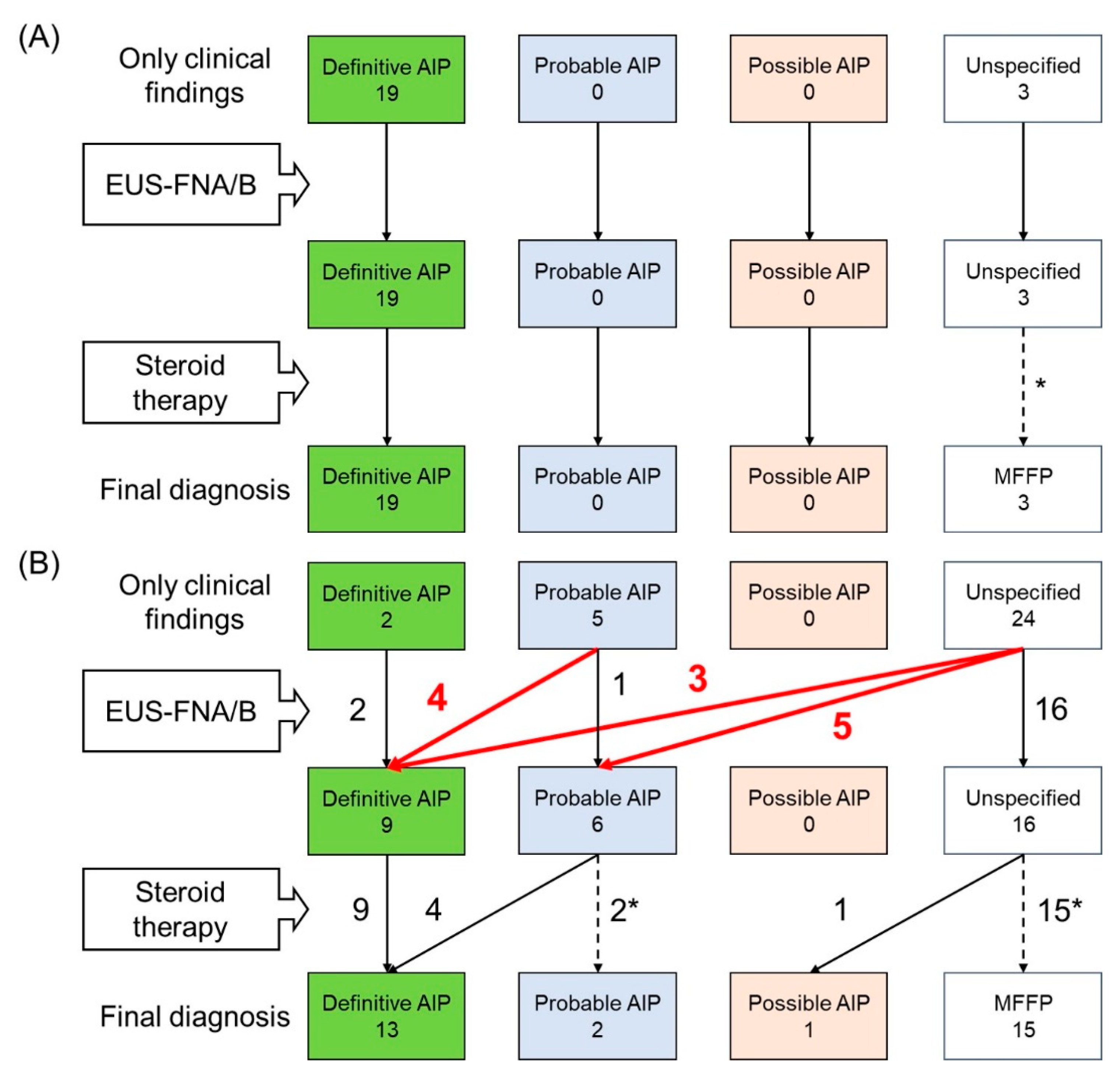
3.2. Contribution of EUS-FNA/B to Diagnosis of AIP

The diagnostic flow of AIP is shown in Figure 1. Among patients with diffuse enlargement of the pancreas, there was no change in the diagnosis after considering the pathological findings of EUS-FNA/B, compared with the diagnosis based on pancreatic imaging, serum IgG4, and other organ involvement (Figure 1A). Thus, EUS-FNA/B provided no contribution to diagnosis of diffuse type AIP.
On the other hand, the diagnosis changed in 12 patients (38.7%) after considering the pathological findings of EUS-FNA/B among patients with focal enlargement of the pancreas: a probable to definitive diagnosis in 4, unspecified to definitive in 3, and unspecified to probable in 5 (Figure 1B). All of these 12 patients were diagnosed as having definitive or probable AIP before the introduction of steroid treatment. Of the five patients whose diagnosis changed from unspecified to probable AIP after EUS-FNA/B, four received steroid therapy, and all patients responded to steroids and were eventually confirmed as having definite AIP.
Comparison of diagnoses with and without considering the pathological findings of EUS-FNA/B is shown in Figure 2, including response of steroid therapy. Without pathological results by EUS-FNA/B, 26 patients were diagnosed as definitive AIP and 5 were diagnosed as probable. When considering the pathological findings of EUS-FNA/B, we could diagnose 32 patients with definitive AIP and 2 with probable. Three patients might not have had the diagnosis of AIP if EUS-FNA/B had not been performed.

3.3. Comparison of the Diagnostic Yield of EUS-FNA/B

Among 34 patients diagnosed with definitive or probable AIP, we compared the diagnostic yield of EUS-FNA (n = 14) and EUS-FNB (n = 20). Figure 3A shows the comparisons of the sensitivity for each typical feature of LPSP. EUS-FNB provided significantly higher sensitivity for prominent lymphoplasmacytic infiltration along with fibrosis (100% vs. 64.3%, p = 0.007), abundant IgG4-positive plasma cells (60.0% vs. 21.4%, p = 0.038), and obstructive phlebitis (70.0% vs. 7.1%, p <0.001). Although there was no statistically significant difference in storiform fibrosis, only EUS-FNB disclosed storiform fibrosis in threepatients (15%).
Among patients who underwent EUS-FNA, no one was diagnosed as pathologically definitive, and only four were diagnosed as pathologically possible. Meanwhile, among patients who underwent EUS-FNB, 10 were diagnosed as pathologically definitive and 7 were diagnosed as pathologically possible. Thus, EUS-FNB provided a significantly higher diagnostic yield than EUS-FNA (Figure 3B).
Among FNB needles, we further compared the diagnostic yield of 22-gauge Franseen needles (n = 10) and that of 20-gauge forward-bevel needles (n = 7). A pathologically possible diagnosis was obtained in 5 patients by Franseen needles and in only 1 by forward-bevel needles (50.0% vs. 14.3%, p = 0.304), and a pathologically definite diagnosis was obtained in 5 by Franseen needles and in 3 by forward-bevel needles (50.0% vs. 42.9%, p = 1).

3.4. Adverse Events of EUS-FNA/B

Adverse events of EUS-FNA/B are shown in Table 2. Both patients complicated by pancreatic fistula were treated conservatively. All of the four patients who developed adverse events had focal enlargement of the pancreas and underwent EUS-FNB. Although there was no significant difference in the adverse event rates between EUS-FNA and FNB (0% vs. 12.5%, p = 0.143), there were no adverse events in patients who underwent EUS-FNA. Among the four patients with adverse events of EUS-FNB, however, pathological findings were helpful in the diagnosis of AIP in two of them—possible to definitive AIP in one, and from an unspecified diagnosis to probable AIP in another.

4. Discussion

In this retrospective analysis on the role of EUS-FNA/B for the diagnosis of AIP, we revealed that EUS-FNA/B effectively contributed to diagnosis prior to the introduction of steroid therapy in 40% of patients with focal enlargement of the pancreas, though in none who had AIP with diffuse enlargement. Finally, 10% of patients with focal enlargement of the pancreas could not be diagnosed as AIP without the pathological findings by EUS-FNA/B, even after a positive response to steroid therapy. EUS-FNB provided a higher diagnostic yield than EUS-FNA, at the cost of potential increase in adverse event rates.
Several studies reported the diagnostic difficulty of AIP by EUS-FNA with the limited sensitivity of 0–19% for pathologically definite diagnosis of LPSP, because EUS-FNA could not provide sufficient tissue samples for histological evaluation [26,27,33,34,35]. Iwashita et al. reported the improved sensitivity of 43% using a 19-gauge FNA needle [29]. Kanno et al. emphasized the importance of quick needle motion for adequate tissue acquisition, and they reported the sensitivity of 41–56% even using 22-gauge FNA needles [30,31]. Recently, Kurita et al. documented for the first time the utility of EUS-FNB using a Franseen needle in the diagnosis of AIP, with a high sensitivity of 56% for pathologically definite diagnosis of LPSP [36].
The primary endpoint of the above studies was sensitivity for pathological diagnosis of AIP by EUS-FNA/B. However, different from pancreatic cancer, the diagnosis of AIP is based on the combination of clinical and pathological features as well as response to steroid therapy. Only a few studies have evaluated the contribution of EUS-FNA/B to the diagnosis of AIP. Additional diagnosis of LPSP by histology ranged from 55.6% to 94.7% in patients without diagnosis of AIP according to the ICDC prior to histological evaluation [26,34,36]. According to the recent multicentric Italian survey on daily practice for AIP, EUS-FNA/B was judged diagnostic for AIP in 75.4% [39]. However, it has not been elucidated who can benefit from EUS-FNA/B in the diagnosis of AIP. Our study revealed that EUS-FNA/B provided useful histological information and changed the diagnosis only in patients with focal enlargement of the pancreas, but not in those with diffuse enlargement. This is consistent with the recent multicentric study by Notohara et al. [40]. They also evaluated the usefulness of histological diagnoses in the context of the diagnostic criteria and illustrated the diagnostic algorithms. They reported that the histological diagnosis contributed to the final diagnosis in only 1 out of 28 cases with diffuse pancreatic swelling, but the histological diagnosis was essential in 25 out of 45 cases with focal swelling. Thus, EUS-FNA/B should be performed in patients with clinical suspicion of AIP with focal enlargement.
In this study, we used the Japanese diagnostic criteria for AIP [37] because it is described in the ICDC that AIP can be diagnosed only on resection or core biopsy specimen. When using the ICDC, we also confirmed that EUS-FNA/B provided significant contribution only among patients with focal disease. Among 31 patients with focal enlargement of the pancreas, 8 were diagnosed with definite type 1 AIP and 23 were diagnosed with unspecified pancreatitis based on pancreatic imaging, serum IgG4 levels, and other organ involvement. After considering the pathological findings by EUS-FNA/B, diagnosis changed in two patients (6.4%), from an unspecified diagnosis to definite AIP. Thus, the contribution of EUS-FNA/B in AIP diagnosis was estimated to be less when using the ICDC as opposed to the JPS2018. This is because histology of the pancreas is originally regarded as less important in the ICDC.
In our analysis, EUS-FNB provided a higher diagnostic yield than EUS-FNA. Sensitivity for obstructive phlebitis was much higher in EUS-FNB compared with EUS-FBA (70.0% vs. 7.0%, p <0.001). Fifty percent of patients were diagnosed with pathologically definite LPSP in patients who underwent EUS-FNB, though none were diagnosed of those who underwent EUS-FNA (50.0% vs. 0%, p = 0.002). FNB needles can acquire a larger amount of tissue with better preservation of tissue architecture than FNA needles [17,19,41,42]. Thus, special staining, such as Elastica van Gieson staining and IgG4 immunostaining, can be performed and is speculated to improve diagnostic yield of EUS-FNB. Notohara et al. referred to the importance of elastic stain on the evaluation of obliterative phlebitis in the guidance for diagnosing AIP with biopsy tissues [38]. According to two recent randomized trials, end-cutting needles can acquire a larger amount of tissue with better preservation of tissue architecture than side-fenestrated needles in autoimmune pancreatitis and for the sampling of solid pancreatic lesions [36,43]. In our study, the number of cases using each needle was too small to detect the statistical significance, though.
As for procedure-related adverse events, EUS-FNB was associated with a higher adverse event rate than EUS-FNA in our study, though this was not statistically significantly (20.0% vs. 0%, p = 0.143). Previous studies comparing EUS-FNA and EUS-FNB for solid pancreatic mass reported no significant difference in adverse event rates [16,19,44]. Since the adverse event rate of EUS-FNB in our cohort appeared too high, further investigation with a larger cohort is warranted.
The limitations of our study are the retrospective nature and the small number of patients enrolled. The selection of the needles was at the discretion of the endoscopist.

5. Conclusions

In conclusion, EUS-FNA/B was helpful in the diagnosis of focal type AIP, and steroid therapy could be introduced after the diagnosis was confirmed. EUS-FNB provided a higher diagnostic yield than EUS-FNA in AIP.

Author Contributions

K.N. performed the literature review and manuscript drafting; Y.N. S.M., K.H., S.K., Y.S., A.I., T.S. (Tatsuya Sato)., R.H., K.I., K.S., T.S. (Tomotaka Saito)., T.H., N.T., H.K., H.I., and K.K. provided topic outlining, expertise, and manuscript editing. All authors have read and agreed to the published version of the manuscript.

Funding

This research received no external funding.

Conflicts of Interest

The authors declare no conflict of interest.

References

  1. Kamisawa, T.; Okazaki, K.; Kawa, S.; Ito, T.; Inui, K.; Irie, H.; Nishino, T.; Notohara, K.; Nishimori, I.; Tanaka, S.; et al. Amendment of the Japanese Consensus Guidelines for Autoimmune Pancreatitis, 2013 III. Treatment and prognosis of autoimmune pancreatitis. J. Gastroenterol. 2014, 49, 961–970. [Google Scholar] [CrossRef] [PubMed] [Green Version]
  2. Kawa, S.; Okazaki, K.; Kamisawa, T.; Kubo, K.; Ohara, H.; Hasebe, O.; Fujinaga, Y.; Irisawa, A.; Notohara, K.; Ito, T.; et al. Amendment of the Japanese Consensus Guidelines for Autoimmune Pancreatitis, 2013 II. Extrapancreatic lesions, differential diagnosis. J. Gastroenterol. 2014, 49, 765–784. [Google Scholar] [CrossRef] [PubMed] [Green Version]
  3. Okazaki, K.; Kawa, S.; Kamisawa, T.; Ito, T.; Inui, K.; Irie, H.; Nishino, T.; Notohara, K.; Nishimori, I.; Tanaka, S.; et al. Amendment of the Japanese Consensus Guidelines for Autoimmune Pancreatitis, 2013 I. Concept and diagnosis of autoimmune pancreatitis. J. Gastroenterol. 2014, 49, 567–588. [Google Scholar] [CrossRef] [PubMed] [Green Version]
  4. Yoshida, K.; Toki, F.; Takeuchi, T.; Watanabe, S.; Shiratori, K.; Hayashi, N. Chronic pancreatitis caused by an autoimmune abnormality. Proposal of the concept of autoimmune pancreatitis. Dig. Dis. Sci. 1995, 40, 1561–1568. [Google Scholar] [CrossRef]
  5. Shimosegawa, T.; Chari, S.T.; Frulloni, L.; Kamisawa, T.; Kawa, S.; Mino-Kenudson, M.; Kim, M.H.; Kloppel, G.; Lerch, M.M.; Lohr, M.; et al. International consensus diagnostic criteria for autoimmune pancreatitis: Guidelines of the International Association of Pancreatology. Pancreas 2011, 40, 352–358. [Google Scholar] [CrossRef]
  6. Hirano, K.; Tada, M.; Isayama, H.; Sasahira, N.; Umefune, G.; Akiyama, D.; Watanabe, T.; Saito, T.; Takagi, K.; Takahara, N.; et al. Outcome of Long-term Maintenance Steroid Therapy Cessation in Patients With Autoimmune Pancreatitis: A Prospective Study. J. Clin. Gastroenterol. 2016, 50, 331–337. [Google Scholar] [CrossRef]
  7. Hirano, K.; Tada, M.; Isayama, H.; Yagioka, H.; Sasaki, T.; Kogure, H.; Nakai, Y.; Sasahira, N.; Tsujino, T.; Yoshida, H.; et al. Long-term prognosis of autoimmune pancreatitis with and without corticosteroid treatment. Gut 2007, 56, 1719–1724. [Google Scholar] [CrossRef] [Green Version]
  8. Kamisawa, T.; Okazaki, K.; Kawa, S.; Shimosegawa, T.; Tanaka, M.; Society, R.C.f.I.P.D.a.J.P. Japanese consensus guidelines for management of autoimmune pancreatitis: III. Treatment and prognosis of AIP. J. Gastroenterol. 2010, 45, 471–477. [Google Scholar] [CrossRef]
  9. Hirano, K.; Tada, M.; Sasahira, N.; Tsujino, T.; Isayama, H.; Kawabe, T.; Omata, M. Are all pancreatic lesions responsive to steroid therapy [corrected] autoimmune pancreatitis? Gut 2009, 58, 1031–1032; author reply 1032. [Google Scholar]
  10. Lohr, J.M.; Beuers, U.; Vujasinovic, M.; Alvaro, D.; Frokjaer, J.B.; Buttgereit, F.; Capurso, G.; Culver, E.L.; de-Madaria, E.; Della-Torre, E.; et al. European Guideline on IgG4-related digestive disease—UEG and SGF evidence-based recommendations. United Eur. Gastroenterol. J. 2020, 8, 637–666. [Google Scholar] [CrossRef]
  11. Hewitt, M.J.; McPhail, M.J.; Possamai, L.; Dhar, A.; Vlavianos, P.; Monahan, K.J. EUS-guided FNA for diagnosis of solid pancreatic neoplasms: A meta-analysis. Gastrointest. Endosc. 2012, 75, 319–331. [Google Scholar] [CrossRef] [PubMed]
  12. Volmar, K.E.; Vollmer, R.T.; Jowell, P.S.; Nelson, R.C.; Xie, H.B. Pancreatic FNA in 1000 cases: A comparison of imaging modalities. Gastrointest. Endosc. 2005, 61, 854–861. [Google Scholar] [CrossRef]
  13. Williams, D.B.; Sahai, A.V.; Aabakken, L.; Penman, I.D.; van Velse, A.; Webb, J.; Wilson, M.; Hoffman, B.J.; Hawes, R.H. Endoscopic ultrasound guided fine needle aspiration biopsy: A large single centre experience. Gut 1999, 44, 720–726. [Google Scholar] [CrossRef] [PubMed] [Green Version]
  14. Wilson, J.L.; Kalade, A.; Prasad, S.; Cade, R.; Thomson, B.; Banting, S.; Mackay, S.; Desmond, P.V.; Chen, R.Y. Diagnosis of solid pancreatic masses by endoscopic ultrasound-guided fine-needle aspiration. Intern. Med. J. 2009, 39, 32–37. [Google Scholar] [CrossRef]
  15. Abdelfatah, M.M.; Grimm, I.S.; Gangarosa, L.M.; Baron, T.H. Cohort study comparing the diagnostic yields of 2 different EUS fine-needle biopsy needles. Gastrointest. Endosc. 2018, 87, 495–500. [Google Scholar] [CrossRef]
  16. Bang, J.Y.; Hawes, R.; Varadarajulu, S. A meta-analysis comparing ProCore and standard fine-needle aspiration needles for endoscopic ultrasound-guided tissue acquisition. Endoscopy 2016, 48, 339–349. [Google Scholar] [CrossRef]
  17. Bang, J.Y.; Hebert-Magee, S.; Hasan, M.K.; Navaneethan, U.; Hawes, R.; Varadarajulu, S. Endoscopic ultrasonography-guided biopsy using a Franseen needle design: Initial assessment. Dig. Endosc. 2017, 29, 338–346. [Google Scholar] [CrossRef]
  18. Bang, J.Y.; Hebert-Magee, S.; Navaneethan, U.; Hasan, M.K.; Hawes, R.; Varadarajulu, S. Randomized trial comparing the Franseen and Fork-tip needles for EUS-guided fine-needle biopsy sampling of solid pancreatic mass lesions. Gastrointest. Endosc. 2018, 87, 1432–1438. [Google Scholar] [CrossRef]
  19. Ishigaki, K.; Nakai, Y.; Oyama, H.; Kanai, S.; Suzuki, T.; Nakamura, T.; Sato, T.; Hakuta, R.; Saito, K.; Saito, T.; et al. Endoscopic Ultrasound-Guided Tissue Acquisition by 22-Gauge Franseen and Standard Needles for Solid Pancreatic Lesions. Gut Liver 2020. [Google Scholar] [CrossRef]
  20. Iwashita, T.; Nakai, Y.; Mukai, T.; Togawa, O.; Matsubara, S.; Hatano, Y.; Hara, A.; Tanaka, M.; Shibahara, J.; Fukayama, M.; et al. A 19-Gauge Histology Needle Versus a 19-Gauge Standard Needle in Endoscopic Ultrasound-Guided Fine-Needle Aspiration for Solid Lesions: A Multicenter Randomized Comparison Study (GREATER Study). Dig. Dis. Sci. 2018, 63, 1043–1051. [Google Scholar] [CrossRef]
  21. Kandel, P.; Tranesh, G.; Nassar, A.; Bingham, R.; Raimondo, M.; Woodward, T.A.; Gomez, V.; Wallace, M.B. EUS-guided fine needle biopsy sampling using a novel fork-tip needle: A case-control study. Gastrointest. Endosc. 2016, 84, 1034–1039. [Google Scholar] [CrossRef] [PubMed]
  22. Nayar, M.K.; Paranandi, B.; Dawwas, M.F.; Leeds, J.S.; Darne, A.; Haugk, B.; Majumdar, D.; Ahmed, M.M.; Oppong, K.W. Comparison of the diagnostic performance of 2 core biopsy needles for EUS-guided tissue acquisition from solid pancreatic lesions. Gastrointest. Endosc. 2017, 85, 1017–1024. [Google Scholar] [CrossRef] [PubMed]
  23. Nishioka, N.; Ogura, T.; Kurisu, Y.; Imanishi, M.; Onda, S.; Takagi, W.; Sano, T.; Okuda, A.; Miyano, A.; Amano, M.; et al. Prospective histological evaluation of a 20G core trap with a forward-cutting bevel needle for EUS-FNA of pancreatic lesions. Surg. Endosc. 2018, 32, 4125–4131. [Google Scholar] [CrossRef] [PubMed]
  24. van Riet, P.A.; Larghi, A.; Attili, F.; Rindi, G.; Nguyen, N.Q.; Ruszkiewicz, A.; Kitano, M.; Chikugo, T.; Aslanian, H.; Farrell, J.; et al. A multicenter randomized trial comparing a 25-gauge EUS fine-needle aspiration device with a 20-gauge EUS fine-needle biopsy device. Gastrointest. Endosc. 2019, 89, 329–339. [Google Scholar] [CrossRef] [PubMed]
  25. Bhattacharya, A.; Cruise, M.; Chahal, P. Endoscopic ultrasound guided 22 gauge core needle biopsy for the diagnosis of Autoimmune pancreatitis. Pancreatology 2018, 18, 168–169. [Google Scholar] [CrossRef] [PubMed]
  26. Cao, L.; Wang, Y.; Wang, J.; Guo, Q.; Chen, Q.; Wu, X.; Tang, S.J.; Cheng, B. The role of EUS-guided fine needle aspiration in autoimmune pancreatitis: A single center prospective study. Scand. J. Gastroenterol. 2018, 53, 1604–1610. [Google Scholar] [CrossRef]
  27. Imai, K.; Matsubayashi, H.; Fukutomi, A.; Uesaka, K.; Sasaki, K.; Ono, H. Endoscopic ultrasonography-guided fine needle aspiration biopsy using 22-gauge needle in diagnosis of autoimmune pancreatitis. Dig. Liver Dis. 2011, 43, 869–874. [Google Scholar] [CrossRef]
  28. Ishikawa, T.; Itoh, A.; Kawashima, H.; Ohno, E.; Matsubara, H.; Itoh, Y.; Nakamura, Y.; Hiramatsu, T.; Nakamura, M.; Miyahara, R.; et al. Endoscopic ultrasound-guided fine needle aspiration in the differentiation of type 1 and type 2 autoimmune pancreatitis. World J. Gastroenterol. 2012, 18, 3883–3888. [Google Scholar] [CrossRef]
  29. Iwashita, T.; Yasuda, I.; Doi, S.; Ando, N.; Nakashima, M.; Adachi, S.; Hirose, Y.; Mukai, T.; Iwata, K.; Tomita, E.; et al. Use of samples from endoscopic ultrasound-guided 19-gauge fine-needle aspiration in diagnosis of autoimmune pancreatitis. Clin. Gastroenterol. Hepatol. 2012, 10, 316–322. [Google Scholar] [CrossRef]
  30. Kanno, A.; Ishida, K.; Hamada, S.; Fujishima, F.; Unno, J.; Kume, K.; Kikuta, K.; Hirota, M.; Masamune, A.; Satoh, K.; et al. Diagnosis of autoimmune pancreatitis by EUS-FNA by using a 22-gauge needle based on the International Consensus Diagnostic Criteria. Gastrointest. Endosc. 2012, 76, 594–602. [Google Scholar] [CrossRef]
  31. Kanno, A.; Masamune, A.; Fujishima, F.; Iwashita, T.; Kodama, Y.; Katanuma, A.; Ohara, H.; Kitano, M.; Inoue, H.; Itoi, T.; et al. Diagnosis of autoimmune pancreatitis by EUS-guided FNA using a 22-gauge needle: A prospective multicenter study. Gastrointest. Endosc. 2016, 84, 797–804.e791. [Google Scholar] [CrossRef] [PubMed] [Green Version]
  32. Kanno, A.; Masamune, A.; Shimosegawa, T. Endoscopic approaches for the diagnosis of autoimmune pancreatitis. Dig. Endosc. 2015, 27, 250–258. [Google Scholar] [CrossRef] [PubMed] [Green Version]
  33. Mizuno, N.; Bhatia, V.; Hosoda, W.; Sawaki, A.; Hoki, N.; Hara, K.; Takagi, T.; Ko, S.B.; Yatabe, Y.; Goto, H.; et al. Histological diagnosis of autoimmune pancreatitis using EUS-guided trucut biopsy: A comparison study with EUS-FNA. J. Gastroenterol. 2009, 44, 742–750. [Google Scholar] [CrossRef] [PubMed]
  34. Morishima, T.; Kawashima, H.; Ohno, E.; Yamamura, T.; Funasaka, K.; Nakamura, M.; Miyahara, R.; Watanabe, O.; Ishigami, M.; Shimoyama, Y.; et al. Prospective multicenter study on the usefulness of EUS-guided FNA biopsy for the diagnosis of autoimmune pancreatitis. Gastrointest. Endosc. 2016, 84, 241–248. [Google Scholar] [CrossRef] [PubMed]
  35. Sugimoto, M.; Takagi, T.; Suzuki, R.; Konno, N.; Asama, H.; Watanabe, K.; Nakamura, J.; Kikuchi, H.; Waragai, Y.; Takasumi, M.; et al. Endoscopic Ultrasonography-Guided Fine Needle Aspiration Can Be Used to Rule Out Malignancy in Autoimmune Pancreatitis Patients. J. Ultrasound. Med. 2017, 36, 2237–2244. [Google Scholar] [CrossRef]
  36. Kurita, A.; Yasukawa, S.; Zen, Y.; Yoshimura, K.; Ogura, T.; Ozawa, E.; Okabe, Y.; Asada, M.; Nebiki, H.; Shigekawa, M.; et al. Comparison of a 22-gauge Franseen-tip needle with a 20-gauge forward-bevel needle for the diagnosis of type 1 autoimmune pancreatitis: A prospective, randomized, controlled, multicenter study (COMPAS study). Gastrointest. Endosc. 2020, 91, 373–381.e372. [Google Scholar] [CrossRef] [PubMed]
  37. Kawa, S.; Kamisawa, T.; Notohara, K.; Fujinaga, Y.; Inoue, D.; Koyama, T.; Okazaki, K. Japanese Clinical Diagnostic Criteria for Autoimmune Pancreatitis, 2018: Revision of Japanese Clinical Diagnostic Criteria for Autoimmune Pancreatitis, 2011. Pancreas 2020, 49, e13–e14. [Google Scholar] [CrossRef] [Green Version]
  38. Notohara, K.; Kamisawa, T.; Fukushima, N.; Furukawa, T.; Tajiri, T.; Yamaguchi, H.; Aishima, S.; Fukumura, Y.; Hirabayashi, K.; Iwasaki, E.; et al. Guidance for diagnosing autoimmune pancreatitis with biopsy tissues. Pathol. Int. 2020, 70, 699–711. [Google Scholar] [CrossRef]
  39. Barresi, L.; Tacelli, M.; Crino, S.F.; Attili, F.; Petrone, M.C.; De Nucci, G.; Carrara, S.; Manfredi, G.; Capurso, G.; De Angelis, C.G.; et al. Multicentric Italian survey on daily practice for autoimmune pancreatitis: Clinical data, diagnosis, treatment, and evolution toward pancreatic insufficiency. United Eur. Gastroenterol. J. 2020, 8, 705–715. [Google Scholar] [CrossRef]
  40. Notohara, K.; Kamisawa, T.; Kanno, A.; Naitoh, I.; Iwasaki, E.; Shimizu, K.; Kuraishi, Y.; Motoya, M.; Kodama, Y.; Kasashima, S.; et al. Efficacy and limitations of the histological diagnosis of type 1 autoimmune pancreatitis with endoscopic ultrasound-guided fine needle biopsy with large tissue amounts. Pancreatology 2020, 20, 834–843. [Google Scholar] [CrossRef]
  41. Laquiere, A.; Lefort, C.; Maire, F.; Aubert, A.; Gincul, R.; Prat, F.; Grandval, P.; Croizet, O.; Boulant, J.; Vanbiervliet, G.; et al. 19 G nitinol needle versus 22 G needle for transduodenal endoscopic ultrasound-guided sampling of pancreatic solid masses: A randomized study. Endoscopy 2019, 51, 436–443. [Google Scholar] [CrossRef] [PubMed] [Green Version]
  42. Polkowski, M.; Jenssen, C.; Kaye, P.; Carrara, S.; Deprez, P.; Gines, A.; Fernandez-Esparrach, G.; Eisendrath, P.; Aithal, G.P.; Arcidiacono, P.; et al. Technical aspects of endoscopic ultrasound (EUS)-guided sampling in gastroenterology: European Society of Gastrointestinal Endoscopy (ESGE) Technical Guideline—March 2017. Endoscopy 2017, 49, 989–1006. [Google Scholar] [CrossRef] [PubMed] [Green Version]
  43. Crino, S.F.; Le Grazie, M.; Manfrin, E.; Conti Bellocchi, M.C.; Bernardoni, L.; Granato, A.; Locatelli, F.; Parisi, A.; Di Stefano, S.; Frulloni, L.; et al. Randomized trial comparing fork-tip and side-fenestrated needles for EUS-guided fine-needle biopsy of solid pancreatic lesions. Gastrointest. Endosc. 2020, 92, 648–658.e2. [Google Scholar] [CrossRef] [PubMed]
  44. Lee, Y.N.; Moon, J.H.; Kim, H.K.; Choi, H.J.; Choi, M.H.; Kim, D.C.; Lee, T.H.; Cha, S.W.; Cho, Y.D.; Park, S.H. Core biopsy needle versus standard aspiration needle for endoscopic ultrasound-guided sampling of solid pancreatic masses: A randomized parallel-group study. Endoscopy 2014, 46, 1056–1062. [Google Scholar] [CrossRef] [PubMed]
Figure 1. Diagnostic flow of autoimmune pancreatitis according to Japanese Clinical Diagnostic Criteria for Autoimmune Pancreatitis 2018 in patients with diffuse enlargement of the pancreas (A) and in those with focal enlargement (B). AIP, autoimmune pancreatitis; EUS-FNA/B, endoscopic ultrasonography-guided fine needle aspiration/biopsy; MFFP, mass-forming focal pancreatitis. * They did not receive steroid therapy.
Diagnostics 10 00954 g001
Figure 2. Comparison of diagnosis with and without considering the pathological findings of EUS-FNA/B. EUS-FNA/B, Endoscopic ultrasonography-guided fine needle aspiration/biopsy.
Diagnostics 10 00954 g002
Figure 3. Comparison of the diagnostic yield of EUS-FNA/B among patients with probable and definitive AIP. (A) Comparison of the sensitivity for each typical histological finding of lymphoplasmacytic sclerosing pancreatitis. (B) Comparison of the sensitivity for pathological diagnosis. AIP, autoimmune pancreatitis; EUS-FNA/B, endoscopic ultrasonography-guided fine needle aspiration/biopsy.
Diagnostics 10 00954 g003
Table 1. Patients characteristics and final diagnosis.
n = 53
Sex: male30 (56.6%)
Age * (year)68 (17–83)
Enlargement of the pancreas: diffuse/focal22/31 (41.5%/58.5%)
Elevated levels of serum IgG4 (≥ 135 mg/dl)33 (62.3%)
Other organ involvement21 (40.0%)
Locus of the target of EUS-FNA/B
Head14 (26.4%)
Body25 (47.2%)
Tail14 (26.4%)
Diameter of the lesion * (mm)25 (20–36)
Needles: FNA/FNB21/32 (39.6%/60.4%)
Final diagnosis
Definitive AIP32 (60.4%)
Probable AIP2 (3.8%)
Possible AIP1 (1.9%)
Mass-forming focal pancreatitis18 (34.0%)
Values are expressed as numbers (%) otherwise indicated. * Values are expressed as medians (ranges). EUS-FNA/B, endoscopic ultrasonography-guided fine needle aspiration/biopsy; AIP, autoimmune pancreatitis.
Table 2. Adverse events of endoscopic ultrasonography-guided fine needle aspiration/biopsy.
Enlargement of the PancreasNeedleAdverse EventDiagnosis without EUS-FNA/BDiagnosis with EUS-FNA/B
1FocalFNB
22-gauge
Franseen
Pancreatic fistulaUnspecified diagnosisMFFP
2FocalFNB
22-gauge
Franseen
Pancreatic fistulaUnspecified diagnosisProbable AIP
3FocalFNB
20-gauge
forward-bevel
Mild pancreatitisUnspecified diagnosisMFFP
4FocalFNB
20-gauge
forward-bevel
Mild pancreatitisPossible AIPDefinitive AIP
EUS-FNA/B, endoscopic ultrasonography-guided fine needle aspiration/biopsy; MFFP, mass-forming focal pancreatitis; AIP, autoimmune pancreatitis.
Publisher’s Note: MDPI stays neutral with regard to jurisdictional claims in published maps and institutional affiliations.

