Article

Roof-Inhabiting Cousins of Rock-Inhabiting Fungi: Novel Melanized Microcolonial Fungal Species from Photocatalytically Reactive Subaerial Surfaces

by
Constantino Ruibal
1,†,
Laura Selbmann
2,3,†,
Serap Avci
4,
Pedro M. Martin-Sanchez
4,5 and
Anna A. Gorbushina
4,6,*
1
Department of Pharmacology, Pharmacognosy, and Botany, Faculty of Pharmacy, Universidad Complutense de Madrid, 28040 Madrid, Spain
2
Department of Ecological and Biological Sciences (DEB), University of Tuscia, 01100 Viterbo, Italy
3
Italian National Antarctic Museum, Mycological Section, 16128 Genoa, Italy
4
Department 4 Materials and Environment, Bundesanstalt für Materialforschung und -prüfung (BAM), 12205 Berlin, Germany
5
Section for Genetics and Evolutionary Biology (EVOGENE), Department of Biosciences, University of Oslo, 0371 Oslo, Norway
6
Department of Earth Sciences & Department of Biology, Chemistry, Pharmacy, Freie Universität Berlin, 14195 Berlin, Germany
*
Author to whom correspondence should be addressed.
These authors contributed equally to this work.
Submission received: 1 May 2018 / Revised: 10 July 2018 / Accepted: 11 July 2018 / Published: 15 July 2018
(This article belongs to the Special Issue Fungi from Extreme Environments)

Abstract

:
Subaerial biofilms (SAB) are an important factor in weathering, biofouling, and biodeterioration of bare rocks, building materials, and solar panel surfaces. The realm of SAB is continually widened by modern materials, and the settlers on these exposed solid surfaces always include melanized, stress-tolerant microcolonial ascomycetes. After their first discovery on desert rock surfaces, these melanized chaetothyrialean and dothidealean ascomycetes have been found on Mediterranean monuments after biocidal treatments, Antarctic rocks and solar panels. New man-made modifications of surfaces (e.g., treatment with biocides or photocatalytically active layers) accommodate the exceptional stress-tolerance of microcolonial fungi and thus further select for this well-protected ecological group. Melanized fungal strains were isolated from a microbial community that developed on highly photocatalytic roof tiles after a long-term environmental exposure in a maritime-influenced region in northwestern Germany. Four of the isolated strains are described here as a novel species, Constantinomyces oldenburgensis, based on multilocus ITS, LSU, RPB2 gene phylogeny. Their closest relative is a still-unnamed rock-inhabiting strain TRN431, here described as C. patonensis. Both species cluster in Capnodiales, among typical melanized microcolonial rock fungi from different stress habitats, including Antarctica. These novel strains flourish in hostile conditions of highly oxidizing material surfaces, and shall be used in reference procedures in material testing.

1. Introduction

Melanized, slow-growing and stress-tolerant microcolonial ascomycetes are considered the most persistent inhabitants of bare rock surfaces [1,2]. After their first discovery on desert rocks covered by rock varnish [3], melanized ascomycetes from Chaetothyriales and Dothideomycetidae have been found on Mediterranean monuments and natural rock outcrops (e.g., [4,5,6,7,8,9]), Acropolis marble after biocidal treatments [10], Antarctic rocks [11,12] and solar panels [13]. Atmosphere-exposed surfaces, as hostile environments open to sun irradiation and fluctuating water and nutrient availability, offer themselves as a perfect niche selecting for these survival specialists.
Manmade materials like building surfaces, monuments, solar panels, etc. have to be protected from the growth of these organisms that might cause pitting [6], color modification [14,15,16] and physical damage of the underlying substrate [17]. Therefore, diverse microbial control technologies are employed to protect these surfaces by modifications such as biocides as well as surface treatments with photocatalytically active layers. These growth-controlling amendments might, however, lead to development of resistance in SAB and thus further select for stress-tolerant microorganisms, including melanized microcolonial fungi.
This investigation was based on a roof-tile exposure case study in Northwestern Germany (Edewecht). Several melanized microcolonial isolates were obtained from tiles with a photocatalytically active surface that were exposed from November 2006 to May 2014. Four of them have been morphologically characterized and here described as a new species in the genus Constantinomyces based on a three-gene multilocus phylogeny that includes both melanized rock-inhabiting and plant pathogenic capnodialean fungi.

2. Materials and Methods

2.1. Sampling and Isolation

Roof tiles covering a residential house were exposed at an angle of 45° in a northerly direction. Exposure started in November 2006, and conventional clay tiles of traditional red color but with different types of surface modifications were juxtaposed. Material with photocatalytic surface properties covered part of the roof while the remaining roof surface comprised unmodified conventional clay tiles. Both tile types formed complete rows from the roof top to the edge.
The roof surface and the growth/soiling development on it was observed with documentation by photography from 2006 to 2014. Unfortunately, the specific roof topography influenced sun irradiation quality and duration, therefore reducing the photocatalytic activity of the surface layer. To concentrate our study on differences in colonization between tiles with different surface qualities, a direct comparison was ensured by placing photocatalytically active tiles among the control ones.
Two tiles with surface modifications were exposed from 2006 to 2009 in a shaded area of the roof. After 2.5 years of exposure (in July 2009), these two tiles with photocatalytic surfaces were placed on the control part of the roof (Figure 1a) where shading by architectural elements could be completely avoided. Thus for the period from July 2009 to May 2014 environmental conditions for the control and photocatalytically active tiles remained exactly the same. All sampled tiles were therefore subjected to semistandardized and reciprocally comparable conditions of a typical subaerial exposure in the maritime-influenced, moderate climate for an extended period of almost five years. The sampling was performed in May 2014 after 7.5 years of total exposure, and concentrated on three tiles—two with photocatalytically active surface (Figure 1) and one control.
Surface colonization phenomena were documented by photography prior to removing fragments to Petri dishes for subsequent cultivation. Photocatalytically active tiles have obviously different subaerial biofilm that is more discoloring (Figure 1a). Both sampled photocatalytically active tiles demonstrate an expressed subaerial biofilm growth in the form of dark pigmented fungal colonies (Figure 1b). Especially prominent were the colonies on the elevated surface area—where time of wetness is additionally reduced, leading to less pronounced algal growth.
The sampling goal was to isolate fungal strains from the visible dark colonies (Figure 1b) that were obviously dominating the atmosphere-exposed surface of the material. Fungal colonies that were sampled with sterile implements to isolate dominant subaerial settlers are indicated by the blue frame in Figure 1b.
Several developed colonies from the surface (Figure 1b) were sampled with sterile implements and parts of them were directly transferred to agar media [5]. The same procedure was also applied to the adjacent control tiles. The material removed from the surface and used for obtaining strains was also directly microscopically observed and only dark-pigmented fungal cell clusters were detected in situ, with admixture of green algae in lower-situated tile areas. The resulting melanized microcolonial strains were isolated and subcultured on malt extract agar (MEA) and subsequently stored at 4 °C.

2.2. DNA Extraction, PCR Amplification, and DNA Sequencing

Genomic DNA of each strain was extracted from its mycelia grown on MEA [13]. Mycelia were harvested and transferred to a 2-mL Eppendorf tube containing 500 μL Tris-NaCl-EDTA (TNE) buffer (10 mM Tris-HCl, 100 mM NaCl, 1 mM EDTA; pH 8) and glass beads of three different diameters (0.5, 2 and 5 mm). The mixture was shaken at 4.5 m/s for 1 min in a Fast Prep RiboLyser cell disrupter (Thermo Hybaid GmbH, Heidelberg, Germany). The DNA was purified through phenol/chloroform extraction and ethanol precipitation. The extracted DNA was resuspended in 100 µL sterile ultrapure water, and DNA concentration was quantified using NanoDrop 2000C (Thermo Fisher Scientific, Wilmington, DE, USA), following the manufacturer’s instructions.
Three molecular markers were amplified by conventional PCR for all fungal strains, the rDNA regions comprising ITS1, 5.8S rRNA gene, ITS2 and partial 28S rRNA gene (ITS + LSU), using the universal primers ITS5 [18] and LR5 [19], and a portion of the RNA polymerase II’s second largest subunit (RPB2) gene using the primers fRPB2-5f and fRPB2-7cr [20]. PCR reactions were performed in a BioRad C1000 Thermal Cycler (BioRad, Hercules, CA, USA). Cycling parameters for ITS+28S were 95 °C for 5 min, followed by 35 cycles of 95 °C for 30 s, 50 °C for 30 s, and 72 °C for 2 min, with a final extension at 72 °C for 10 min. For RPB2, cycling parameters were 95 °C for 5 min, followed by 35 cycles of 95 °C for 1 min, 55 °C for 2 min, and 72 °C for 1.5 min, with a final step at 72 °C for 10 min. Reactions were performed in duplicate, and negative controls (containing no DNA) were included in each PCR trial. All PCR products were checked by electrophoresis in 1.5% (w/v) agarose gels stained with GelRed dye (Genaxxon Bioscience GmbH, Ulm, Germany) and visualized under UV light. Positive amplification products were purified by using a QIAquick PCR Purification Kit (Qiagen GmBH, Hilden, Germany) and sequenced in duplicate by Macrogen Europe Company (Amsterdam, The Netherlands) with the same primer sets used for PCR. After edition, final sequences were submitted to the European Nucleotide Archive (ENA, EMBL-EBI) under the accession numbers listed in Table 1.

2.3. Alignment and Tree Reconstruction

ITS, LSU and RPB2 nucleotide sequences were compared in GenBank using BLASTN search [21], and RPB2 protein sequences were additionally checked using BLASTP; both algorithms were run through the NCBI website. The most similar sequences, as well as reference sequences from a selection of rock-inhabiting and plant pathogenic fungi in the order Capnodiales, were exported and iteratively aligned using the MUSCLE option [22] in MEGA6 [23]; final alignment was improved manually. The one-gene alignments were concatenated using FABOX [24] to combine sequences for the three-loci dataset. Optimized alignment was exported and the best-fit substitution model was determined using MODELTEST MrAIC 1.4.3 [25] as implemented in PHYML [26]. The phylogenetic tree was reconstructed by the maximum likelihood method, using TREEFINDER [27]. The robustness of the phylogenetic inference was estimated using a bootstrap method [28] with 1000 pseudoreplicates generated and analyzed with TREEFINDER.

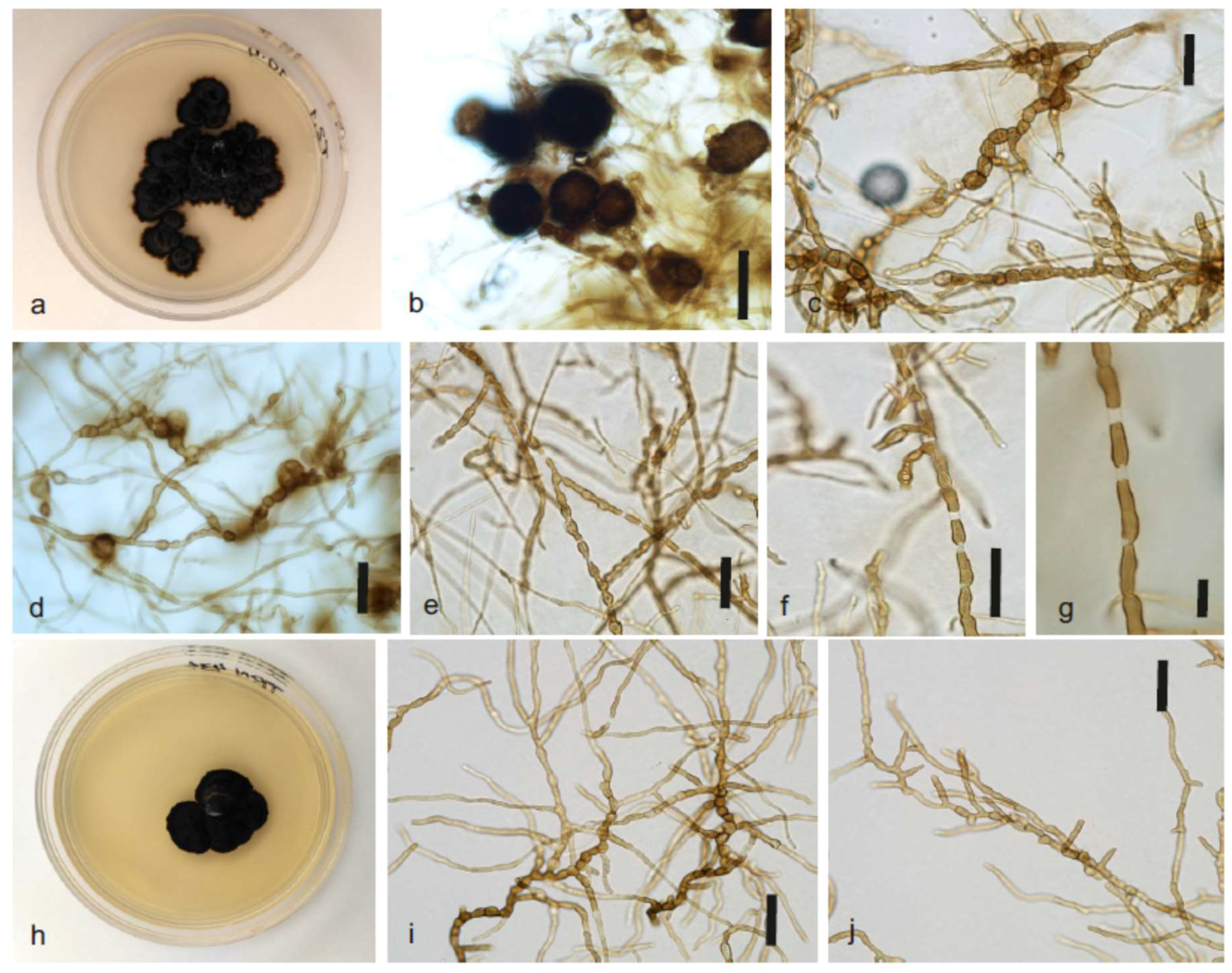
2.4. Morphological Characterization

Microscopic features were characterized after 12 and 35 weeks of growth on 4% MEA incubated at 15°C. Mycelium and other fungal structures were then transferred onto slides mounted in 5% KOH and observed with light microscopy. Digital images were acquired using an Eclipse 80i microscope equipped with a DS-Fi1 digital camera and a Digital Sight DS-L2 image acquisition system (Nikon, Badhoevedrop, the Netherlands). Colony descriptions are based on cultures grown onto 4% MEA incubated at 15°C for 35 weeks.

3. Results

During the period of 2006–2014, a whole series of changes occurred on the air-exposed (subaerial) surface of the investigated roof. Alterations in the material surface color were the most apparent signs of the environmental exposure and accompanying colonization, and attracted our attention to a prominent difference between the photocatalytically active and control roof tiles (Figure 1). These color changes result from a combination of airborne deposition and biogenic conditioning by the subaerial biofilms. In this study, we aimed at characterizing the organisms forming subaerial biofilms on these surfaces.

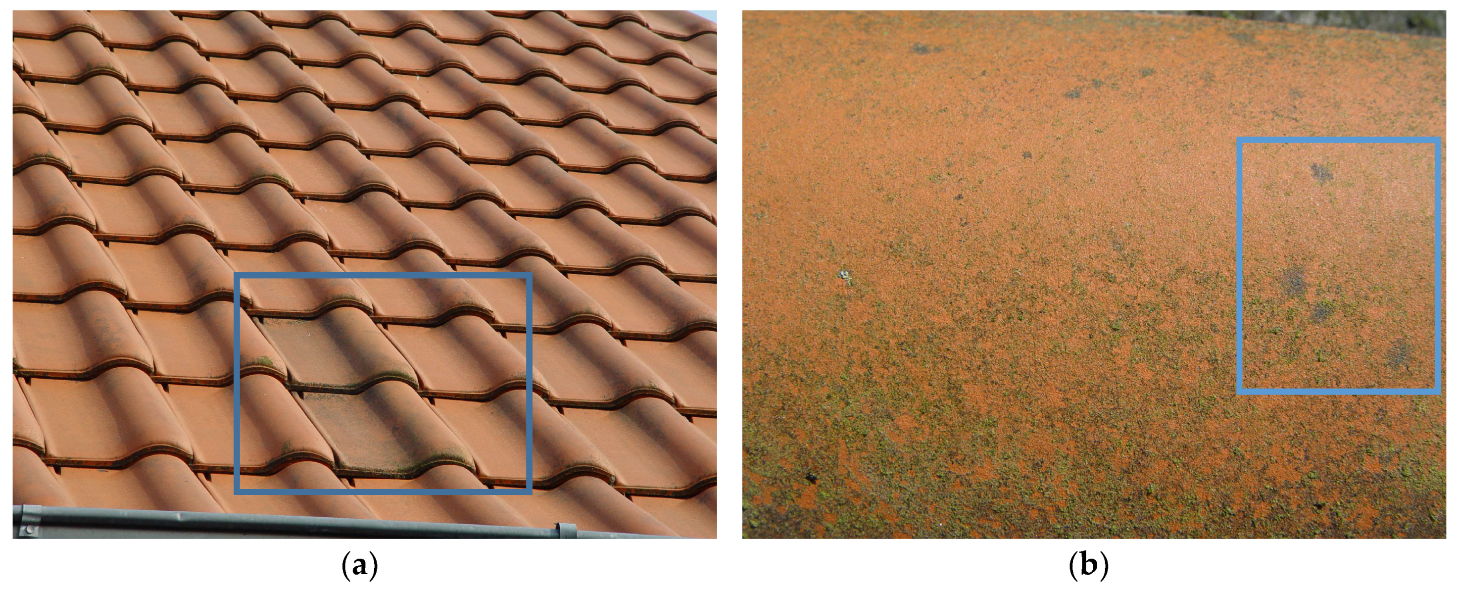
3.1. Subaerial Biofilm Development and Isolation

The biofilms on the experimental roof developed through different phases: the first contrast was achieved in 2008, when the photocatalytically active part was seemingly free from the fouling biofilm, while the conventional tiles were overgrown with an intensely green-colored biofilm (results not shown). It is interesting to mention that from 2006 until 2009, photocatalytically active tiles were visually biofilm-free, while the control tiles just a couple of m away were covered in biofilm (data not shown). Exactly at that moment, two tiles with a photocatalytically active surface not visibly overgrown by a biofilm were transferred to the unshaded control part of the roof. Through the transfer of the photocatalytically active tiles, the following goals were accomplished: (i) any shading was avoided, thus ensuring the same environmental exposure for all tiles under study and (ii) the only difference influencing colonization was the type of surface.
In the following years, the colonization of tiles changed; two photocatalytically active tiles after almost five years of exposure demonstrated clearly different and visible biofilm growth (Figure 1b). Subaerial biofilms formed by melanized microcolonial fungi and some green algae were covering the sample as well as all other photocatalytically active tiles. Also, with the help of light microscopy of surface swabs material, only dark-pigmented fungal cell clusters were observed in situ, with admixture of the green algae in lower-situated tile areas. In May 2014, these biofilms were intensively colored and dominated by the dark fungal colonies, which were frequently single and especially prominent on the elevated areas of the roof tiles where time of wetness is additionally reduced (Figure 1b).
As we were mainly interested in organisms capable of withstanding the stress of subaerial exposure with the additional stress of photocatalytic oxidation, we sampled selectively the strains that were developing visible colonies on the material surface. Though the same cultivation procedures were applied to the photocatalytic tiles as well as control tiles, isolation from control tiles did not deliver any melanized microcolonial strains.

3.2. Strains Analyzed

Melanized microcolonial strains T2.1, T2.3, T2.4 and T2.5 were analyzed and compared to strain TRN431 (Table 1). Phylogenetic analyses conducted in this study showed that the four strains isolated from photocatalytically active roof tiles represent a taxonomic novelty within the class Dothideomycetes, order Capnodiales. As is frequently the case with microcolonial melanized ascomycetes, the observed strains were scarcely differentiated from a morphological point of view, and therefore taxonomic decisions were based primarily on partial sequences of nucLSU, RPB2, and on the complete ITS regions of rDNA.

3.3. Sequences and BLAST Search

Ribosomal sequences (complete ITS plus partial LSU) obtained for the four strains from roof tiles were 1382-4 bp length. Their sequence variability was rather limited but with a few differences located in 10 nucleotide positions, which mostly support the separation of two groups of strains (T2.1/T2.5 and T2.3/T2.4). All four strains showed the same closest related sequence using BLASTN search: Teratosphaeria sp. CPC13917 (EU707885; 94% similarity) isolated from Protea nitida leaves, followed by diverse Colletogloeopsis and Teratosphaeria species (93%).
The partial RPB2 sequences were 1037 bp length. RPB2 sequences of strains T2.1 and T2.5 were identical, but significantly different than T2.3 (93.2% similarity) and T2.4 (92.3%). Likewise, the similarity between T2. 3 and T2.4 was 93.2%. However, after protein translation, the four strains showed identical sequences of amino acids. With regard to BLAST results, using nucleotide DNA sequences (BLASTN) the four strains showed the same closest related sequence Davidiellaceae sp. CBS117950 (GU371755; 90%); using protein sequences (BLASTP), the best hits were Davidiellaceae sp. CBS117950 (ADB93966; 99% aa) and Teratosphaeriaceae sp. CBS117937 (ADB93965; 90% aa), both rock isolates.

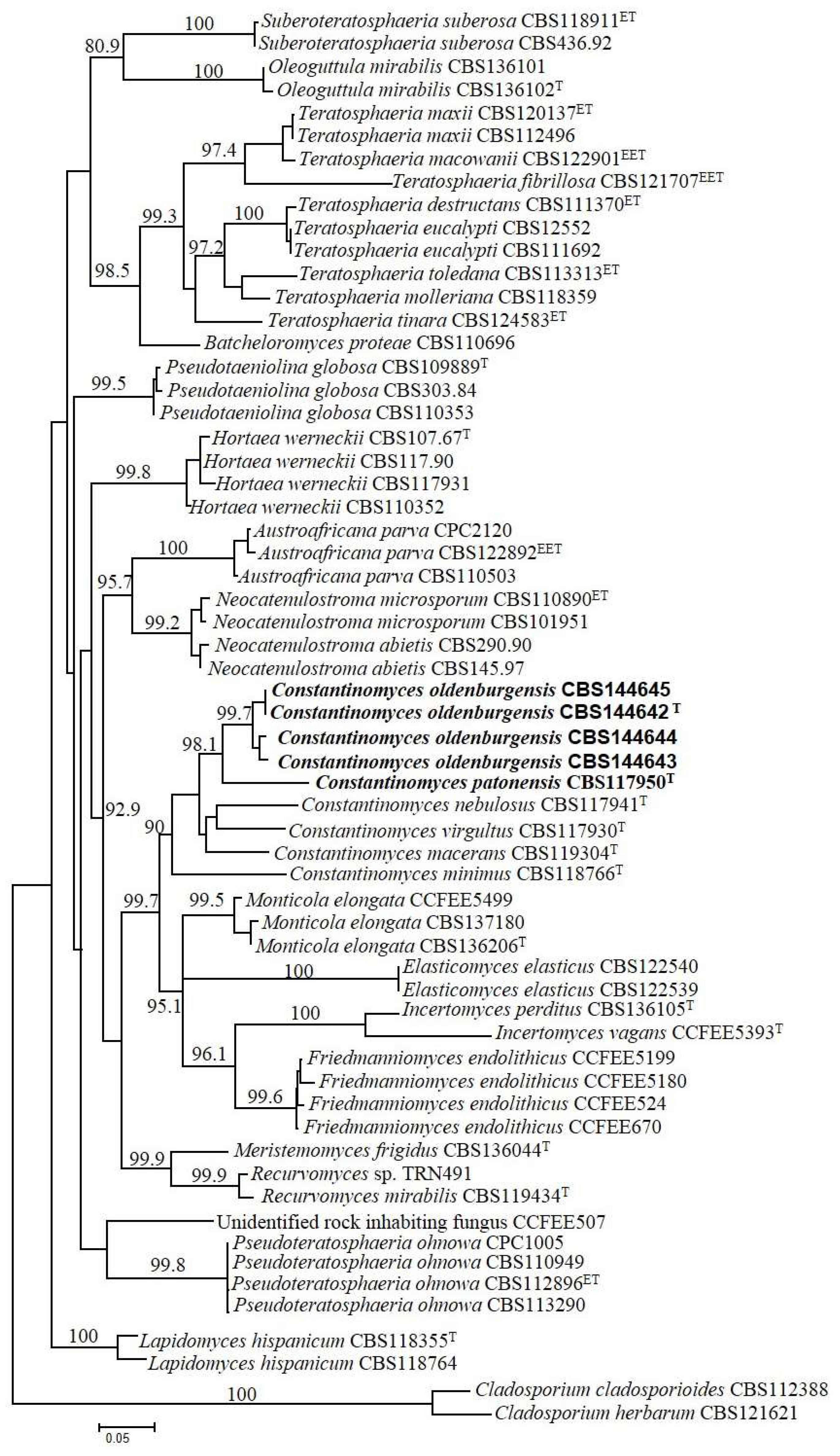
3.4. Alignment and Tree Reconstruction

Phylogeny was based on the concatenated 3-genes dataset that included 59 strains in Teratosphaeriaceae whose sequences could be confidently aligned with sequences of the studied strains; the combined dataset was based on 1681 positions (ITS: 1–525; LSU: 526–1408; RPB2: 1409–1681) including gaps. The specimens T2.1, T2.3, T2.4, T2.5 here analyzed shared five positions in ITS sequences that were different in the other close relatives analyzed; the positions and substitutions in the analyzed specimens/closest relatives are reported as follows: position 26: T/C; position 30: T/C; position 54: T/A or C; position 171–172: TT/AC or AA or AT or TC. The multilocus phylogenetic tree was generated using a GTR + G(4) model selected using the Akaike’s information criterion with a maximum likelihood (ML) approach. The tree was rooted with the strains Cladosporium cladosporioides CBS112388 and Cladosporium herbarum CBS121621 (Cladosporiaceae) and is shown in Figure 2.
The sequences analyzed belonged to strains of both rock-inhabiting and plant pathogenic fungi and also some known extremophiles, such as the halophilic species Hortaea werneckii; all strains analyzed sit in the Teratosphaeriaceae, order Capnodiales. The base frequencies were as follows: T = 0.2163, C = 0.2628, A = 0.2320, G = 0.2887.
The topology of the tree and positions of the groups, even if based on a much more restricted selection to focus on the genus Constantinomyces and relatives, is in accordance with wider recent phylogenetic studies on the family Teratosphaeriaceae [29]. Although the species C. nebulosus was placed far from C. macerans and in a misguided position in the reference study, all the species hitherto described in the genus Constantinomyces cluster together in our phylogeny. All genera highlighted in the tree were well separated and, including the genus Constantinomyces supported with 90% bootstrap. The four isolates from photocatalytic surfaces, here described as C. oldenburgensis, pooled together in a cluster with 99.7% bootstrap support; the rock-inhabitant strain TRN431 was clearly outside this group, rather isolated on a longer branch. Based on these phylogenetic evidences and different ecology, we have described this strain as a separate new species C. patonensis.

3.5. Taxonomy of Constantinomyces oldenburgensis sp. nov. and Constantinomyces patonensis sp. nov.

Phylogenetic analyses conducted in this study showed that the strains isolated from photocatalytically active roof tiles represent a taxonomic novelty within the class Dothideomycetes, order Capnodiales. The observed strains were morphologically scarcely differentiated from a rock-surface isolate, TRN431, from the Iberian penninsula. Therefore taxonomic decisions were based primarily on partial sequences of nucLSU, RPB2, and on the complete ITS regions of rDNA.
Descriptions based on 35-week-old cultures grown on MEA at 15 °C.
Constantinomyces oldenburgensis Gorbushina, Martin-Sanchez, Selbmann & Ruibal, sp. nov. MycoBank MB825220, Figure 3a–g.
Holotypus: CBS144642 = T2.1 = CCFEE6311, from a photocatalytically active roof tile, Edewecht, Germany. Culture preserved in liquid nitrogen and in dried condition.
Etymology: named after Oldenburg, the German city near the isolation source where the work on black fungi from monuments and building materials started in the 1990s.
Diagnosis: Descriptions based on 35-week-old cultures grown on MEA at 15 °C. Colonies growing very slowly in irregularly shaped colonies, velvety dark olive green to dark brown to black, dark in reverse, with regular margin, sometimes developing radiated, submerged margin at long cultivation times, raised centrally, flat near the margin. Hyphae pale to dark brown, septate, thick-walled, with apical germination producing elongated, cylindrical, irregular hyphae; in later stages torulose hyphae often present at sections, composed of swollen cells with or without transverse septa, brown, thick-walled, smooth. Chlamydospore-like cells sometimes present. Dark, spherical multicellular bodies sometimes present. Conidiophores micronematous, conidiogenous cells intercalary, occasionally with thallic-arthric conidia, liberating sparcely. Teleomorph unknown.
Note: Arthroconidia are generated by hyphae fragmentation but secession is not always completed, and adjacent cells remain connected by intercalary elongations of narrow pale connectives. Multicellular bodies are typical of the species C. oldenburgensis since they have not been reported in any of the other hiterto described species in the genus (C. virgultus, C. macerans; C. minimus; C. nebulosus). A similar trend in hyphal progression was observed in Elasticomyces elasticus, another globally distributed microcolonial black fungus developing on rocks.
Constantinomyces patonensis Ruibal & Selbmann, sp. nov. MycoBank MB825222, Figure 3h–k.
Holotypus: CBS117950 = TRN431, from granite, Patones, Central Mountain System. Culture preserved in liquid nitrogen and in dried condition.
Etymology: named after Patones, the locality in the Spanish Mountain System where the strain was isolated.
Diagnosis: Descriptions based on 35-week-old cultures grown on MEA at 15 °C. Colonies growing very slowly in irregularly shaped colonies, velvety dark olive green to dark brown to black, dark in reverse, with regular margin, raised centrally, flat near the margin. Hyphae pale to dark brown, septate, thick-walled, with apical germination producing elongated, cylindrical, regular hyphae; in later stages torulose hyphae often present at sections, composed of swollen cells with or without transverse septa, brown, thick-walled, smooth. Teleomorph unknown.
Note: No conidiogenesis of any type has been observed for this species. None of the structures observed in C. oldenburgensis, such as multicellular bodies or chlamydospore-like cells, have been found in C. patonensis. All these differences, together with those regarding its biogeography and mainly the molecular divergence of its nucITS DNA fragment, account for its definition as a different species from C. oldenburgensis.

4. Discussion

Microcolonial black fungi are specialized in exploiting different stressing niches, from saltpans to acidic sites, from hot deserts to Antarctic rocks [7,8,9,30,31,32,33,34]. They show a stunning ability to resist and overcome a number of different challenges as high and low temperatures, drought, osmotic stress, high solar and UV radiation or prolonged period of water deficiency [32,35,36,37]. Among other characteristics, the presence of melanins in the cell wall enable them to cope with different stressors, including UV- and quite intense ionizing radiations [38].
The working hypothesis in this study was that microcolonial melanized ascomycetes, usually forming resistant biofilms on subaerial rock and material surfaces, flourish in hostile conditions of highly oxidizing material surfaces and that, under these conditions, the occurrence of novel species is promoted. We also hypothesize that in additionally stressed systems that are designed for growth avoidance—like on photocatalytically active surfaces and in biocide-treated materials—stress-tolerant microcolonial ascomycetes are going to be dominant.
Four presented isolates of Constantinomyces oldenburgensis were components of a developed subaerial biofilm that formed in 7.5 years under the influence of normal environmental exposure. Growth of these material-inhabiting melanized ascomycetes was not inhibited on photocatalytically active surfaces of environmentally exposed roof tiles. It is even possible that the additional stress of photocatalytic oxidation selects for melanized, stress-tolerant strains, because (i) no microcolonial isolates could be obtained from the control tile surfaces and (ii) subaerial biofilms on the control tiles were significantly less pigmented (Figure 1a). Even if we assume that photocatalysis is not exceptionally active during mild cloudy winters of Northern Germany, differences between the control and photocatalytic surface were sufficient to ensure that microcolonial black fungi were prevailing isolates only on roof-tile surfaces with photocatalytic oxidation stress.
The genus Constantinomyces includes typical rock-inhabiting fungi, possessing two important qualities (i) occurrence on bare solid surfaces in the Mediterranean area and (ii) characteristic morphology and properties of microcolonial surface-inhabiting fungi. Ubiquitous findings of these strains underline the similarities existing between SAB niches on man-made materials (that are recent anthropogenic additions to Earth ecosystems) to naturally existing rock surfaces that were the first terrestrial environments of this planet [1].
The intraspecific variability within the C. oldenburgensis clade accounted to 1.5%. This is not as high as found for other black microcolonial fungi, as Friedmanniomyces endolithicus or Elasticomyces elasticus, where the intraspecific variability was calculated as high as 4–4.5%. This may be explained as a sampling effect, since the cluster here analyzed is composed of four strains only, while the two examples reported above included up to 10 representatives [31]. Alternatively it could be a consequence of the restricted sampling location: in fact the strains here examined came from photocatalitically active surfaces of two adjacent tiles (Figure 1d), while strains of the examples above were from distant locations (for E. elasticus even different continents). Based on the phylogenetic position of TRN431 (rather isolated compared to all the other species, on a long branch and support of the common branch 98.6%) as well as considering its different ecology (C. oldenburgensis is from roof tiles and TRN431 from natural rocks) we described TRN431 as a separate species, C. patonensis.
Melanized rock- and material-inhabiting (essentially subaerial-biofilm-inhabiting) ascomycetes form a resource of climate-dependent and anthropogenically influenced biodiversity. Characteristic properties of melanized rock-inhabiting fungi are their stress tolerance and substrate interaction abilities. These aptitudes enable these organisms to form visible and biodeterioration-involved subaerial biofilms on exposed modern materials—exactly in the same fashion as their ancestors once conquered the surfaces of desert boulders. As reactions of these melanized microcolonial fungi to growth-avoiding measures will give us clues to design material protection measures, these organisms are a focus of applied research. Melanized fungi that persist under realistic material exposure conditions are also promising candidates as new reference organisms in material testing procedures.

5. Conclusions

Progress in biodeterioration sciences depends on reflecting the constantly evolving biodiversity of the biofilms associated with specific biodeterioration phenomena. The diversity on bare material surfaces is high, largely unexplored and highly dynamic. Forces that influence this biodiversity are the introduction of new materials along with climatic impacts and climate change. Awareness of this diversity is necessary for advancement in the biodeterioration field and necessitates application of new isolation techniques and state-of-art characterization of resulting strains, followed by employing these strains in novel material testing procedures. Isolation, characterization and description of new material-associated strains will support (i) broadening our knowledge of the ecosystem associated with material biodeterioration and (ii) designing new material testing procedures with dominant organisms.
For exposed material surfaces from rock to roof tiles to solar panels, melanized fungal ascomycetes arise as a group of special interest. To understand the diversity of microbial settlers on bare material surfaces, coordinated efforts of biodeterioration scientists, culture collections and “black-fungi-experienced” taxonomy and phylogeny specialists are indispensable.

Author Contributions

C.R. contributed C. patonensis strain TRN431 as well as comparative morphological analysis tools, wrote the taxonomical description as well as deposited the strains in the CBS culture collection; L.S. used the whole set of molecular data for phylogenetic analysis, selected a relevant comparative framework of strains, contributed tree-construction and -analysis tools and wrote the phylogenetic part of the paper; S.A. isolated fungal strains from the tile samples into a case-specific pure culture collection; P.M.M.-S. analyzed this original strain collection, singled out potential novel species, performed amplification and sequencing of ITS, LSU and RPB2 genes, and critically contributed to method development and writing the paper; A.A.G. designed the tile monitoring experiment, performed the sampling and biofilm development description and wrote the paper using coauthors’ contributions.

Acknowledgments

This work in part of lab consumables for isolates growth, DNA extraction and PCR amplification as well as staff positions was financially supported by internal funds of the BAM. During this study CR’s position was funded by the Spanish Ministerio de Economía y Compeptitividad (CGL2013-42498-P). Wolfgang Krumbein and BIOGEMA are thanked for their support in developing and monitoring the test site. The Italian National Antarctic Museum is acknowledged for funding the Culture Collection of Fungi from Extreme Environments where the strains were deposited and the related sequences database used for implementing the phylogenetic study.

Conflicts of Interest

The authors declare no conflict of interest.

References

  1. Gorbushina, A.A. Life on the rocks. Environ. Microbiol. 2007, 9, 1613–1631. [Google Scholar] [CrossRef] [PubMed] [Green Version]
  2. Gadd, G.M. Fungi, rocks, and minerals. Elements 2017, 13, 171–176. [Google Scholar] [CrossRef]
  3. Staley, J.T.; Palmer, F.; Adams, J.B. Microcolonial fungi: Common inhabitants on desert rocks? Science 1982, 215, 1093–1095. [Google Scholar] [CrossRef] [PubMed]
  4. Urzì, C.; Krumbein, W.E.; Warscheid, T. On the question of biogenic color changes of Mediterranean monuments (Coatings, Crust, Microstromatolite, Patina, Scialbatura, Skin, Rock Varnish). In Proceedings of the 2nd International Symposium “The Conservation of Monuments in the Mediterranean Basin, Genève, Switzerland, 19–21 November 1991. [Google Scholar]
  5. Wollenzien, U.; De Hoog, G.S.; Krumbein, W.E.; Urzi, C. On the isolation of microcolonial fungi occurring on and in marble and other calcareous rocks. Sci. Total Environ. 1995, 167, 287–294. [Google Scholar] [CrossRef]
  6. Sterflinger, K.; Krumbein, W.E. Dematiaceous fungi as a major agent for biopitting on Mediterranean marbles and limestones. Geomicrobiol. J. 1997, 14, 219–230. [Google Scholar] [CrossRef]
  7. Ruibal, C.; Platas, G.; Bills, G.F. Isolation and characterization of melanized fungi from limestone formations in Mallorca. Mycol. Prog. 2005, 4, 23–38. [Google Scholar] [CrossRef]
  8. Ruibal, C.; Platas, G.; Bills, G.F. High diversity and morphological convergence among melanized fungi from rock formations in the Central Mountain System of Spain. Persoonia 2008, 21, 93–110. [Google Scholar] [CrossRef] [PubMed]
  9. Isola, D.; Zucconi, L.; Onofri, S.; Caneva, G.; De Hoog, G.S.; Selbmann, L. Extremotolerant rock inhabiting black fungi from Italian monumental sites. Fungal Divers. 2016, 76, 75–96. [Google Scholar] [CrossRef]
  10. Gorbushina, A.A.; Diakumaku, E.; Krumbein, W.E.; (Geomicrobiology, Carl von Ossietzky University, Oldenburg, Germany). Personal communication, 1996.
  11. Selbmann, L.; Zucconi, L.; Isola, D.; Onofri, S. Rock black fungi: Excellence in the extremes. From the Antarctic to Space. Curr. Genet. 2015, 61, 335–345. [Google Scholar] [CrossRef] [PubMed]
  12. Selbmann, L.; Onofri, S.; Zucconi, L.; Isola, D.; Rottigni, M.; Ghiglione, C.; Piazza, P.; Alvaro, M.C.; Schiaparelli, S. Distributional records of Antarctic fungi based on strains preserved in the Culture Collection of Fungi from Extreme Environments (CCFEE) Mycological Section associated with the Italian National Antarctic Museum (MNA). MycoKeys 2015, 10, 57–71. [Google Scholar] [CrossRef] [Green Version]
  13. Martin-Sanchez, P.M.; Gebhardt, C.; Toepel, J.; Barry, J.; Munzke, N.; Günster, J.; Gorbushina, A.A. Monitoring microbial soiling in photovoltaic systems: A qPCR-based approach. Int. Biodeterior. Biodegrad. 2018, 129, 13–22. [Google Scholar] [CrossRef]
  14. Gorbushina, A.A.; Krumbein, W.E.; Hamman, C.H.; Panina, L.; Soukharjevski, S.; Wollenzien, U. Role of black fungi in color change and biodeterioration of antique marbles. Geomicrobiol. J. 1993, 11, 205–221. [Google Scholar] [CrossRef]
  15. Sterflinger, K.; De Baere, R.; De Hoog, G.S.; De Wachter, R.; Krumbein, W.E.; Haase, G. Coniosporium perforans and C. apollinis, two new rock-inhabiting fungi isolated from marble in the Sanctuary of Delos (Cyclades, Greece). Antonie van Leeuwenhoek 1997, 72, 349–363. [Google Scholar] [CrossRef] [PubMed]
  16. Gorbushina, A.A.; Krumbein, W.E.; Volkmann, M. Rock surfaces as life indicators: New ways to demonstrate life and traces of former life. Astrobiology 2002, 2, 203–213. [Google Scholar] [CrossRef] [PubMed]
  17. Dornieden, T.; Gorbushina, A.A.; Krumbein, W.E. Aenderungen der physikalischen Eigenschaften von Marmor durch Pilzbewuchs. Int. J. Restor. Build. Monum. 1997, 3, 441–456. [Google Scholar]
  18. White, T.J.; Bruns, T.; Lee, S.; Taylor, J. Amplification and direct sequencing of fungal ribosomal RNA genes for phylogenetics. In PCR Protocols: A Guide to Methods and Applications; Innis, M.A., Gelfand, D.H., Sninsky, J.J., White, T.J., Eds.; Academic Press: San Diego, CA, USA, 1990; pp. 315–322. [Google Scholar]
  19. Vilgalys, R.; Hester, M. Rapid genetic identification and mapping of enzymatically amplified ribosomal DNA from several Cryptococcus species. J. Bacteriol. 1990, 172, 4239–4246. [Google Scholar] [CrossRef]
  20. Liu, Y.J.; Whelen, S.; Hall, B.D. Phylogenetic relationships among Ascomycetes: Evidence from an RNA Polymerase II subunit. Mol. Biol. Evol. 1999, 16, 1799–1808. [Google Scholar] [CrossRef] [PubMed]
  21. Altschul, S.F.; Madden, T.L.; Schaffer, A.A.; Zhang, J.; Zhang, Z.; Miller, W.; Lipman, D.J. Gapped BLAST and PSI-BLAST: A new generation of protein database search programs. Nucleic Acids Res. 1997, 25, 3389–3402. [Google Scholar] [CrossRef] [PubMed]
  22. Edgar, R.C. MUSCLE: Multiple sequence alignment with high accuracy and high throughput. Nucleic Acids Res. 2004, 32, 1792–1797. [Google Scholar] [CrossRef] [PubMed]
  23. Tamura, K.; Stecher, G.; Peterson, D.; Filipski, A.; Kumar, S. MEGA6, Molecular Evolutionary Genetics Analysis Version 6.0. Mol. Biol. Evol. 2013, 30, 2725–2729. [Google Scholar] [CrossRef] [PubMed]
  24. Villesen 2017. Available online: http://users-birc.au.dk/biopv/php/fabox/alignment_joiner.php (accessed on 22 November 2017).
  25. Nylander, J.A.A. Mr AIC.pl. Programme Distributed by the Author; Evolutionary Biology Centre, Uppsala University: Uppsala, Sweden, 2004. [Google Scholar]
  26. Guindon, S.; Gascuel, O. A simple, fast, and accurate algorithm to estimate large phylogenies by maximum likelihood. Syst. Biol. 2003, 52, 696–704. [Google Scholar] [CrossRef] [PubMed]
  27. Jobb, G.A.; von Haeseler, A.; Strimmer, K. TREEFINDER: A powerful graphical analysis environment for molecular phylogenetics. BMC Evol. Biol. 2004, 4, 18. [Google Scholar] [CrossRef] [PubMed] [Green Version]
  28. Felsenstein, J. Confidence limits on phylogenies: An approach using the bootstrap. Evolution 1985, 40, 783–791. [Google Scholar] [CrossRef] [PubMed]
  29. Quaedvlieg, W.; Binder, M.; Groenewald, J.Z.; Summerell, B.A.; Carnegie, A.J.; Burgess, T.I.; Crous, P.W. Introducing the Consolidated Species Concept to resolve species in the Teratosphaeriaceae. Persoonia 2014, 33, 1–40. [Google Scholar] [CrossRef] [PubMed] [Green Version]
  30. Plemenitaš, A.; Gunde-Cimerman, N. Cellular responses in the halophilic black yeast Hortaea werneckii to high environmental salinity. In Adaptation to Life at High Salt Concentrations in Archaea, Bacteria, and Eukarya; Gunde-Cimerman, N., Oren, A., Plemenitaš, A., Eds.; Springer: Dordrecht, The Netherlands, 2005; pp. 455–470. [Google Scholar]
  31. Selbmann, L.; de Hoog, G.S.; Mazzaglia, A.; Friedmann, E.I.; Onofri, S. Fungi at the edge of life: Cryptoendolithic black fungi from Antarctic deserts. Stud. Mycol. 2005, 51, 1–32. [Google Scholar]
  32. Selbmann, L.; de Hoog, G.S.; Zucconi, L.; Isola, D.; Ruisi, S.; Gerrits van den Ende, A.H.G.; Ruibal, C.; De Leo, F.; Urzì, C.; Onofri, S. Drought meets acid: Three new genera in a dothidealean clade of extremotolerant fungi. Stud. Mycol. 2008, 61, 1–20. [Google Scholar] [CrossRef] [PubMed]
  33. Selbmann, L.; Isola, D.; Egidi, E.; Zucconi, L.; Gueidan, C.; de Hoog, G.S.; Onofri, S. Mountain tips as reservoirs for new rock-fungal entities: Saxomyces gen. nov. and four new species from the Alps. Fungal Divers. 2014, 65, 167–182. [Google Scholar] [CrossRef]
  34. Egidi, E.; de Hoog, G.S.; Isola, D.; Onofri, S.; Quaedvlieg, W.; de Vries, M.; Verkley, G.J.M.; Stielow, J.B.; Zucconi, L.; Selbmann, L. Phylogeny and taxonomy of meristematic rock-inhabiting black fungi in the dothidemycetes based on multi-locus phylogenies. Fungal Divers. 2014, 65, 127–165. [Google Scholar] [CrossRef]
  35. Sterflinger, K. Black Yeasts and Meristematic Fungi: Ecology, Diversity and Identification. In Biodiversity and Ecophysiology of Yeasts; Péter, G., Rosa, C., Eds.; Springer: Berlin/Heidelberg, Germany, 2006; pp. 501–514. [Google Scholar]
  36. Selbmann, L.; Isola, D.; Zucconi, L.; Onofri, S. Resistance to UV-B induced DNA damage in extreme-tolerant cryptoendolithic Antarctic fungi: Detection by PCR assays. Fungal Biol. 2011, 115, 937–944. [Google Scholar] [CrossRef] [PubMed]
  37. Zakharova, K.; Tesei, D.; Marzban, G.; Dijksterhuis, J.; Wyatt, T.; Sterflinger, K. Microcolonial fungi on rocks: A life in constant drought? Mycopathologia 2013, 175, 537–547. [Google Scholar] [CrossRef] [PubMed]
  38. Pacelli, C.; Selbmann, L.; Zucconi, L.; Raguse, M.; Moeller, R.; Shuryak, I.; Onofri, S. Survival, DNA integrity and ultrastructural damage in Antarctic cryptoendolithic eukaryotic microorganisms exposed to ionizing radiation. Astrobiology 2017, 17, 126–135. [Google Scholar] [CrossRef] [PubMed]
Figure 1. The roof of a residential house with indicated sampled tiles at the moment of sampling in May 2014—after 7.5 years of total exposure and 59 months of exposure beside each other. This area is free from shading by architectural elements and thus photocatalytic activity might occur at least during sun irradiation periods. (a) The position of the two sampled photocatalytically active tiles on the control part of the roof was “home” to those tiles for almost five years prior to the sampling. The photocatalytically active tiles have the visibly different subaerial biofilm that is more discoloring. (b) Sampled tile surface in a close-up showing dark-pigmented fungal colonies on the elevated surface area—the area where time of wetness is additionally reduced, leading to less pronounced algal growth. Fungal colonies that were sampled with sterile implements to isolate dominant subaerial settlers are indicated by the blue frame.
Life 08 00030 g001
Figure 2. ML 3-genes multilocus phylogeny based on a selection of rock-inhabiting and plant pathogenic fungi in the order Capnodiales showing the phylogenetic placement of the new species described. The tree, based on 59 sequences and 1681 nucleotide positions, has been generated using a GTR + G(4) model calculated using ML in the software MrAIC. Bootstrap values above 80%, calculated from 1000 resampled data sets, are shown.
Life 08 00030 g002
Figure 3. Appearance of the Constantinomyces oldenburgensis and Constantinomyces patonensis. (ag) Constantinomyces oldenburgensis, strain T2.1. (a) Colony appearance. (b) Dark, spherical multicellular bodies. Scale bar 50 µm. (c) Irregular toruloid brown hyphae. Scale bar 25 µm. (d) Irregular toruloid brown hyphae with development of chlamydospore-like cells. Scale bar 25 µm. (e) Micronematous conidiophores and arthroconidia. Scale bar 25 µm. (fg) Micronematous conidiophores and arthroconidia, details. Scale bars 10 µm. (hj) Constantinomyces patonensis, strain TRN431. (h) Colony appearance. (ij) Irregular toruloid brown hyphae. Scale bars 25 µm.
Life 08 00030 g003
Table 1. Strains used in this study.
SpeciesStrainOriginLocationGenBank Accession Number
ITSLSURPB2
Austroafricana parvaCBS122892 = CPC 12421 EETEucalyptus globulusAustraliaKF901514KF901832KF902193
Austroafricana parvaCBS110503 = CMW 14459Eucalyptus globulusAustraliaKF901513KF901831KF902189
Austroafricana parvaCPC2120Protea repensSouth AfricaAY260091--
Batcheloromyces proteaeCBS110696 = CPC1518 = CPC18701Protea cynaroidesSouth Africa-KF901833KF902195
Constantinomyces maceransCBS119304 = TRN440 TGranite, Patones, Central Mountain SystemPatones, SpainAY843139KF310005KF310081
Constantinomyces nebulosusCBS117941 = TRN262 TGranite, Atazar, Central Mountain SystemAtazar, SpainAY843109KF310014KF310068
Constantinomyces oldenburgensisTCBS144642 = CCFEE6311 = T2.1Photocatalytically active roof tilesEdewecht, GermanyLT976552 *LT976552 *LT976526
Constantinomyces oldenburgensisCBS144643 = CCFEE6310 = T2.3Photocatalytically active roof tilesEdewecht, GermanyLT976553 *LT976553 *LT976527
Constantinomyces oldenburgensisCBS144644 = CCFEE6309 = T2.4Photocatalytically active roof tilesEdewecht, GermanyLT976554 *LT976554 *LT976528
Constantinomyces oldenburgensisCBS144645 = CCFEE6305 = T2.5Photocatalytically active roof tilesEdewecht, GermanyLT976555 *LT976555 *LT976529
Constantinomyces minimusCBS118766 = TRN159Granite, La Cabrera, Central Mountain SystemLa Cabrera, SpainAY843066KF310003KF310077
Constantinomyces patonensisTCBS117950 = TRN431Granite, Patones, Central Mountain SystemPatones, SpainAY843129KF310004KF310080
Constantinomyces virgultusCBS117930 = TRN79 TLimestone, Cala Sant VicençMallorca, SpainAY559339GU323964KF310082
Elasticomyces elasticusCBS122539 = CCFEE5319Lecanora sp.AntarcticaFJ415475GU250375Unpublished
Elasticomyces elasticusCBS122540 = CCFEE5320Usnea antarcticaAntarcticaFJ415476GU250376-
Friedmanniomyces endolithicusCCFEE5199RockAntarcticaJN885547KF310007KF310093
Friedmanniomyces endolithicusCCFEE5180RockAntarcticaJN885544GU250367-
Friedmanniomyces endolithicusCBS119426 = CCFEE670RockAntarcticaJN885542GU250366KF310056
Friedmanniomyces endolithicusCBS119427 = CCFEE 524RockAntarcticaJN885541GU250364KF310054
Hortaea werneckiiCBS107.67 TTinea nigra: manPortugalAJ238468EU019270-
Hortaea werneckiiCBS117.90Salted fish, Osteoglossum bicirrhosumBrazilAJ238472--
Hortaea werneckiiCBS117931 = TRN122RockCala Sant Vicenç, Mallorca, SpainAY559357GU323969KF310058
Hortaea werneckiiCBS110352Hollow treeKarthoumUnpublished--
Incertomyces perditusCBS136105 = CCFEE 5385 TRockAlps, ItalyKF309977KF310008KF309977
Incertomyces vagansCCFEE5393 TRockAlps, ItalyKF309964KF310009KF309964
Lapidomyces hispanicusCBS118355 = TRN500RockPuebla la Sierra, SpainAY843182KF310017-
Lapidomyces hispanicusCBS118764 = TRN126Cala Sant VicençMallorca, SpainAY559361KF310016KF310076
Meristemomyces frigidusCBS136044 = CCFEE5401 TRockAlps, ItalyKF309966GU250383KF310105
Monticola elongataCCFEE5499RockAlps, ItalyKF309969GU250398KF310065
Monticola elongataCBS137180 = CCFEE 5492RockAlps, ItalyKF309968KF309994KF310064
Monticola elongataCBS136206 = CCFEE5394 TRockAlps, ItalyKF309965KF309995KF309965
Neocatenulostroma abietisCBS145.97Sandstone of cathedralGermanyAJ244265--
Neocatenulostroma abietisCBS290.90Skin lesionNetherlandsAJ244267--
Neocatenulostroma microsporumCBS110890 = CPC1832 ETProtea cynaroidesSouth AfricaAY260097EU019255-
Neocatenulostroma microsporumCBS101951 = CPC1960 ETProtea cynaroidesSouth AfricaAY260097EU019255-
Oleoguttula mirabilisCBS136101 = CCFEE5522RockAntarctic PeninsulaKF309972KF310019KF310070
Oleoguttula mirabilisCBS136102 = CCFEE5523 TRockAntarctic PeninsulaKF309973KF310031Unpublished
Pseudoteratosphaeria ohnowaCBS112896 = CPC1004 ETEucalyptus grandisSouth AfricaKF901620KF901946KF902348
Pseudoteratosphaeria ohnowaCPC1005Eucalyptus grandisSouth AfricaAF173299GU214511-
Pseudoteratosphaeria ohnowaCBS113290 = CMW9102Eucalyptus smithiiSouth AfricaKF937236-KF937270
Pseudoteratosphaeria ohnowaCBS110949 = CPC1006Eucalyptus grandisSouth AfricaAY725575--
Pseudotaeniolina globosaCBS109889 TRockItalyNR136960EU019283-
Pseudotaeniolina globosaCBS303.84RockItalyAJ244268--
Pseudotaeniolina globosaCBS110353Human aorta at autopsyGermanyUnpublished--
Recurvomyces mirabilisCBS119434 = CCFEE5264 TSandstoneAntarcticaFJ415477GU250372KF310059
Recurvomyces sp.CBS117957 = TRN491QuarzitePuebla de la Sierra, SpainAY1843175--
Suberoteratosphaeria suberosaCBS11891 = CPC12085 ETEucalyptus sp.UruguayKF901786KF902144-
Suberoteratosphaeria suberosaCBS436.92 = CPC515 ETEucalyptus dunniiBrazilKF901623KF901949KF902404
Teratosphaeria destructansCBS111370 = CPC1368 ETEucalyptus grandisIndonesiaKF901574KF901898KF902427
Teratosphaeria eucalyptiCBS111692 = CMW14910 = CPC1582Eucalyptus sp.New Zealand-KF902119-
Teratosphaeria eucalyptiCPC 12552Eucalyptus nitensTasmaniaKF901576KF901900KF902429
Teratosphaeria fibrillosaCBS121707 = CPC13960 EETProtea sp.South AfricaKF901728KF902075-
Teratosphaeria macowaniiCBS122901 = CPC13899 EETProtea nitidaSouth AfricaKF937241-KF937276
Teratosphaeria maxiiCBS120137 = CPC12805 ETProtea repensSouth AfricaKF937243-KF937278
Teratosphaeria maxiiCBS112496 = CPC3322Protea sp.Australia-KF937242KF937277
Teratosphaeria mollerianaCBS118359 = CMW11560Eucalyptus globulusTasmaniaKF901764KF902120KF902451
Teratosphaeria tinaraCBS124583 = MUCC666 ETCorymbia sp.AustraliaKF901599KF901923KF902491
Teratosphaeria toledanaCBS113313 = CMW14457 ETEucalyptus spSpainKF901734KF902081KF902492
Unidentified rock inhabiting fungusCCFEE507Powdered rocksAntarcticaUnpublished--
CBS: Westerdijk Fungal Biodiversity Institute, Utrecht, The Netherlands; CCFEE: Culture Collection of Fungi from Extreme Environments, DEB, University of Tuscia, Viterbo, Italy; CMW: Culture collection of the Forestry and Agricultural Biotechnology Institute (FABI) of the University of Pretoria, Pretoria, South Africa; CPC: Collection Pedro Crous; MUCC: Murdoch University Culture Collection, Murdoch, Australia; NZFS: Forest Research Culture Collection, Private Bag 3020, Rotorua, New Zealand; STE-U: Department of Plant Pathology, University of Stellenbosch, South Africa; TRN: C. Ruibal private collection; EET: ex-epitype; ET: ex-type. ITS: internal transcribed Spacers; LSU: Large SubUnit; RPB2: RNA Polymerase II Large Subunit. * ITS plus LSU submitted as single sequence in the GenBank.

