Case Report

Intensive Management of a Patient with HIV, Active Tuberculosis, and COVID-19: A Multidisciplinary Approach in the Intensive Care Unit

by
Brayan Ricardo Mosquera-Arias
1,
Valeria Sanclemente-Cardoza
2 and
Jose Luis Estela-Zape
1,3,*
1
Faculty of Health, Universidad Santiago de Cali, Cali 760035, Colombia
2
Faculty of Health, School of Public Health, Universidad del Valle, Cali 760043, Colombia
3
Health and Movement Research Group, Universidad Santiago de Cali, Cali 760035, Colombia
*
Author to whom correspondence should be addressed.
Life 2025, 15(9), 1435; https://doi.org/10.3390/life15091435
Submission received: 22 August 2025 / Revised: 7 September 2025 / Accepted: 11 September 2025 / Published: 13 September 2025
(This article belongs to the Section Medical Research)

Abstract

Coinfection with HIV, active tuberculosis, and COVID-19 is rare but markedly increases mortality risk and complicates treatment due to the interactions between these infections. Management requires a multidisciplinary approach that integrates antiretroviral therapy, antituberculous drugs, antibiotics, and supportive care for COVID-19. We report the case of a 28-year-old male with HIV (viral load 30 copies, CD4 count 303), active tuberculosis, and a history of resolved syphilis, who presented with severe respiratory decompensation and hypoxemia (SpO2 55%), requiring orotracheal intubation. Initial treatment included broad-spectrum antibiotics, antiretrovirals, and antituberculous therapy. Despite the critical illness, the patient demonstrated progressive clinical improvement, was successfully extubated after a spontaneous breathing trial, and continued recovery under supplemental oxygen. This case underscores the clinical complexity of triple coinfection and highlights the potential for favorable outcomes when management is timely and multidisciplinary.

1. Introduction

The management of patients with HIV, active tuberculosis (TB), and COVID-19 presents a significant clinical challenge due to the complex interactions between these infections. Coinfection with these three diseases is uncommon but leads to a substantial increase in morbidity and mortality [1,2,3]. HIV weakens immune function, making individuals more susceptible to opportunistic infections like TB, which remains a major cause of death among HIV-positive individuals [4]. The COVID-19 pandemic further complicates this by impairing respiratory function and modifying immune responses, which increase the risk of severe complications in coinfected patients [5].
The combination of HIV and active TB in patients with COVID-19 significantly raises mortality, with a 38% increased risk of death compared to those without these comorbidities [6]. Additionally, active TB exacerbates pulmonary dysfunction, increasing the likelihood of respiratory failure in the context of COVID-19, which complicates clinical management [7].
The management of these patients requires a multidisciplinary approach that integrates antiretroviral therapy (ART), antituberculous treatment, and appropriate COVID-19 care [8,9,10]. Therapeutic strategies must be tailored to minimize drug interactions and adverse effects. This case report outlines the intensive clinical management of a patient with HIV, active TB, and COVID-19, emphasizing the application of an integrated therapeutic approach and multidisciplinary intervention in the context of complex comorbidities. The report is presented in accordance with the SCARE 2023 guidelines [11], ensuring methodological rigor and transparency in case reporting.

2. Case Report

A 28-year-old male patient with a history of resolved syphilis, HIV (viral load 30 copies, CD4 count 303), and active TB (first phase of treatment initiated on 9 August 2024, with expected completion by 13 October 2024) is currently undergoing the second phase of TB treatment (biconjugate therapy, initiated on 14 October 2024).
He was presented with decompensation for his underlying condition, characterized by 5 h of respiratory distress, multiple episodes of vomiting, and stool type 4 according to the Bristol Stool Chart [12].
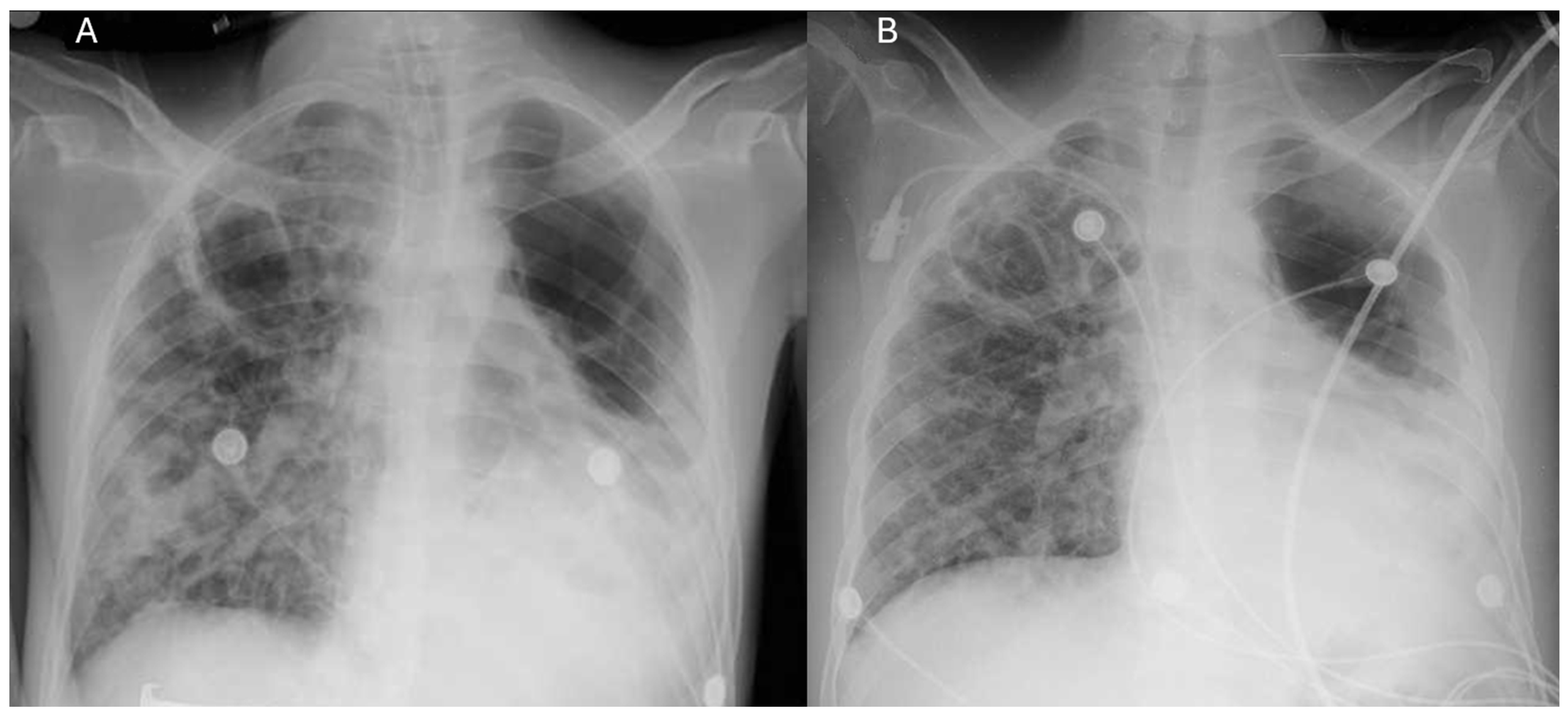
Upon admission, the patient exhibited progressive respiratory deterioration, utilizing accessory muscles and presenting an oxygen saturation (SpO2) of 55% despite a 50% Venturi mask, prompting orotracheal intubation. Sedation with midazolam and fentanyl, continuous cisatracurium infusion for neuromuscular blockade, and dual vasopressor support (norepinephrine and vasopressin) were required. Baseline chest imaging at the initiation of antituberculous therapy revealed left pneumothorax (~30%), multiple bilateral bullae, interstitial-alveolar infiltrates, and left basal opacification, consistent with cavitary pulmonary TB. Orotracheal intubation was performed, and diagnostic studies (Table 1) were ordered, including blood tests, a nasal swab for SARS-CoV-2 PCR-RT (Table 2), a chest angiotomography (Angio-CT) (Figure 1), and a chest X-ray (Figure 2).
In the intensive care unit (ICU), the patient developed persistent tachycardia and a tendency toward hypotension (mean arterial pressure [MAP] < 60 mmHg). Dual vasopressor support was initiated with norepinephrine (1.0 mcg/kg/min) and vasopressin (2.4 U/h). Laboratory results showed impaired oxygenation and elevated C-reactive protein (CRP), confirming the diagnosis of COVID-19 (Table 2), along with leukocytosis. Treatment with dexamethasone (6 mg/24 h) was started according to the RECOVERY protocol [13], and empirical antibiotic coverage was initiated with meropenem (2 g/8 h) and linezolid (600 mg/12 h), given the risk of secondary infections in an immunocompromised patient.
Chest imaging revealed cavities and bronchiectasis, leading to the initiation of vancomycin (1 g/12 h), liposomal amphotericin B (250 mg/24 h), and trimethoprim/sulfamethoxazole (80/400 mg/8 h). Clarification was required: The indication, duration, and discontinuation dates for each agent needed to be specified to support the therapeutic rationale. In particular, the TMP/SMX dose corresponds to prophylaxis; therefore, it must be clarified whether it was administered as prophylaxis or treatment, with the dose adjusted accordingly. These decisions were made in the absence of microbiological cultures or fungal markers, which should be reported or, alternatively, discussed within the framework of preventative strategies in profound immunosuppression.
Persistent respiratory acidosis necessitated continuous cisatracurium infusion (5 mcg/kg/min) to facilitate mechanical ventilation and maintain an RASS score of −4. Midazolam (0.05 mg/kg/h) and fentanyl (0.05 mcg/kg/h) were used for sedation and analgesia.
Antiretroviral and antituberculosis regimens were continued with emtricitabine (200 mg/4 h), tenofovir (300 mg/24 h), dolutegravir (50 mg/24 h), and rifampicin/isoniazid (4 tablets/24 h, continuation phase). It should also be clarified whether pyrazinamide and ethambutol were included during the initial intensive phase, with corresponding dates, to complete the therapeutic timeline.
After treatment adjustment, oxygenation and hemodynamic status improved, allowing the discontinuation of vasopressin, cisatracurium, and midazolam, with norepinephrine reduced to 0.06 mcg/kg/min. Propofol (1–4 mg/kg/h) was introduced to assess extubation readiness. A 120 min spontaneous breathing trial with a negative inspiratory force (NIF) of −40 cmH2O confirmed readiness, and the patient was successfully extubated to a 35% Venturi mask. Persistent hypoxemia (PaO2/FiO2 < 150) required escalation to high-flow nasal cannula (HFNC) at 50%, followed by de-escalation to low-flow nasal cannula at 3 L/min and subsequent discontinuation.
Auscultation revealed bilateral vesicular sounds with apical rhonchi, leading to the addition of ipratropium bromide (3 inhalations every 6 h). With subsequent improvement, oxygen support was progressively weaned from HFNC to nasal cannula at 3 L/min, which was later discontinued, and the patient was transferred to the general ward once oxygenation parameters stabilized.

3. Discussion

Coinfection with HIV, active TB, and COVID-19 is rare but markedly increases morbidity and mortality. This overlap presents major diagnostic and therapeutic challenges because of similar clinical features and complex drug interactions. HIV-related immunosuppression increases susceptibility to TB and complicates both the diagnosis and treatment decisions. The overlapping symptoms of HIV and TB often lead to diagnostic delays and inadequate management. In addition, immune dysfunction in HIV can result in false-negative TB tests, emphasizing the need for more sensitive diagnostic methods, such as molecular assays, for early detection [14].
Coinfection with COVID-19 further complicates the scenario, as respiratory manifestations may obscure TB-related findings, hinder clinical evaluation, and increase the risk of severe complications [15]. Early and accurate diagnosis is therefore essential to initiate timely therapy that adequately addresses all three infections. TB treatment also requires adjustment to account for interactions with ART. For example, rifampicin reduces the efficacy of several ART agents, making dose modification or substitution with alternatives such as rifabutin necessary [9,14,16,17].
Patients coinfected with HIV and TB who initiate antiretroviral therapy (ART) remain at significant risk of immune reconstitution inflammatory syndrome (IRIS). This immunopathological response, triggered by ART-induced immune recovery, can unmask or worsen TB, contributing to morbidity and complicating treatment. Consequently, close clinical surveillance is necessary to enable early recognition and timely therapeutic adjustments that mitigate its impact [2,3].
Management of HIV–TB coinfection also requires careful coordination to minimize pharmacological interactions. Rifampicin, an essential component of TB therapy, induces hepatic metabolism of several antiretroviral agents, reducing their plasma concentrations and therapeutic efficacy. In such cases, either dose modification or substitution with rifabutin becomes necessary to maintain virological suppression while preserving TB treatment effectiveness [17]. These adjustments are central to preventing opportunistic infections and avoiding therapeutic failure.
The timing of ART initiation in active TB remains a critical clinical decision. Current guidelines recommend initiating ART within two weeks for patients with CD4 counts below 50 cells/mm3, yet this must be balanced against the heightened risk of IRIS. Thus, treatment decisions require individualized assessment, considering both the benefits of early immune recovery and the potential complications arising from immune dysregulation [18,19].
The coexistence of COVID-19 further amplifies these challenges. Overlapping respiratory manifestations may accelerate pulmonary compromise, especially in immunocompromised hosts, increasing the risk of acute respiratory failure. In such scenarios, therapeutic strategies must remain flexible, integrating ART, antituberculous therapy, and COVID-19 management while anticipating the need for advanced respiratory support, including invasive ventilation [5,7].
Mortality in patients coinfected with HIV and TB is significantly higher when associated with COVID-19. Recent studies [3,4,6,9,20] indicate that TB may exacerbate the severity and mortality of COVID-19, emphasizing the need for the integrated management of these concurrent infections. In such cases, broad-spectrum antibiotics such as vancomycin, antifungals like liposomal amphotericin B, and antibacterial agents such as trimethoprim-sulfamethoxazole are important to prevent secondary infections, which are frequent in immunocompromised patients [4,9,14,18] and may contribute to additional pulmonary complications and increased mortality.
ART was continued with emtricitabine (200 mg once daily), tenofovir disoproxil fumarate (300 mg once daily), and dolutegravir (50 mg once daily), with dosing adjustments for rifampicin co-administration. Active TB was managed with an intensive phase of quadruple therapy including isoniazid (300 mg/day), rifampicin (600 mg/day), pyrazinamide (1500 mg/day), and ethambutol (1200 mg/day) for two months, followed by a continuation phase with isoniazid (300 mg/day) and rifampicin (600 mg/day). Adjunctive corticosteroid therapy was administered in the setting of extensive cavitary TB, recognizing that outcomes in immunocompromised patients are variable and may only partially mitigate inflammatory complications [14,20].
Managing respiratory failure was a central component of treatment. The patient developed severe respiratory decompensation that required intubation and invasive mechanical ventilation. Sedation with midazolam and analgesia with fentanyl were combined with continuous cisatracurium infusion to facilitate protective ventilation. Ventilatory management included high oxygen requirements (FiO2 70%) and PEEP of 10–12 cm H2O during the initial phase, with plateau pressures maintained at <30 cmH2O. Gasometric analysis on admission confirmed severe acidosis and hypoxemia (PaO2/FiO2 127), with gradual improvement under these settings. Dexamethasone, administered according to the RECOVERY protocol [12], contributed to the control of COVID-19-related pulmonary inflammation, though its immunosuppressive effect may have interfered with the immune response to tuberculosis. Antiviral therapy with remdesivir was not initiated because the patient did not meet the eligibility criteria. Prophylactic anticoagulation with low-molecular-weight heparin, adjusted to body weight and renal function, was maintained to reduce the risk of thromboembolic events.
Intensive monitoring of oxygenation, along with high-flow oxygen therapy, was essential to maintain respiratory stability. After a period of intubation and management of respiratory failure, the patient was successfully extubated and transitioned to high-flow nasal cannula oxygen therapy [21]. Although mild hypoxemia persisted, proper adjustment of oxygen therapy facilitated improved spontaneous breathing and supported recovery [13].
This case also emphasizes the importance of resource-appropriate antimicrobial stewardship and infection-control measures, particularly in the context of HIV, TB, and COVID-19. Rational antimicrobial use and standardized infection-prevention protocols are essential to minimize resistance, avoid unnecessary exposure, and optimize outcomes in resource-limited settings.

New Insights for Clinical Practice

The early implementation of rapid molecular diagnostics facilitates timely identification of coinfections and accelerates therapeutic intervention. Individualized adjustment of ART in the context of rifampicin co-administration reflects the evolving need for dynamic optimization of pharmacological regimens to minimize interactions and complications. In cases of severe respiratory failure, flexible ventilatory strategies and meticulous corticosteroid titration emerge as key elements to balance the benefits of inflammation control in COVID-19 with the risks of TB progression. Furthermore, the coordination of multidisciplinary teams and the promotion of infection-control measures remain essential pillars for effective care, particularly in settings with constrained resources [15,16,19].

4. Conclusions

Intensive management of a patient with HIV, active TB, and COVID-19 requires a multidisciplinary approach, prompt diagnosis, coordinated treatment to manage drug interactions, and close monitoring for complications such as respiratory failure and IRIS to optimize outcomes and reduce mortality.

Author Contributions

Conceptualization, V.S.-C. and J.L.E.-Z.; methodology, V.S.-C. and J.L.E.-Z.; validation, V.S.-C., B.R.M.-A. and J.L.E.-Z.; formal analysis, V.S.-C., B.R.M.-A. and J.L.E.-Z.; investigation, V.S.-C., B.R.M.-A. and J.L.E.-Z.; resources, V.S.-C.; data curation, V.S.-C. and J.L.E.-Z.; writing—original draft preparation, V.S.-C., B.R.M.-A. and J.L.E.-Z.; writing—review and editing, V.S.-C., B.R.M.-A. and J.L.E.-Z.; visualization, J.L.E.-Z.; supervision, J.L.E.-Z.; project administration, J.L.E.-Z.; funding acquisition, B.R.M.-A. and J.L.E.-Z. All authors have read and agreed to the published version of the manuscript.

Funding

This research was funded by Dirección General de Investigaciones of Universidad Santiago de Cali under call No. DGI-01-2025.

Institutional Review Board Statement

This case report did not require any institutional review board statement.

Informed Consent Statement

Written informed consent for publication of clinical data and images was obtained from a family member. All identifying information was anonymized in accordance with ethical and legal standards.

Data Availability Statement

The authors declare that all data supporting the report are available upon request from the corresponding author.

Conflicts of Interest

The authors declare no conflicts of interest.

References

  1. Amegashie, E.A.; Asamoah, P.; Ativi, L.E.A.; Adusei-Poku, M.; Bonney, E.Y.; Tagoe, E.A.; Paintsil, E.; Torpey, K.; Quaye, O. Clinical outcomes and immunological response to SARS-CoV-2 infection among people living with HIV. Exp. Biol. Med. 2024, 249, 10059. [Google Scholar] [CrossRef]
  2. Hanson, H.A.; Kim, E.; Badowski, M.E. A Systematic Review: Impact of SARS-CoV-2 Infection on Morbidity, Mortality, and Viral Suppression in Patients Living With HIV. SN Compr. Clin. Med. 2023, 5, 144. [Google Scholar] [CrossRef]
  3. François Anicet, O.A.; Kouanfack, C.; Dama, U.; Nkfusai, C.N.; Abanda, J.N.; Tchoffo, D.; Mbu, P.N.; Yoniene, P.Y. Epidemiological, and Clinical Profile of Patients Coinfected with Human Immunodeficiency Virus and Tuberculosis in the Coronavirus Disease 2019 Context in Health Facilities in the East Region, Cameroon. Int. J. Matern. Child Health AIDS 2024, 13, e006. [Google Scholar] [CrossRef]
  4. du Bruyn, E.; Stek, C.; Daroowala, R.; Said-Hartley, Q.; Hsiao, M.; Schafer, G.; Goliath, R.T.; Abrahams, F.; Jackson, A.; Wasserman, S.; et al. Effects of tuberculosis and/or HIV-1 infection on COVID-19 presentation and immune response in Africa. Nat. Commun. 2023, 14, 188. [Google Scholar] [CrossRef]
  5. Aiello, A.; Najafi-Fard, S.; Goletti, D. Initial immune response after exposure to Mycobacterium tuberculosis or to SARS-CoV-2: Similarities and differences. Front. Immunol. 2023, 14, 1244556. [Google Scholar] [CrossRef] [PubMed]
  6. Bertagnolio, S.; Thwin, S.S.; Silva, R.; Nagarajan, S.; Jassat, W.; Fowler, R.; Haniffa, R.; Reveiz, L.; Ford, N.; Doherty, M.; et al. Clinical features of, and risk factors for, severe or fatal COVID-19 among people living with HIV admitted to hospital: Analysis of data from the WHO Global Clinical Platform of COVID-19. Lancet HIV 2022, 9, e486–e495. [Google Scholar] [CrossRef]
  7. Visca, D.; Ong, C.W.M.; Tiberi, S.; Centis, R.; D’Ambrosio, L.; Chen, B.; Mueller, J.; Mueller, P.; Duarte, R.; Dalcolmo, M.; et al. Tuberculosis and COVID-19 interaction: A review of biological, clinical and public health effects. Pulmonology 2021, 27, 151–165. [Google Scholar] [CrossRef] [PubMed]
  8. Adepoju, P. Tuberculosis and HIV responses threatened by COVID-19. Lancet HIV 2020, 7, e319–e320. [Google Scholar] [CrossRef] [PubMed]
  9. Mazzitelli, M.; Trunfio, M.; Sasset, L.; Leoni, D.; Castelli, E.; Lo Menzo, S.; Gardin, S.; Putaggio, C.; Brundu, M.; Garzotto, P.; et al. Factors Associated with Severe COVID-19 and Post-Acute COVID-19 Syndrome in a Cohort of People Living with HIV on Antiretroviral Treatment and with Undetectable HIV RNA. Viruses 2022, 14, 493. [Google Scholar] [CrossRef]
  10. Grifoni, A.; Alonzi, T.; Alter, G.; Noonan, D.M.; Landay, A.L.; Albini, A.; Goletti, D. Impact of aging on immunity in the context of COVID-19, HIV, and tuberculosis. Front. Immunol. 2023, 14, 1146704. [Google Scholar] [CrossRef]
  11. Sohrabi, C.; Mathew, G.; Maria, N.; Kerwan, A.; Franchi, T.; Agha, R.A. The SCARE 2023 guideline: Updating consensus surgical CAse REport (SCARE) guidelines. Int. J. Surg. 2023, 109, 1136. [Google Scholar] [CrossRef]
  12. Harvey, S.; Matthai, S.; King, D.A. How to use the Bristol Stool Chart in childhood constipation. Arch. Dis. Child.-Educ. Pract. 2023, 108, 335–339. [Google Scholar] [CrossRef]
  13. RECOVERY Collaborative Group. Dexamethasone in Hospitalized Patients with COVID-19. N. Engl. J. Med. 2021, 384, 693–704. [Google Scholar] [CrossRef] [PubMed]
  14. Vu, P.H.; Tran, V.D.; Duong, M.C.; Cong, Q.T.; Nguyen, T. Predictive value of the negative inspiratory force index as a predictor of weaning success: A crosssectional study. Acute Crit. Care 2020, 35, 279–285. [Google Scholar] [CrossRef] [PubMed]
  15. Udoakang, A.J.; Djomkam Zune, A.L.; Tapela, K.; Nganyewo, N.N.; Olisaka, F.N.; Anyigba, C.A.; Tawiah-Eshun, S.; Owusu, I.A.; Paemka, L.; Awandare, G.A.; et al. The COVID-19, tuberculosis and HIV/AIDS: Ménage à Trois. Front. Immunol. 2023, 14, 1104828. [Google Scholar] [CrossRef]
  16. Bhimraj, A.; Morgan, R.L.; Shumaker, A.H.; Baden, L.R.; Cheng, V.C.C.; Edwards, K.M.; Gallagher, J.C.; Gandhi, R.T.; Muller, W.J.; Nakamura, M.M.; et al. Infectious Diseases Society of America Guidelines on the Treatment and Management of Patients With COVID-19 (September 2022). Clin. Infect. Dis. 2024, 78, e250–e349. [Google Scholar] [CrossRef]
  17. Tamuzi, J.L.; Ayele, B.T.; Shumba, C.S.; Adetokunboh, O.O.; Uwimana-Nicol, J.; Haile, Z.T.; Inugu, J.; Nyasulu, P.S. Implications of COVID-19 in high burden countries for HIV/TB: A systematic review of evidence. BMC Infect. Dis. 2020, 20, 744. [Google Scholar] [CrossRef]
  18. Le, X.; Shen, Y. Advances in Antiretroviral Therapy for Patients with Human Immunodeficiency Virus-Associated Tuberculosis. Viruses 2024, 16, 494. [Google Scholar] [CrossRef] [PubMed]
  19. Hogan, A.B.; Jewell, B.L.; Sherrard-Smith, E.; Vesga, J.F.; Watson, O.J.; Whittaker, C.; Hamlet, A.; Smith, J.A.; Winskill, P.; Verity, R.; et al. Potential impact of the COVID-19 pandemic on HIV, tuberculosis, and malaria in low-income and middle-income countries: A modelling study. Lancet Glob. Health 2020, 8, e1132–e1141. [Google Scholar] [CrossRef]
  20. Tolossa, T.; Tsegaye, R.; Shiferaw, S.; Wakuma, B.; Ayala, D.; Bekele, B.; Shibiru, T. Survival from a Triple Co-Infection of COVID-19, HIV, and Tuberculosis: A Case Report. Int. Med. Case Rep. J. 2021, 14, 611–615. [Google Scholar] [CrossRef]
  21. Payán Salcedo, H.A.; Cabrera Barandica, M.C.; Estela Zape, J.L. Estrategias de destete y desmonte de cánula nasal de alto flujo en adultos. Respirar 2024, 16, 151–159. [Google Scholar] [CrossRef]
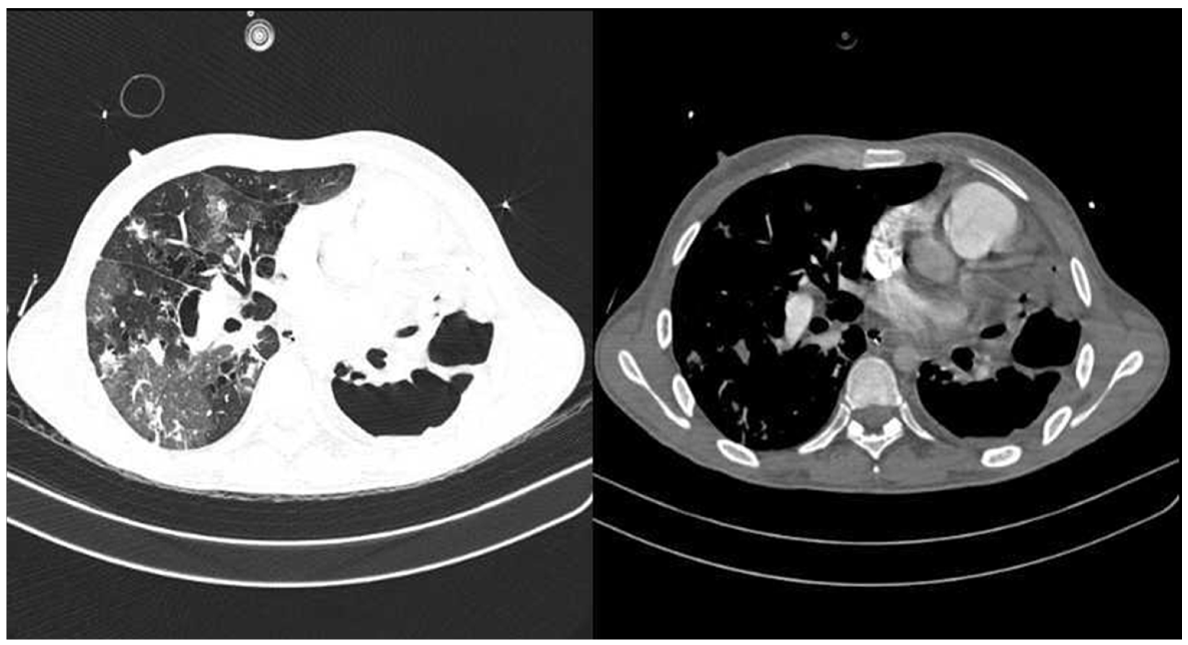
Figure 1. Chest angiotomography on admission. Axial contrast-enhanced chest CT images showing extensive bilateral pulmonary involvement with multiple cavitary lesions predominantly in the right lung, associated with areas of consolidation and parenchymal destruction, consistent with severe infectious processes.
Life 15 01435 g001
Figure 2. Sequential chest radiographic follow-up. (A) Endotracheal intubation: Multiple bullae in both lung fields, alveolar infiltrates in the right lung field, and bilateral perihilar atelectatic bands. (B) Post endotracheal intubation: The clouding of the left lung base persists, as do the multiple bullae in the right apex.
Life 15 01435 g002
Table 1. Longitudinal biochemical assessment during emergencies, hospitalization, and intensive care.
Laboratory
Parameters
Patient’s Values
Emergencies Date of
Entry
ICUHospitalization
15 December 2415 December 2417 December 2421 December 2422 December 2426 December 244 January 25
Hematology
Hemoglobin
(13.5–17.5 g/dL)
10.511.310.39.28.99.411.9
Hematocrit
(40–50%)
35%39%36%31%29%32%40%
Platelets
(150–450 × 103/µL)
237372248242210234307
Leukocytes
(4.5–11.0 × 10³/µL)
22.022.012.04.23.23.65.6
C-reactive protein (less than 2 mg/L)39.5
Lactate
(0.5–2.2 mmol/L)
1.41.32.91.81.2
Renal Function
Creatinine
(0.7–1.3 mg/dL)
1.10.430.450.520.500.69
Blood urea nitrogen (7–20 mg/dL) 13.55.94.26.812.113.4
Coagulation
Prothrombin time
(11.7–15.5 s)
12.6
Partial thromboplastin time
(24–45 s)
36.1
Biochemistry
Electrolytes
Sodium (135–145 mmol/L)137136126141142138
Potassium
(3.5–4.5 mmol/L)
5.15.44,93.43.43.63.9
Chloride
(95–105 mmol/L)
105.8102.099.9106.2108.7100.9
Arterial blood gases, mmHg
Supplemental oxygen supportVenturi maskInvasive mechanical ventilationHigh-flow nasal cannulaSimple nasal cannulaAmbient breathing
FiO250%70%40%50%50%28%21%
Flow per minute 50 L/min
pH
(7.35–7.45)
6.93 (pre-intubation)7.107.147.397.407.417.30
pCO2
(35–45 mmHg)
113.072684444.04646
PaO2
(75–100 mmHg)
128.08997931588766
HCO3
(22–26 mmol/L)
15.122.423.126.62729.222
BE
(–2 to +2 mmol/L)
−12.5−7.4−6.61.52.24.0−3.8
PaO2/FiO2 > 400
(ratio)
256127242186316310314
ROX Index
(SpO2/FiO2)/RR
15 (indicative of high probability of HFNC success, threshold > 5)
Table 2. Microbiological and virological results.
TestSampleResultInterpretation
Respiratory panelNasopharyngeal swabNegativeNo respiratory viral or bacterial pathogens detected
SARS-CoV-2 RT-PCRNasopharyngeal swabPositiveConfirmed COVID-19 infection
TB drug susceptibility (DST)Sputum (SOT)Sensitive patternSensitive pattern
Blood culture 1BloodNegativeNo bacterial growth
Blood culture 2BloodNegativeNo bacterial growth
Disclaimer/Publisher’s Note: The statements, opinions and data contained in all publications are solely those of the individual author(s) and contributor(s) and not of MDPI and/or the editor(s). MDPI and/or the editor(s) disclaim responsibility for any injury to people or property resulting from any ideas, methods, instructions or products referred to in the content.