Article

Proteomics Analysis of Plasma Biomarker of Cognitive Frailty in Elders Who Locally Reside in Chiang Mai Province of Thailand

by
Paitoon Aobchey
1,
Parunya Chaiyawat
2,
Mathuramat Seesen
3 and
Jetsada Ruangsuriya
1,4,*
1
Functional Food Research Center for Well-Being, The Multidisciplinary Research Institute, Chiang Mai University, Chiang Mai 50200, Thailand
2
Center of Multidisciplinary Technology for Advanced Medicine, Faculty of Medicine, Chiang Mai University, Chiang Mai 50200, Thailand
3
Department of Community Medicine, Faculty of Medicine, Chiang Mai University, Chiang Mai 50200, Thailand
4
Department of Biochemistry, Faculty of Medicine, Chiang Mai University, Chiang Mai 50200, Thailand
*
Author to whom correspondence should be addressed.
Life 2025, 15(8), 1330; https://doi.org/10.3390/life15081330
Submission received: 18 July 2025 / Revised: 16 August 2025 / Accepted: 19 August 2025 / Published: 21 August 2025
(This article belongs to the Section Proteins and Proteomics)

Abstract

Cognitive frailty in elders has become very common in Thailand society and is extremely difficult to manage in clinical practice due to poor detection and diagnosis. Plasma proteomic studies may be able to provide promising protein markers specific to this condition in order to improve the quality of life in the elderly. The purpose of this study was to differentiate and identify a potential protein marker of cognitive frailty in plasma samples by proteomic approaches. Nine plasma samples from elders with cognitive frailty or non-frailty were pooled and subjected to 2D gel separation. Conventional analysis was performed with the stained gel, and the selected protein spot was identified by liquid chromatography–tandem mass spectrometry coupled to a SCIEX TripleTOF 6600+. It was found that fibrinogen gamma (FGG) chain protein was suggested. FGG was further validated with a commercial ELISA kit using both pooled and individual plasma samples. It was found that both pooled and individual samples showed a significant reduction in FGG levels in elders with cognitive frailty. The results from this study suggest that FGG chain may be a potentially useful plasma biomarker for early detection, diagnosis, and therapeutic applications of cognitive frailty in elders in order to achieve sustainable health in an aging society.

1. Introduction

An aging society has become the forefront of management policy in all countries around the world, especially regarding healthcare for elders, for whom a huge budget must be allocated to ensure their well-being. It is projected that the number of people aged 60 years or over worldwide will grow by 56 percent, from 901 million to 1.4 billion, between 2025 and 2030. By 2050, the global population of older persons is expected to more than double, reaching nearly 2.1 billion. Moreover, the number of people aged 80 years or over, the “oldest-old” persons, is increasing even faster than the overall older population [1]. Since 2005, Thailand has entered an aging society, with the proportion accounting for 14.57%, and in 2020, a completely aging society (20%) was officially announced [2]. The proportion of the late-elderly population (aged 80 years and above) shows a clear increasing trend. This has impacted the prevalence of chronic diseases, requiring increased long-term care and higher associated costs, and also affected socioeconomic conditions, particularly related to healthcare. The number of workers to support the aging population is decreasing; in Thailand, only two people of working age are available to care for each elderly person [3].
Due to increased vulnerability in all aspects among elders, health promotion and disease prevention in the elderly population have become serious issues for governments worldwide. It was found that 69.3% of the population aged 60–69 years had chronic diseases, and the prevalence increased to 83.3% in the age group of 80 years and over. For example, joint pain and chronic back pain have a prevalence of up to 1 in 3 other health problems [4], and the prevalence of neurodegenerative diseases like dementia in the elderly was found to be 12.4% along with common clinical syndromes, which contribute to frailty [5]. Frailty is associated with increasing age and chronic diseases and is a major cause of disability, risk of falls, and burden for families and communities. Therefore, promoting and maintaining the health of the elderly are necessary to improve their quality of life and foster a sustainable community in an aging society [6].
Frailty is defined as an impaired ability to endure and respond to stress and can be determined using five phenotypic indicators: unintentional weight loss, slow gait speed, weakened grip strength, exhaustion, and physical inactivity [4]. It was found that 10.7% of adults aged 65–74 years are frail [7]. As mentioned above, frail older adults face higher risks of disabilities, incidents of falls, hospitalization, and death [8]. Clinically, physical frailty is a syndrome with multiple causes and contributing factors characterized by multisystem impairment that increases vulnerability to stress while decreasing physiological reserve [4]. Various methods have been proposed to evaluate frailty, including assessments of physical function, cognition, activities of daily living, comorbidities, deficits, and physiological dysfunctions. Nevertheless, the most widely accepted diagnostic method for frailty relies on the presence of three or more of the five aforementioned indicators, known as Fried’s criteria [4].
Cognitive frailty, which can potentially lead to other neurodegenerative diseases if left untreated, is a geriatric clinical syndrome characterized by the coexistence of mild cognitive impairment (MCI) and physical frailty in the absence of diagnosed dementia [9]. Epidemiological data from Asian populations indicate prevalence rates of 2.7% in Japan and 1.6% in Singapore [10,11], whereas higher rates of approximately 4.4% have been reported in Western countries [12]. Although relatively uncommon in community-dwelling older adults, cognitive frailty has been consistently associated with adverse health outcomes, including increased risks of disability, reduced health-related quality of life, falls, and death [13,14,15]. As a potentially reversible condition involving both physical and cognitive decline, early detection and targeted interventions for modifiable risk factors are critical to mitigating the risk of dementia and preserving functional capacity in older adults.
Using proteomics techniques to study differences in protein expression patterns in cognitive frailty among elder adults allows for the rapid identification of potential plasma biomarkers due to the capability to detect slight changes between groups [16]. The proteomic approach can also facilitate the creation of specific diagnostic models and treatment modalities to improve the quality of life in the elderly. However, specific molecular biomarkers for diagnosis and therapeutic targets of cognitive frailty, an early condition leading to other neurodegenerative diseases, are still lacking.
In this study, we aimed to comparatively identify plasma protein expression of elderly people with cognitive frailty using proteomic approaches. The results suggested in this study could be further developed as a plasma biomarker for the early detection and diagnosis of cognitive frailty in older adults. Ultimately, this will contribute to building a sustainably healthy community in an aging society.

2. Materials and Methods

2.1. Participants

Participants in this study were randomly selected from groups as reported by Seesen and colleagues in 2021 [17]. With the incorporation of Khua Mung Health Center in Saraphi District, Chiang Mai Province, Thailand, this research was made possible. The study was advertised through the staff at the health center and was opened to all participants who permanently resided in this district and were aged 65–84 years. There were 934 people eligible for this study. After primary screening by the volunteer health center staff to exclude individuals with neurodegenerative diseases such as dementia, psychological conditions such as depression, kidney diseases, hepatic diseases, autoimmune diseases, cancer, acute trauma or illness, and those taking steroidal drugs, 494 adults were selected, and 24 people declined to participate. All participants were assessed for physical frailty using Fried’s criteria [4] and for cognitive impairment using the Thai version of The Montreal Cognitive Assessment-Basic (MoCA-B) [17]. Finally, there were 48 robust and 135 cognitively frail individuals. According to Seesen and colleagues’ observation, 61 cognitively frail people were randomly selected for biochemical analyses, while 9 people were randomly sampled in this study based on the prevalence (13%) in the studied population. Therefore, 9 robust (non-frailty) and 9 cognitive frailty individuals were randomly selected. The Committee for Human Research Ethics of the Faculty of Medicine, Chiang Mai University granted permission to conduct this research under the approval number COA 050/2022.

2.2. Plasma Sample Collection and Protein Preparation

A total of 5 mL of fasting blood was drawn from each participant in a heparin-coated tube. A total of 1 mL of whole blood was separated by centrifugation at 3000× g for 10 min, and 0.2 mL of plasma was subsequently obtained. The plasma samples were stored at −80 °C for further analyses. Nine plasma samples from each group were pooled by mixing 10 μL from each individual plasma.
Protein quantification was performed using the Pierce Coomassie (Bradford) Protein Assay Kit Prod# 23236 (Thermo Scientific, Waltham, MA, USA) by adding 100× diluted plasma sample into 200 μL of working Bradford solution. The protein concentration was calculated using a standard curve prepared with Pierce Albumin Standard Ampules, 2 mg/mL Prod# 23209 (Thermo Scientific, Waltham, MA, USA).

2.3. Two-Dimensional Polyacrylamide Gel Electrophoresis or 2-DE

A total of 500 μg of protein was added to immobilized pH gradient (IPG) buffer (0.5%, pH 4–7) in 65 mM dithioerythritol (DTE) and heated at 95 °C for 5 min. Each sample was mixed with a lysis buffer containing 7 M urea, 4% 3-[(3-cholamidopropyl) dimethylammonio]-1-propanesulfonate) (CHAP), 2 M thiourea, and 0.02% bromophenol blue (sodium salt) in a total volume of 350 μL. After incubation at room temperature (RT) for at least one hour, centrifugation at 12,000× g for 20 min was performed. The whole mixture was loaded onto dry IPG strips (Immobiline DryStrip, 18 cm, pH 4–7, GE Healthcare, Chicago, IL, USA). The first-dimension separation was performed using an IPGphore system (Ettan IPGphor3, GE Healthcare, Uppsala, Sweden) under the following conditions. IPG strip rehydration was conducted at 50 V and 25 °C for 14 h. After rehydration, isoelectric focusing (IEF) was performed sequentially at 100 V (1 h), 250 V (1 h), 500 V (1 h), 1000 V (1 h), 3000 V (1 h), and 8000 V until 65 kVh was reached.
Afterward, the IPG strips were equilibrated in equilibrating buffer containing 50 mM Tris-HCl, pH 8.8, 6 M Urea, 30% glycerol, 2% (w/v) DTE, and a trace of bromophenol blue for 15 min. Subsequently, the equilibrated strip was treated with 2.5% (w/v) iodoacetamide (IAA) in the equilibrating buffer for 15 min.
Second dimension separation was then performed by placing the IPG strip on the top of the 12.5% polyacrylamide gel (18 × 18 cm) and covering it with 0.5% agarose. The separation was carried out at 45 mA per gel, and the gel was fixed with 10% methanol and 7% acetic acid solution for 3 h at RT. Then, the gel was washed three times in deionized water (dH2O) for 15 min each. The washed gel was stained with SYPRO® Ruby gel stain for one hour and washed three more times in dH2O for 15 min each. Finally, the gel was scanned with a Typhoon TRIO image scanner (GE Healthcare, Chicago, IL, USA) and analyzed using conventional methods [18].

2.4. In-Gel Tryptic Digestion

After the selection of protein spots, proteins in each gel spot were completely destained by 50% acetonitrile (ACN) in 10 mM ammonium bicarbonate (Sigma-Aldrich, Saint Louis, MO, USA). The excised protein spot was treated with 4 mM dithiothreitol (DTT) at 65 °C for 30 min, followed by treatment with 50 mM IAA (Sigma-Aldrich, Saint Louis, MO, USA) at RT with light protection for 30 min. Then, 4 mM DTT was added to stop the reaction by incubation at RT for 5 min. All solutions were removed, and gel pieces were dehydrated by adding 100% ACN. The gels were dried at RT, and protein digestion was performed in 10 mM ammonium bicarbonate containing trypsin (Thermo Scientific, Waltham, MA, USA) overnight at 37 °C. The peptides were extracted by adding 100% ACN to each tube and incubating for 15 min. The supernatant was transferred into a new microcentrifuge tube. All supernatants were dried using a centrifugal concentrator (TOMY, Tokyo, Japan). Samples were stored at −20 °C prior to mass spectrometric analysis [19].

2.5. Liquid Chromatography–Tandem Mass Spectrometry Analysis

Analysis of the digested peptides was performed using an Ultimate 3000 ultrahigh pressure system coupled with a SCIEX TripleTOF 6600+. Separation of the peptides was carried out in reversed-phase mode using a Pepmap100 C18 column (75 µm internal diameter × 150 mm length × 1.9 µm particle size) at 55 °C. Mobile phase A for liquid chromatography contained 0.05% formic acid (FA) in water, and mobile phase B contained 80% acetonitrile in 0.04% FA. After the samples were loaded into the column, the first linear gradient separation was operated for 95 min from 3 to 35% of mobile phase B at a flow rate of 300 nL/min. Regeneration of the column was performed with 90% mobile phase B for 10 min, followed by re-equilibration with 5% mobile phase B for 15 min. Precursor masses of 400–1500 m/z were collected within 250 ms under “high sensitivity” mode. Further fragmentation of each precursor spectrum was performed with a maximum of 30 precursors per cycle.

2.6. Data Analysis for Protein Identification

The MS spectra results recorded in wiff format were analyzed using Paragon™ Algorithm in ProteinPilot™ Software version 5.0.2 (Sciex, Concord, ON, Canada) installed with the Uniprot protein database of human (Taxonomy ID: 9606), retrieved from UniProtKB and assembled in FASTA format. The data were downloaded in August 2023, and the threshold of [Unused ProtScore (Conf)] ≥ 0.05 with 1% false discovery rate (FDR) was set to detect protein data with ≥10 peptides/protein.

2.7. ELISA for Plasma Fibrinogen Gamma Chain (FGG)

ELISA was performed to measure the plasma concentration of fibrinogen gamma chain in 18 plasma samples using a single-wash 90 min sandwich ELISA protocol provided by the manufacturer (Abcam, Cambridge, UK). Briefly, all plasma samples were subjected to a 2000× dilution in sample diluent, and 50 μL of either diluted plasma or the fibrinogen gamma chain standards (35–2300 pg/mL) were loaded into each well of the ELISA plate, along with 50 μL of the antibody cocktail into each well. Subsequently, the plate was sealed and incubated for an hour at RT on a plate shaker set to 400 rpm. After removing the liquid and performing a triple wash with 350 μL of the wash buffer, 100 μL of the TMB development solution was added and incubated in the dark for 15 min on a plate shaker set to 400 rpm. Each well received 100 μL stop solution, and the plate was shaken for a minute to mix prior to immediately recording the 450 nm absorbance. Each sample was run in duplicate, and the concentrations of plasma FGG between non-frailty and cognitive frailty were compared.

2.8. Statistical Analysis

Data are presented as mean ± standard deviation, and statistical analyses were executed using Graph Pad version 5.0. The Fisher’s exact test was used for categorical variables, and an unpaired t-test (two-tailed) was used for comparison of two sets of continuous variables. A p-value < 0.05 was considered statistically significant.

3. Results

3.1. Demographic Data of Participants

The summary details of the participants from each group are summarized in Table 1. It was found that there was a significant difference between groups in terms of age, weight, height, and MoCA score. The number of participants categorized by sex and educational background was still closely comparable.

3.2. 2-DE of Human Plasma Samples

A comparison of differential protein expression profiles between plasma samples from non-frail and cognitively frail elderly individuals revealed distinct differences in protein patterns within the molecular weight range of 10 to 250 kDa (Figure 1). Notably, several protein spots within a narrow pH range showed significant variation, particularly at approximately 29 kDa, where the protein spot intensity in non-frail plasma was increased by approximately two-fold compared to cognitively frail plasma. Two additional protein candidates are presented in Table S1 of the Supplementary Materials.

3.3. Protein Identification

There were more than 100 spots shown on SYPRO® Ruby-stained 2D gels in both non-frailty and cognitive frailty. In non-frailty, an overexpressed protein was spotted as compared to the cognitive frailty group. The spot was identified by SCIEX TripleTOF 6600+ system analysis. The details of the suggested protein identified by mass spectroscopy are shown in Table 2, the fibrinogen gamma chain (FGG), matched with UniProtKB ID of P02679 form FGG gene belonging to secreted localization. Details on biological processes, biological classes, and regulatory subclasses of the fibrinogen gamma chain are presented in Table 3.

3.4. Validation of FGG in Human Plasma by ELISA Analysis

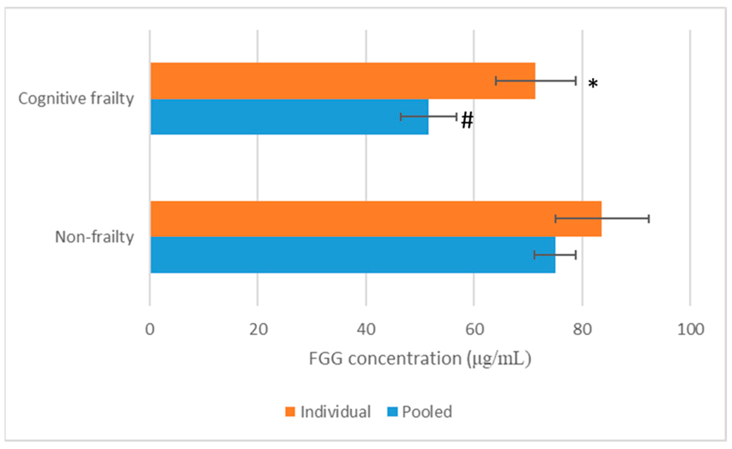
To validate whether the suggested protein was FGG protein, the expression levels of FGG in non-frailty and cognitive frailty elder plasma samples were measured using an ELISA kit. In plasma-pooled samples, the expression level of FGG in non-frailty was 75.05 ± 3.86 μg/mL, while it was 51.60 ± 5.14 μg/mL in cognitive frailty (Figure 2).
In addition, individual expression levels of the FGG chain in non-frailty and cognitive frailty plasma samples were also detected, and it was found that FGG in non-frailty (83.70 ± 8.69 μg/mL) was significantly higher (p < 0.01) than that in cognitive frailty (71.42 ± 7.45 μg/mL) (Figure 2).

4. Discussion

Cognitive frailty is a clinical condition that necessitates the development of reliable diagnostic tools for early detection because it is a silent harm leading to severe neurodegenerative diseases. Proteomic approaches offer a promising strategy for the identification of candidate biomarkers and the investigation of quantitative protein alterations in plasma and other biological specimens. In this study, we employed gel-based proteomic techniques, including two-dimensional electrophoresis (2-DE) and mass spectrometry (MS), to analyze and compare plasma protein profiles between cognitively frail and non-frail elderly.
We found that fibrinogen gamma (FGG) chain was markedly downregulated in cognitive frailty plasma samples compared with non-frailty elders. Our study showed that the plasma FGG chain level in non-frailty was more than 2.0-fold higher than in cognitive frailty, as observed by 2D gel electrophoresis. Secreted FGG was found in plasma of non-frailty at 83.70 (73.78–97.05) μg/mL, which was significantly higher than that in cognitive frailty measured at 71.42 (58.15–82.69) μg/mL.
FGG is a subunit of fibrinogen, which is a large complex glycoprotein with MW of 340 kDa containing α, β, and γ subunits encoded by FGA, FGB, and FGG genes, respectively [20]. The role of FGG in fibrinogen is to bind to the cell surface through integrin using its C-terminal region that plays a crucial role in thrombosis, angiogenesis, and inflammation [21,22]. Previous studies have reported that fibrinogenemia, characterized by significant deviations from the normal plasma fibrinogen concentration (2–4 mg/mL) [23,24,25], is frequently observed in patients with malignant tumors and is closely associated with tumor invasion, metastasis [26,27,28], angiogenesis [29], and tumor growth processes [30]. As fibrinogen is made of FGG, the reduction in FGG expression may affect the level of fibrinogen in plasma.
FGG is primarily synthesized and secreted into the bloodstream by the liver [31,32]. As a component of fibrinogen, FGG functions as a positive acute phase protein, with its plasma levels increasing during inflammation responses. FGG plays a critical role in coagulation, inflammation [33,34], and carcinogenesis [35,36] and has been proposed as a potential diagnostic marker in various diseases, including non-small-cell lung cancer [37] and bladder cancer [38]. Fibrinogen, a key element of the hemostatic system, is involved in both primary and secondary hemostatic responses. Upon thrombin-catalyzed cleavage of fibrinopeptides (Fp) A and B, fibrinogen is converted into fibrin, which spontaneously polymerizes into double-stranded protofibrils. These protofibrils further assemble into branched fibrin fibers, forming the fibrin clot [32,39]. Structurally, fibrinogen is a glycoprotein composed of three pairs of non-identical polypeptide chains (α, β, and γ), with FGG representing the γ-chain. The FGG gene also produces transcript variant isoforms via alternative splicing, including the FGG A precursor isoform [40].
Functionally, FGG is essential for fibrin polymerization and cross-linking, initiation of fibrinolysis, and regulation of factor XIII activity. It also mediates high-affinity binding to integrin expressed on platelets and leukocytes and facilitates thrombin binding to fibrin, exhibiting an inhibitory function formerly referred to as “anti-thrombin I” [41]. Frameshift mutations of FGG cause hypofibrinemia, a low serum fibrinogen level, which strongly indicates that FGG regulates fibrinogen secretion [42]. FGG has also been shown to inhibit platelet adhesion to fibrinogen via its interaction with hepatitis B splicing protein [43].
Dysregulation of FGG expression has been reported in multiple malignancies, including liver cancer [44], stomach cancer [23], and prostate cancer [45], suggesting its potential as a tumor-associated biomarker. Elevated FGG is related to gastric cancer [46], and plasma FGG levels have been proposed as predictors of prostate cancer progression [45]. Furthermore, FGG has been utilized to distinguish pancreatic cancer from normal serum samples, highlighting its potential clinical utility in cancer diagnostics [47]. Additionally, urine FGG levels could be used as a diagnostic marker for NSCLC [37]. Zhang et al. [48] found that FGG was highly expressed in the lung tissue of chronic obstructive pulmonary disease (COPD), and the expression level of FGG mRNA was related to the pulmonary function of patients. In addition, Zhang et al. [37] reported that FGG found in urinary proteins was a promising marker for non-small-cell lung cancer (NSCLC) due to the significant upregulation of FGG expression in 112 NSCLC patients detected by ELISA. Likewise, FGG was identified in the urine of IgA nephropathy (IgAN) patients using data independent acquisition (DIA)-based liquid chromatography–mass spectrometry (DIA-MS). Moreover, ELISA was also used in a validation cohort. The study suggested the potential use of urinary FGG as a non-invasive biomarker for the determination of mild renal fibrosis in IgAN [49].
Agnihotri et al. [50] conducted proteomic profiling of plasma samples obtained from 10 cognitively frail and 10 non-frail elderly individuals using two-dimensional gel electrophoresis (2-DE) combined with matrix-assisted laser desorption/ionization time-of-flight/time-of-flight mass spectrometry (MALDI-TOF/TOF). A total of 105 protein spots were identified. Among these, 22 spots in the frail group and 12 spots in the non-frail group exhibited differential expression. Mass spectrometric analysis revealed eight upregulated proteins in the frail group: haptoglobin (Hp) isoforms (1-1, 2-1, and 2-2), plasma amyloid A1, trafficking kinesin-binding protein 1 (TRAK1), nuclear body protein SP110, NOD-like receptor family CARD domain-containing protein 3 (NLRC3), matrix metalloproteinase-12 (MMP12), mortalin, and nucleoside diphosphate kinase 3 (NDK3). Additionally, five downregulated protein spots in the frail group were identified as PSRC1, NKG2A, and three unnamed protein products. Lin et al. [51] used proteomic approaches to identify frailty and non-frailty biomarkers in six older adults. It was found that 31 proteins, such as angiotensinogen (ANGT), kininogen-1 (KG), and antithrombin III (AT), in plasma were significantly elevated in frailty, while three proteins, Ig kappa chain V-III region WOL, COX7A2, and albumin, were significantly low. It was suggested that the levels of sestrin1 and sestrin2 in serum were significantly low and associated with frailty syndrome [52].
As cognitive frailty in elders could be worsened by the development of other neurodegenerative diseases, such as dementia and Alzheimer’s disease, if left untreated, proper treatments could reverse cognitive frailty back to normal cognition and or non-frailty. It is worth finding biomarkers to monitor the early development of these severe diseases, as the biomarkers may eventually shift to those specific for the condition. For example, amyloid-beta (Aβ) 42/Aβ40 ratio and phosphorylated tau derived from cerebrospinal fluid provide reliable indicators of Alzheimer’s disease, while emerging biomarkers like microtubule binding region (MTBR)-tau243 offer insights into disease progression. Likewise, matrix metalloproteinases, soluble platelet-derived growth factor receptor-β, and inflammatory markers reflect vascular cognitive impairment and dementia [53]. Moreover, the meta-analysis of various blood biomarkers associated with neuronal function as indicators of vascular cognitive impairment (VCI) suggested that amyloid beta 42 (Aβ42), amyloid beta 40 (Aβ40), Aβ42/Aβ40 ratio, neurofilament light (NfL), and S100B are significant biomarkers of VCI when compared with non-VCI [54].

5. Conclusions

This study employed proteomic approaches to differentially investigate the expression of protein markers in both non-frailty and cognitive frailty plasma samples from elders who resided in Saraphi District, Chiang Mai, Thailand. It was found that FGG chain was significantly downregulated in cognitive frailty plasma. In addition, ELISA was used to validate the expression level of FGG chain in both non-frailty and cognitive frailty. Cognitively frail elders had significantly lower levels of FGG chain in their plasmas when compared with non-frail elders. This study suggests that FGG chain may become one of the potential biomarkers used for early detection of the occurrence of cognitive frailty and to prevent the emergence of this deteriorated condition as well as for further diagnostic and therapeutic applications. However, further studies are still required for assurance in a larger study group or cohort and for understanding the mechanisms on how FGG is involved in cognitive frailty.

Supplementary Materials

The following supporting information can be downloaded at: https://www.mdpi.com/article/10.3390/life15081330/s1. Table S1 List of candidate proteins differentially expressed between non-frailty and cognitive frailty elders and as identified by 2D-PAGE followed by LC-MS/MS.

Author Contributions

Conceptualization, P.A. and J.R.; methodology, P.A. and J.R.; software, P.A.; validation, P.A. and J.R.; formal analysis, M.S. and P.A.; investigation, P.A. and J.R.; resources, M.S. and P.A.; data curation, P.A., P.C., M.S. and J.R.; writing—original draft preparation, P.A.; writing—review and editing, P.A., P.C., M.S. and J.R.; visualization, P.A. and J.R.; supervision, J.R.; project administration, P.A.; funding acquisition, J.R. All authors have read and agreed to the published version of the manuscript.

Funding

This research was funded by the Faculty of Medicine, Chiang Mai University, Thailand, with the study code BIO-2565-08747 and the grant contract number of 98-2565.

Institutional Review Board Statement

This study was conducted in accordance with the Declaration of Helsinki and approved by the Institutional Review Board (or Ethics Committee) of Research Ethics Committee No.5 Faculty of Medicine, Chiang Mai University (COA No. 050/2022 and date of approval: 10 February 2022) for studies involving humans.

Informed Consent Statement

Informed consent was obtained from all subjects involved in this study.

Data Availability Statement

Data are included in the article or Supplementary Materials; further inquiries can be directed to the corresponding author.

Acknowledgments

The researchers are grateful to all the volunteer staff of Khua Mung Health Center in Saraphi District, Chiang Mai Province, Thailand. Much gratitude is offered to all subjects participating in this study. We also thank the Functional Food Research Center for Well-being, the Multidisciplinary Research Institute, Chiang Mai University, Thailand for accomplishing this research as partial support.

Conflicts of Interest

The authors declare that there are no conflict of interest regarding the publication of this paper.

Abbreviations

The following abbreviations are used in this manuscript:
MCIMild cognitive impairment
MoCA-BThe Montreal Cognitive Assessment-Basic
FGGFibrinogen gamma chain

References

  1. United Nations, Department of Economic and Social Affairs, Population Division. World Population Ageing 2015; United Nations: New York, NY, USA, 2015; pp. 1–149. Available online: https://www.un.org/en/development/desa/population/publications/pdf/ageing/WPA2015_Report.pdf (accessed on 23 June 2025).
  2. Medical Department. Manual Screening Assessment for Seniors; Veterans’ Affairs Office Printing Synthesis Organization: Bangkok, Thailand, 2014. [Google Scholar]
  3. Cesari, M.; Leeuwenburgh, C.; Lauretani, F.; Onder, G.; Bandinelli, S.; Maraldi, C.; Guralnik, J.M.; Pahor, M.; Ferrucci, L. Frailty syndrome and skeletal muscle: Results from the Invecchiare in Chianti study. Am. J. Clin. Nutr. 2006, 83, 1142–1148. [Google Scholar] [CrossRef]
  4. Fried, L.P.; Tangen, C.M.; Walston, J.; Newman, A.B.; Hirsch, C.; Gottdiener, J.; Seeman, T.; Tracy, R.; Kop, W.J.; Burke, G.; et al. Frailty in older adults: Evidence for a phenotype. J. Gerontol. Biol. Sci. Med. Sci. 2001, 56, M146–M157. [Google Scholar] [CrossRef]
  5. Rockwood, K.; Song, X.; MacKnight, C.; Bergman, H.; Hogan, D.B.; McDowell, I.; Mitnitski, A. A global clinical measure of fitness and frailty in elderly people. CMAJ. 2005, 173, 489–495. [Google Scholar] [CrossRef]
  6. The Promotion and Development of the Elderly. The Elderly Thailand; S Plus Media Ltd.: Nonthaburi, Thailand, 2013. [Google Scholar]
  7. Collard, R.M.; Boter, H.; Schoevers, R.A.; Oude Voshaar, R.C. Prevalence of frailty in community-dwelling older persons: A systematic review. J. Am. Geriatr. Soc. 2012, 60, 1487–1492. [Google Scholar] [CrossRef]
  8. Clegg, A.; Young, J.; Iliffe, S.; Olde Rikkert, M.; Rockwood, K. Frailty in elderly people. Lancet 2013, 381, 752–762. [Google Scholar] [CrossRef]
  9. Buchman, A.S.; Bennett, D.A. Cognitive frailty. J. Nutr. Health Aging. 2013, 17, 738–739. [Google Scholar] [CrossRef]
  10. Shimada, H.; Makizako, H.; Doi, T.; Yoshida, D.; Tsutsumimoto, K.; Anan, Y.; Uemura, K.; Ito, T.; Lee, S.; Park, H.; et al. Combined prevalence of frailty and mild cognitive impairment in a population of elderly Japanese people. J. Am. Med. Dir. Assoc. 2013, 14, 518–524. [Google Scholar] [CrossRef] [PubMed]
  11. Chye, L.; Wei, K.; Nyunt, M.S.Z.; Gao, Q.; Wee, S.L.; Ng, T.P. Strong relationship between malnutrition and cognitive frailty in the Singapore Longitudinal Ageing Studies (SLAS-1 and SLAS-2). J. Prev. Alzheimers Dis. 2018, 5, 142–148. [Google Scholar] [CrossRef] [PubMed]
  12. Roppolo, M.; Mulasso, A.; Rabaglietti, E. Cognitive frailty in Italian community-dwelling older adults: Prevalence rate and its association with disability. J. Nutr. Health Aging 2016, 21, 631–636. [Google Scholar] [CrossRef]
  13. Feng, L.; Nyunt, M.S.Z.; Gao, Q.; Feng, L.; Lee, T.S.; Tsoi, T.; Chong, M.S.; Lim, W.S.; Collinson, S.; Yap, P.; et al. Physical frailty, cognitive impairment, and the risk of neurocognitive disorder in the Singapore Longitudinal Ageing Studies. J. Gerontol. Biol. Sci. Med. Sci. 2016, 72, 369–375. [Google Scholar] [CrossRef] [PubMed]
  14. Solfrizzi, V.; Scafato, E.; Seripa, D.; Lozupone, M.; Imbimbo, B.; D’Amato, A.; Tortelli, R.; Schilardi, A.; Galluzzo, L.; Gandin, C.; et al. Reversible cognitive frailty, dementia, and all-cause mortality: The Italian Longitudinal Study on Aging. J. Am. Med. Dir. Assoc. 2017, 18, 89.e1–89.e8. [Google Scholar] [CrossRef]
  15. Kim, H.; Awata, S.; Watanabe, Y.; Kojima, N.; Osuka, Y.; Motokawa, K.; Sakuma, N.; Inagaki, H.; Edahiro, A.; Hosoi, E.; et al. Cognitive frailty in community-dwelling older Japanese people: Prevalence and its association with falls. Geriatr. Gerontol. Int. 2019, 19, 647–653. [Google Scholar] [CrossRef]
  16. Magdeldin, S.; Enany, S.; Yoshida, Y.; Xu, B.; Zhang, Y.; Zureena, Z.; Lokamani, I.; Yaoita, E.; Yamamoto, T. Basics and recent advances of two-dimensional polyacrylamide gel electrophoresis. Clin. Proteom. 2014, 11, 16–24. [Google Scholar] [CrossRef] [PubMed]
  17. Seesen, M.; Sirikul, W.; Ruangsuriya, J.; Griffiths, J.; Siviroj, P. Cognitive frailty in Thai community-dwelling elderly: Prevalence and its association with malnutrition. Nutrients 2021, 13, 4239. [Google Scholar] [CrossRef] [PubMed]
  18. Ma, H.T.; Sriyam, S.; Sinchaikul, S.; Tsai, H.Y.; Phutrakul, S.; Chen, S.-T. Proteomics Characterization of Haptoglobin Alpha-2 Subunit in Non-Small Cell Lung Cancer Serum and Various Human Materials. J. Proteomics Bioinform. 2013, 6, 187–196. [Google Scholar]
  19. Havlis, J.; Thomas, H.; Sebela, M.; Shevchenko, A. Fast-response proteomics by accelerated in-gel digestion of proteins. Anal. Chem. 2003, 75, 1300–1306. [Google Scholar] [CrossRef]
  20. Simurda, T.; Brunclikova, M.; Asselta, R.; Caccia, S.; Zolkova, J.; Kolkova, Z.; Loderer, D.; Skornova, I.; Hudecek, J.; Lasabova, Z.; et al. Genetic variants in the FGB and FGG genes mapping in the beta and gamma nodules of the fibrinogen molecule in congenital quantitative fibrinogen disorders associated with a thrombotic phenotype. Int. J. Mol. Sci. 2020, 21, 4616. [Google Scholar] [CrossRef]
  21. Yokoyama, K.; Zhang, X.P.; Medved, L.; Takada, Y. Specific binding of integrin αvβ3 to the fibrinogen γ and αE chain C-terminal domains. Biochemistry 1999, 38, 5872–5877. [Google Scholar] [CrossRef]
  22. Yokoyama, K.; Erickson, H.P.; Ikeda, Y.; Takada, Y. Identification of amino acid sequences in fibrinogen γ-chain and tenascin-C C-terminal domains critical for binding to integrin αvβ3. J. Biol. Chem. 2000, 275, 16891–16898. [Google Scholar] [CrossRef]
  23. Duan, S.; Gong, B.; Wang, P.; Huang, H.; Luo, L.; Liu, F. Novel prognostic biomarkers of gastric cancer based on gene expression microarray: COL12A1, GSTA3, FGA and FGG. Mol. Med. Rep. 2018, 18, 3727–3736. [Google Scholar] [CrossRef]
  24. Wang, M.; Zhang, G.; Zhang, Y.; Cui, X.; Wang, S.; Gao, S.; Wang, Y.; Liu, Y.; Bae, J.H.; Yang, W.-H.; et al. Fibrinogen alpha chain knockout promotes tumor growth and metastasis through integrin–AKT signaling pathway in lung cancer. Mol. Cancer Res. 2020, 18, 943–954. [Google Scholar] [CrossRef]
  25. Peng, H.H.; Wang, J.N.; Xiao, L.F.; Yan, M.; Chen, S.P.; Wang, L.; Yang, K. Elevated serum FGG levels prognosticate and promote the disease progression in prostate cancer. Front. Genet. 2021, 12, 651647. [Google Scholar] [CrossRef] [PubMed]
  26. Lyman, G.H.; Khorana, A.A. Cancer, clots and consensus: New understanding of an old problem. J. Clin. Oncol. 2009, 27, 4821–4826. [Google Scholar] [CrossRef]
  27. Niu, J.Y.; Tian, T.; Zhu, H.Y.; Liang, J.H.; Wu, W.; Cao, L.; Lu, R.-N.; Wang, L.; Li, J.-Y.; Xu, W. Hyperfibrinogenemia is a poor prognostic factor in diffuse large B-cell lymphoma. Ann. Hematol. 2018, 97, 1841–1849. [Google Scholar] [CrossRef]
  28. Huang, W.; Wang, S.; Zhang, H.; Zhang, B.; Wang, C. Prognostic significance of combined fibrinogen concentration and neutrophil-to-lymphocyte ratio in patients with resectable non-small cell lung cancer. Cancer Biol. Med. 2018, 15, 88–96. [Google Scholar]
  29. Shiose, S.; Hata, Y.; Noda, Y.; Sassa, Y.; Takeda, A.; Yoshikawa, H.; Fujisawa, K.; Kubota, T.; Ishibashi, T. Fibrinogen stimulates in vitro angiogenesis by choroidal endothelial cells via autocrine VEGF. Graefe’s Arch. Clin. Exp. Ophthalmol. 2004, 242, 777–783. [Google Scholar] [CrossRef]
  30. Adams, G.N.; Rosenfeldt, L.; Frederick, M.; Miller, W.; Waltz, D.; Kombrinck, K.; McElhinney, K.E.; Flick, M.J.; Monia, B.P.; Revenko, A.S.; et al. Colon cancer growth and dissemination relies upon thrombin, stromal PAR-1, and fibrinogen. Cancer Res. 2015, 75, 4235–4243. [Google Scholar] [CrossRef]
  31. Richardson, S.J. Evolutionary changes to transthyretin: Evolution of transthyretin biosynthesis. FEBS J. 2009, 276, 5342–5356. [Google Scholar] [CrossRef] [PubMed]
  32. Weisel, J.W. Fibrinogen and fibrin. Adv. Protein Chem. 2005, 70, 247–299. [Google Scholar]
  33. Farrell, D.H. Pathophysiologic roles of the fibrinogen gamma chain. Curr. Opin. Hematol. 2004, 11, 151–155. [Google Scholar] [CrossRef] [PubMed]
  34. Mosesson, M.W. Fibrinogen and fibrin structure and functions. J. Thromb. Haemost. 2005, 3, 1894–1904. [Google Scholar] [CrossRef] [PubMed]
  35. Karaosmanoglu, O.; Banerjee, S.; Sivas, H. Identification of biomarkers associated with partial epithelial to mesenchymal transition in the secretome of slug over-expressing hepatocellular carcinoma cells. Cell. Oncol. 2018, 41, 439–453. [Google Scholar] [CrossRef]
  36. Repetto, O.; De Re, V. Coagulation and fibrinolysis in gastric cancer. Ann. N. Y. Acad. Sci. 2017, 1404, 27–48. [Google Scholar] [CrossRef] [PubMed]
  37. Zhang, W.; Gao, Z.; Zeng, G.; Xie, H.; Liu, J.; Liu, N.; Wang, G. Clinical significance of urinary plasminogen and fibrinogen gamma chain as novel potential diagnostic markers for non-small-cell lung cancer. Clin. Chim. Acta 2020, 502, 55–65. [Google Scholar] [CrossRef] [PubMed]
  38. Araújo, J.E.; López-Fernández, H.; Diniz, M.S.; Baltazar, P.M.; Pinheiro, L.C.; da Silva, F.C.; Carrascal, M.; Videira, P.; Santos, H.M.; Capelo, J.L. Dithiothreitol-based protein equalization technology to unravel biomarkers for bladder cancer. Talanta 2018, 180, 36–46. [Google Scholar] [CrossRef]
  39. Doolittle, R.F. Searching for differences between fibrinogen and fibrin that affect the initiation of fibrinolysis. Cardiovasc. Hematol. Agents Med. Chem. 2008, 6, 181–189. [Google Scholar] [CrossRef]
  40. Mosesson, M.W. Antithrombin I. Inhibition of thrombin generation in plasma by fibrin formation. Thromb. Haemost. 2003, 89, 9–12. [Google Scholar] [CrossRef] [PubMed]
  41. Mosesson, M.W. Update on antithrombin I (fibrin). Thromb. Haemost. 2007, 98, 105–108. [Google Scholar] [CrossRef]
  42. Nagata, K.; Arai, S.; Taira, C.; Sugano, M.; Honda, T.; Okumura, N. A novel frameshift mutation in the fibrinogen γC terminal region, FGG c.1169_1170delAT, leading to hypofibrinogenemia. Thromb. Res. 2017, 159, 82–85. [Google Scholar] [CrossRef]
  43. Chen, J.Y.; Chen, W.N.; Liu, L.L.; Lin, W.S.; Jiao, B.Y.; Wu, Y.L.; Lin, J.; Lin, X. Hepatitis B spliced protein (HBSP) generated by a spliced hepatitis B virus RNA participates in abnormality of fibrin formation and functions by binding to fibrinogen γ chain. J. Med. Virol. 2010, 82, 2019–2026. [Google Scholar] [CrossRef]
  44. Gao, H.J.; Chen, Y.J.; Zuo, D.; Xiao, M.M.; Li, Y.; Guo, H.; Zhang, N.; Chen, R.-B. Quantitative proteomic analysis for high-throughput screening of differential glycoproteins in hepatocellular carcinoma serum. Cancer Biol. Med. 2015, 12, 246–254. [Google Scholar]
  45. Davalieva, K.; Kiprijanovska, S.; Maleva Kostovska, I.; Stavridis, S.; Stankov, O.; Komina, S.; Petrusevska, G.; Polenakovic, M. Comparative proteomics analysis of urine reveals down-regulation of acute phase response signaling and LXR/RXR activation pathways in prostate cancer. Proteomes 2018, 6, 1. [Google Scholar] [CrossRef]
  46. Fukumura, D.; Kloepper, J.; Amoozgar, Z.; Duda, D.G.; Jain, R.K. Enhancing cancer immunotherapy using antiangiogenics: Opportunities and challenges. Nat. Rev. Clin. Oncol. 2018, 15, 325–340. [Google Scholar] [CrossRef]
  47. Bloomston, M.; Zhou, J.X.; Rosemurgy, A.S.; Frankel, W.; Muro-Cacho, C.A.; Yeatman, T.J. Fibrinogen gamma overexpression in pancreatic cancer identified by large-scale proteomic analysis of serum samples. Cancer Res. 2006, 66, 2592–2599. [Google Scholar] [CrossRef] [PubMed]
  48. Zhang, H.; Li, C.; Song, X.; Cheng, L.; Liu, Q.; Zhang, N.; Wei, L.; Chung, K.; Adcock, I.M.; Ling, C.; et al. Integrated analysis reveals lung fibrinogen gamma chain as a biomarker for chronic obstructive pulmonary disease. Ann. Transl. Med. 2021, 9, 1765–1777. [Google Scholar] [CrossRef]
  49. Guan, J.; Wang, M.; Zhoa, M.; Ni, W.; Zhang, M. Discovery of fibrinogen γ-chain as a potential urinary biomarker for renal interstitial fibrosis in IgA nephropathy. BMC Nephrol. 2023, 24, 60–69. [Google Scholar] [CrossRef]
  50. Agnihotri, V.; Gupta, A.; Bajpai, S.; Singhal, S.; Dey, A.B.; Dey, S. Serum proteomic approach for differentiation of frail and non-frail elderly. Adv. Gerontol. 2021, 11, 190–198. [Google Scholar] [CrossRef]
  51. Lin, C.-H.; Liao, C.-C.; Huang, C.-H.; Tung, Y.-T.; Chang, H.-C.; Hsu, M.-C.; Huang, C.-C. Proteomics analysis to identify and characterize the biomarkers and physical activities of non-frail and frail older adults. Int. J. Med. Sci. 2017, 14, 231–239. [Google Scholar] [CrossRef] [PubMed]
  52. Rai, N.; Venugopalan, G.; Pradhan, R.; Ambastha, A.; Upadhyay, A.D.; Dwivedi, S.; Dey, A.B.; Dey, S. Exploration of novel anti-oxidant protein sestrin in frailty syndrome in elderly. Aging Dis. 2018, 9, 220–227. [Google Scholar] [CrossRef]
  53. Hosoki, S.; Sachdev, P.S. Advances in biomarkers for Alzheimer’s disease and vascular cognitive impairment and dementia: Current status and clinical implications. Vas-Cog J. 2025, 11, 26–35. [Google Scholar]
  54. Huang, W.; Liao, L.; Liu, Q.; Ma, R.; He, X.; Du, X.; Sha, D. Blood biomarkers for vascular cognitive impairment based on neuronal function: A systematic review and meta-analysis. Front. Neurol. 2025, 16, 1496711. [Google Scholar] [CrossRef] [PubMed]
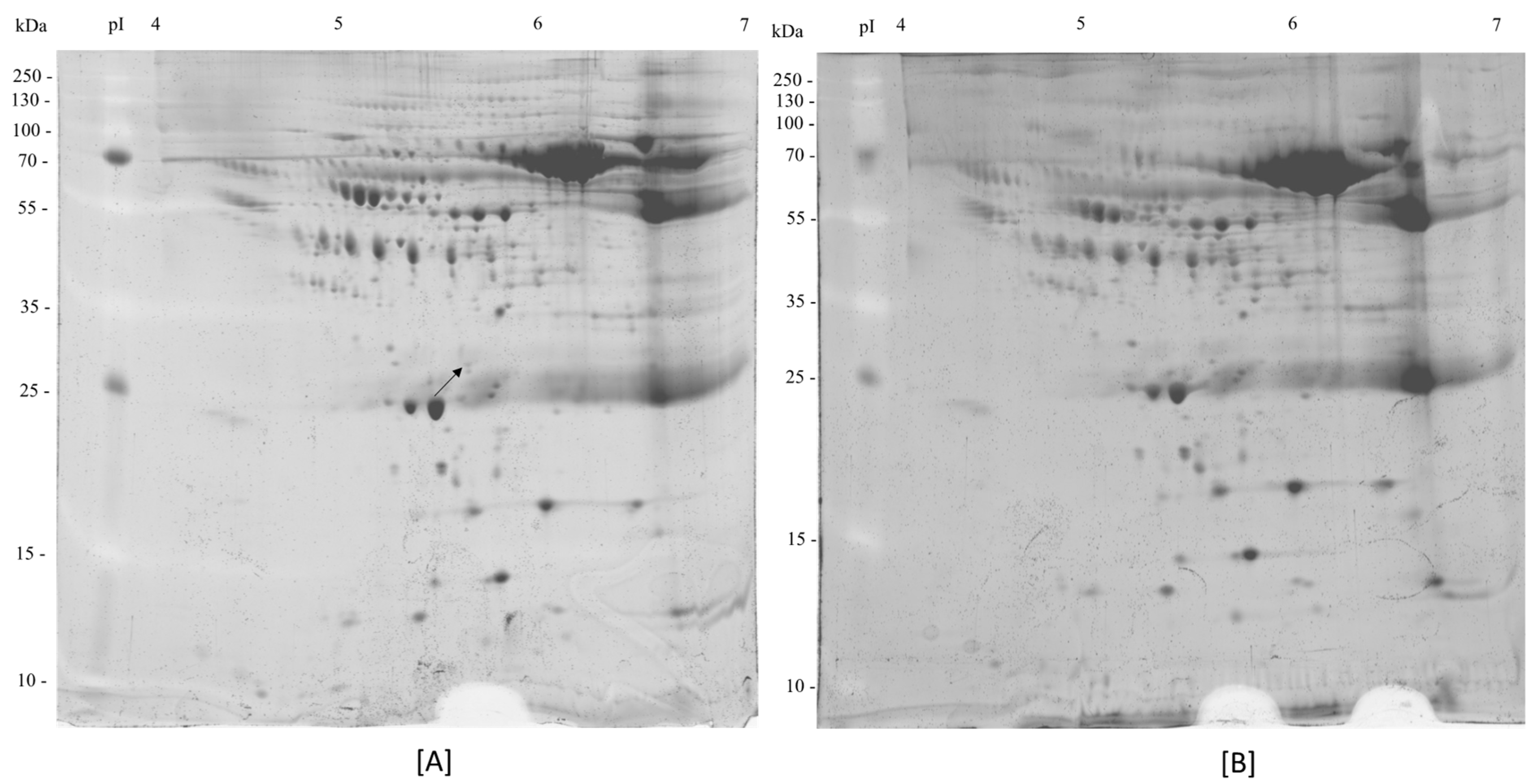
Figure 1. 2-DE gel image profile. It was derived from non-frailty plasma (A) and cognitive frailty plasma samples (B), with IPG strip pH 4–7. The arrow indicates the protein identified in this study.
Life 15 01330 g001
Figure 2. Concentrations of fibrinogen γ chain (FGG) determined by ELISA in plasma of cognitive frailty (n = 9) and non-frailty (n = 9) participants. Two determination methods were individual sample (Individual) and the pooled sample (Pooled) determinations. * and # indicate significant difference at p < 0.01 when compared with the similar determination method in non-frailty.
Life 15 01330 g002
Table 1. Summary of demographic information of the participants.
ParametersNon-Frailty (n = 9)Cognitive Frailty (n = 9)p-Value
Age (years)72.33 ± 5.3469.67 ± 4.420.299
Weight (kg)55.61 ± 8.9260.06 ± 12.880.367
Height (cm)157.22 ± 7.08162.00 ± 8.460.293
Actual MoCA score (points)19.11 ± 3.7221.11 ± 2.930.067
Sex (male/female)3/65/4-
Educational level (Grade 1–3/Grade 4–6/High school or over)6/1/28/0/1-
Table 2. Protein identified from non-frail and cognitive frailty plasma samples by LC–MS/MS analysis.
Protein SpotProtein NameAccession No.aSpot LocationTheoretical MW (kDa)%Cov (95)pIPeptides Matched (95%)Fold Change (FC)
Upregulation in non-frail
1Fibrinogen gamma chainP02679Non-frail51.54.1945.602>2
a Accession no. was obtained in UniProt database: http://www.uniprot.org/ (accessed on 10 June 2024).
Table 3. Biological details of the validated abundant protein.
Protein Identification NumberProtein Name (Gene)Biological RolesBiological Categories
P02679Fibrinogen γ chain (FGG)Many biological roles are suggested as follows:
  • Platelet degranulation, including fibrinolysis, blood coagulation, fibrin clot formation, platelet aggregation, plasminogen activation, platelet activation
  • Innate immune response
  • Response to calcium ion
  • Positive regulation of peptide hormone secretion
  • Positive regulation of heterotypic cell–cell adhesion
  • Involvement of protein polymerization in both cellular protein complex assembly and extracellular matrix (ECM) organization
  • Positive regulation of exocytosis
  • Negative regulation of endothelial cell apoptotic process
  • Positive regulation of vasoconstriction. Positive regulation of substrate adhesion-dependent cell spreading
  • Signaling pathway inhibition of death domain receptors for extrinsic apoptosis and induction of bacterial agglutination
Immune system, regulation, transport and homeostasis, coagulation, fibrinolysis, response, cell and ECM organization, protein metabolism
Disclaimer/Publisher’s Note: The statements, opinions and data contained in all publications are solely those of the individual author(s) and contributor(s) and not of MDPI and/or the editor(s). MDPI and/or the editor(s) disclaim responsibility for any injury to people or property resulting from any ideas, methods, instructions or products referred to in the content.