Case Report

Acute Rheumatic Fever in Caucasians: A Case Report and Systematic Review

1
Mersey & West Lancashire Teaching Hospital NHS Trust, Southport PR8 6PN, UK
2
Centre for Rheumatic Diseases, Weston Education Centre, Cutcombe Road, King's College London, London SE5 9RJ, UK;mrinalini.dey@kcl.ac.uk
*
Author to whom correspondence should be addressed.
Life 2025, 15(7), 1131; https://doi.org/10.3390/life15071131
Submission received: 9 April 2025 / Revised: 4 July 2025 / Accepted: 8 July 2025 / Published: 18 July 2025
(This article belongs to the Special Issue Infection, Inflammation and Rheumatology)

Abstract

Acute Rheumatic Fever (ARF) is more common in children in the developing world. The current incidence in the United Kingdom is reported to be less than 1 in 100,000 children. It is, however, rare in the developed world, particularly in the adult Caucasian population. We present a case of ARF in a 39-year-old Caucasian female who needed multiple hospital admissions before the ARF diagnosis was made. A comprehensive, up-to-date literature review of ARF in Caucasians is lacking. Therefore, a systematic literature review (SLR) of Medline, PubMed, and Google Scholar was conducted to investigate the characteristics, management, and prognostic outcomes of new cases of ARF among Caucasians. A total of 10 cases were reported from six countries between 1990 and 2022. The mean age of patients was 33.2 (range 18–41), and most were females (6, 60%). The most common presenting symptoms were fever, arthralgia, and malaise. All patients met the modified Jones criteria for ARF diagnosis. All patients received antibiotics, with only one patient requiring corticosteroids. Two patients developed rheumatic heart disease (RHD), and none died as a result of ARF. This case-based literature review underscores the critical importance of a high index of clinical suspicion in promptly diagnosing ARF to mitigate long-term sequelae of RHD.

1. Introduction

Acute Rheumatic Fever (ARF) is a severe inflammatory condition that can develop as a complication of untreated or inadequately treated streptococcus pyogenes throat infection or scarlet fever [1]. Although it primarily affects children and adolescents, ARF can occur at any age, leading to long-term damage to the heart, joints, and nervous system, if not promptly diagnosed and treated [2]. The underlying cause of ARF has been demonstrated to be an immunological reaction to group A streptococcus bacterial infection; however, the exact mechanisms by which the immune system attacks healthy tissues following a strep infection are not fully understood [3]. It is believed that the bacteria trigger an autoimmune reaction, causing the immune system to mistakenly target specific tissues in the body, particularly the heart and joints [4]. The diagnosis of ARF is based on a combination of clinical criteria, blood tests, and imaging tests such as echocardiography [5]. Despite the availability of data on rheumatic fever (ARF) cases within the developed world, the existing literature predominantly focuses on non-Caucasian populations, revealing a significant gap in the investigation of ARF cases among Caucasians. Consequently, there is a pressing need to examine the incidence, management strategies, and clinical outcomes related to ARF within this demographic. This systematic literature review (SLR) endeavors to critically analyze and synthesize the clinical characteristics and demographic factors pertaining to newly diagnosed ARF cases in the adult Caucasian population.

2. Case Presentation

This is a case of a 39-year-old female with a past medical history of Bell’s Palsy, who presented to the local accident and emergency (A&E) department at the end of 2022 with a 3-week history of fever, sore throat, arthralgia, myalgia, chest pain, and dyspnea. Her young school-aged daughter had a two-week history of fever, sore throat, generalized body aches, and fatigue, which were resolved spontaneously.
During her initial presentation, her blood tests showed raised C-reactive protein (CRP) at 171 (0–4 mg/L), lymphopenia, and abnormal liver function tests.
The test results are summarized in Table 1.
As the patient’s symptoms were not severe enough to warrant hospitalization, the patient was discharged home from A&E with a suspected viral infection. A week later, the patient returned to the A&E with worsening chest pain, dyspnea, and polyarthritis in the hands and feet. Her ECG was normal. Her blood tests showed worsening of CRP at 191, hemoglobin 97 (115–165 g/L), and gamma-glutamyl transferase (GGT) at 115 (0–39 u/L). Her echocardiogram showed moderate pericardial effusion (Figure 1) and an ejection fraction (EF) of 55% without valvular involvement. The ultrasound scan of her liver and gall bladder was normal. She had negative connective tissue disease, anti-neutrophilic cytoplasmic antibody (ANCA), cyclic citrullinated peptide (CCP) antibodies, and rheumatoid factor. Her complement (C3) was slightly raised, but C4 was normal. The patient was prescribed Ibuprofen for her pericarditis and arthralgia and was discharged.
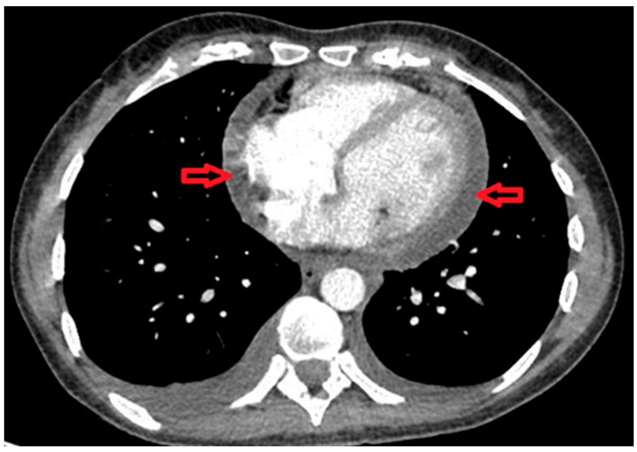
She presented to the A&E for the third time after six days with nausea, heartburn, and vomiting, which were thought to be caused by Ibuprofen. Although Ibuprofen improved her joint symptoms, it was stopped owing to the side effects, and she was started on Omeprazole. Chest computed tomography (CT) revealed moderate pericardial effusion (Figure 2) and small bilateral pleural effusions. Since her clinical features improved, she was discharged.
She returned to A&E for the fourth time 3 days later with palpitations. Her CRP and hemoglobin had improved at this visit, along with other blood parameters. Her repeat echocardiogram showed improved pericardial effusion but no changes in EF. She also had normal HIV, hepatitis C antibody, hepatitis B surface antigen, Lyme serology, Epstein–Barr virus (EBV) immunoglobulin-M, and parvovirus B19 IgM. An anti-streptolysin O titer (ASOT) was elevated at 400 (0–200); however, her throat swab was normal.
The possibility of ARF was considered by the rheumatology team when consulted, given that the patient had two major criteria (carditis and polyarthritis) and two minor criteria (elevated CRP and fever) according to the modified Jones diagnostic criteria for acute ARF (Table 2). After discussion with the infectious disease team, the patient commenced on oral Penicillin-V 500 mg 3 times a day for 10 days, followed by intramuscular Benzylpenicillin 1.2 million units 4 weekly for 10 years.
The patients’ symptoms improved gradually, and she was asymptomatic in a few weeks’ time. Her repeated echocardiogram (Table 3) with the cardiology team after a few months showed complete resolution of pericardial effusion, and EF had improved to >60%. She was completely asymptomatic on consultation.

3. Methods

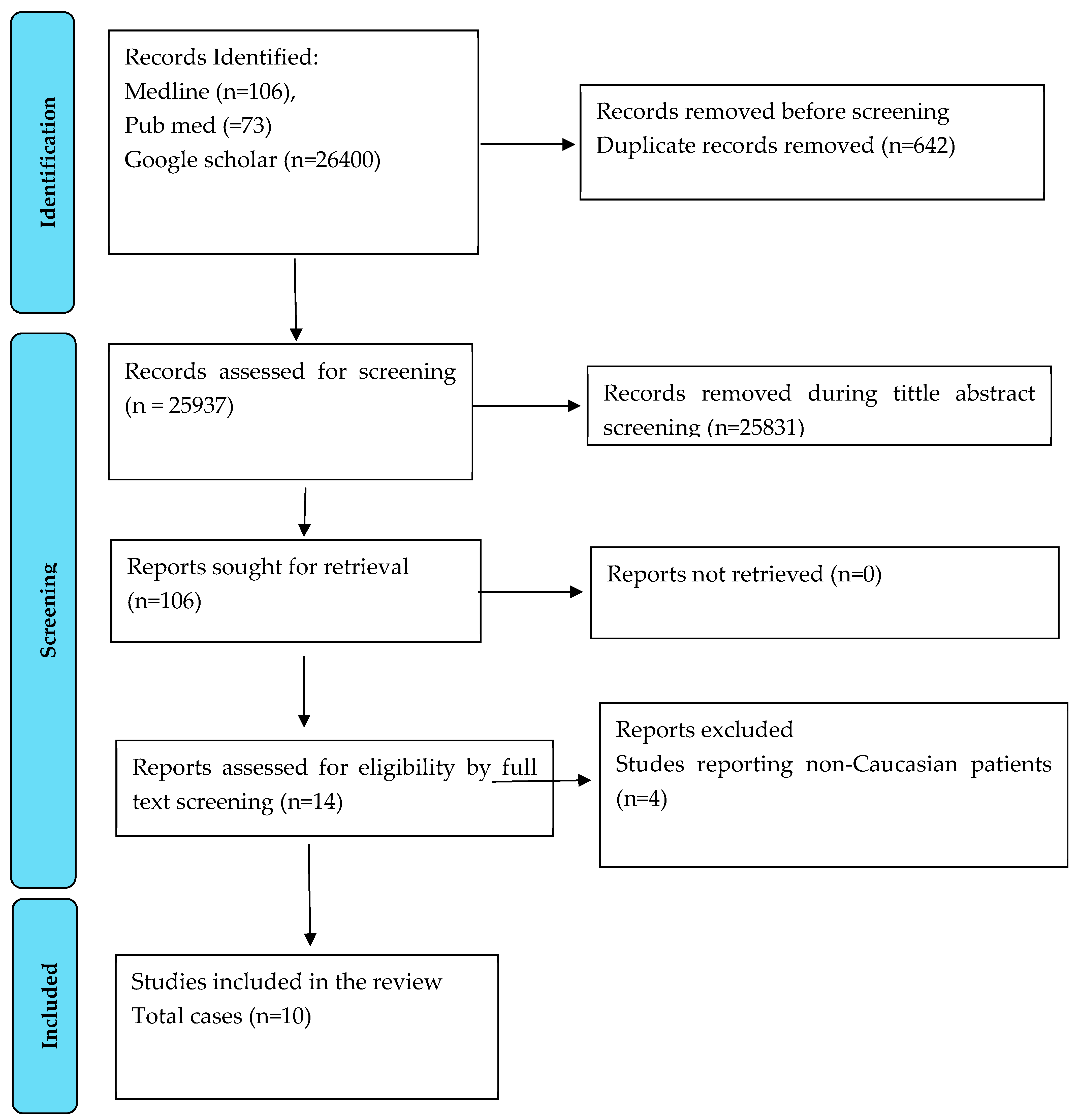
We conducted an SLR to summarize the existing cases within the literature detailing the diagnosis and management of ARF in the Caucasian population. This SLR was conducted in accordance with the Cochrane Handbook and Preferred Reporting Items for Systematic Reviews and Meta-Analysis guidelines [6]. The search strategy is available in the Supplementary Material. The bibliographic databases Medline, PubMed, and Google Scholar were searched on 13th of May 2025, Figure 3.
No time restriction was applied to the search. Only English-language articles were included. We included adult Caucasian patients with a clinician-confirmed diagnosis of ARF. We opted to focus on the Caucasian population due to the relative paucity of data on this patient group, despite the increasing prevalence of ARF in this patient group. Only single case reports were included, with all other article types excluded. Titles and abstracts were then screened by one reviewer, with full articles that met the inclusion criteria examined in detail. Screen and data extraction were validated by a second reviewer.
Data extracted from included articles comprised (but were not limited to): demographics, presenting symptoms, organ involvement, investigations, and clinical outcome. Data were summarized using descriptive statistics.

4. Results

From the 106 articles retrieved (following de-duplication), 10 cases were ultimately included. The mean age was 33.2 years (SD 8.7, range 18–41), with 60% of patients being female. Cases were published between 1990 and 2022, with the following countries of origin: USA (n = 4), UK (n = 2), Australia (n = 1), Cuba (n = 1), Cyprus (n = 1), and Ireland (n = 1) (Table 4).
All patients met the modified Jones’ criteria for the diagnosis of ARF. Where reported, all patients presented with a raised C-reactive protein (range 37–503 mg/L, normal < 7), raised erythrocyte sedimentation rate (58–115 mm/h, normal ≤ 20) and positive ASOT. The most common manifestations on presentation were fever (n = 8), arthralgia (n = 6), and malaise (n = 3). One patient presented with monoarthritis, another developed an advanced atrioventricular block, and another exhibited erythema mariginatum and chorea. Two patients developed rheumatic heard disease (RHD), one of whom required admission to the intensive care unit for inotrope support [7,8].
All included patients survived following treatment with antibiotics, most frequently penicillin (n = 6). Three were given aspirin in addition, and one received prednisolone [7,9,10,11].
Table 4. Summary of case reports of Caucasians diagnosed with acute rheumatic fever.
SNArticleAgeSexCountryPresenting ComplainsMet modified Jones CriteriaEvidence of Rheumatic Heart DiseaseCRP
(mg/L)
ESR
(mm/h)
Raised ASOT Treatment ReceivedITU
Admission
Outcome
1Farrell 1990 [9]40FUKRash, fever, arthralgiaYesNone9270YesPenicillin, AspirinNoSurvived
2Sahi 1993 [12]38FCyprusMalaise, flu-like symptomsYesNoneN/A100YesPenicillinNoSurvived
3Barold 1996 [7]39MUSASore throat, fever, myalgiaYesYes41111YesPenicillin, AspirinNoSurvived
4Grover 2009 [11]25MUSAPolyarthritis, feverYesNoneN/A115YesPenicillin, PrednisoloneNoSurvived
5Ilgenfritz 2013 [10]27MUSAKnee pain and swelling, feverYesNoneN/A58YesPenicillin, AspirinNoSurvived
6Khan 2018 [13]41FAustraliaFever, polyarthritisYesNone116103YesCephalexinNoSurvived
7Wilson 2021 [14]24MUKShoulder pain, shortness of breathYesNone200N/AYesPenicillinNoSurvived
8Batta 2022 [15]41FUSAErythematous papules, FeverYesNone37N/AYesCeftriaxoneNoSurvived
9Our case
2022
39FUKFever, sore, throat, myalgiaYesNone171108YesPenicillinNoSurvived
10Coyle 2025 [8]18FIrelandMalaise, fever, diarrhea and vomiting.YesYes503115YesCeftriaxoneYes (inotrope support)Survived
11Reel 2025 [16]39FCubanFever, sore throat, wrist and chest painYesNone18061YesAzithormycinNoSurvived
CRP: C-reactive protein; ESR: erythrocyte sedimentation rate; ASOT: anti-streptolysin O titer; ITU: intensive care unit; N/A; not available.

5. Discussion

ARF is believed to occur about three weeks after a group A beta-hemolytic streptococcal (GAS) infection causes a type II hypersensitivity cross-reaction between streptococcal and host antigens [17]. It is a condition that exerts deleterious effects on the heart, brain, and joints. If not diagnosed early or untreated, it can graduate to rheumatic heart disease (RHD), with considerable morbidity and mortality. Its diagnosis is based on the presence of major and minor 2015 revised Jones criteria (Table 2) [18]. Our patient met the revised Jones criteria for ARF. For diagnosis, two major or one major and two minor features are needed. Our patient had arthritis and carditis as major features and two minor criteria, along with the evidence of throat infection. Incidentally, she developed ARF during the outbreak of Streptococcal-A infection in the UK.
Our patient’s case was challenging because she did not meet the criteria for ARF diagnosis at a couple of presentations to the A&E department. Arthralgia and fever falsely led clinicians to the diagnosis of a viral infection. A diagnosis of ARF was arrived at about three weeks after her first presentation, when she manifested two major criteria (carditis and polyarthritis) and two minor criteria (elevated CRP and fever). Perhaps the diagnosis could have been aided by the knowledge that there was a simultaneous Streptococcal-A infection outbreak in the UK and that her daughter recently recovered from some symptoms. The fact that her throat swab was normal was not remarkable, as only 25% of patients had a positive throat culture test at the time of diagnosis, 10+ days later [19]. It has also been shown that the ASO titer may persist for 3 months after the subsidence of the causal infection [20].
This case did not follow the typical presentation in a couple of ways. The latency of disease evolution and the initial lack of some typical clinical features made diagnosis challenging. However, such variability in typical Jones criteria has been reported in the medical literature [6]. However, and importantly, for an atypical ARF presentation, arthralgia, carditis, elevated ESR, and persistent low-grade fever are the most common symptoms [15,21]. It has been noted that arthralgia and low-grade fever may represent a milder form of ARF. Furthermore, it has been suggested that exceptions are made for the presentation of minor Jones criteria, especially when ARF is endemic in the population.
Our patient presented with carditis, which is thought to be a consequence of the overstimulation of vagal nerve endings by the rheumatic toxin [22]. This is somewhat surprising because the progression of polyarthritis or arthralgia to carditis is more common in children than adults [23]. Scientific opinions do not support the use of minor criteria like monoarthralgia and fever for the diagnosis of ARF because these symptoms are shared by other disease conditions [19].
Our SLR demonstrated that ARF remains rare in Caucasians, although incidence has been rising in recent years across regions such as Europe and North America. An observational analysis over 29 years in EU15+ countries demonstrated an increasing trend in RHD, specifically by +0.4% to + 24.7% for males and +0.6% to +11.4% for females [24]. Speculatively, this rising trend has been attributed to disparities in healthcare access for migrants, amongst other socioeconomic factors. Similar trends were observed in a 10-year review of pediatric data pertaining to acute ARF and RHD in children in the USA, with those living in more deprived communities being at more risk of severe disease [25]. Rising socioeconomic inequalities and health disparities in developed regions may account for the rising prevalence, albeit modest, in these populations. However, based on our review of cases in the literature, penicillin remains an effective mainstay of treatment, with good outcomes and rare requirements for intensive care and/or adjunctive therapies such as corticosteroids. Robust antibiotic prophylaxis for recurrent episodes has undoubtedly contributed to an overall continuing low incidence of adverse ARF outcomes in developed regions; however, lack of access to healthcare and medication for marginalized populations and the socioeconomically deprived likely in part explains the modest rise in the incidence of acute ARF and RHD in certain patient groups.
It is unusual that ARF presents for the first time in an adult, in this case at 39 years of age [15,21,26]. ARF primarily affects children from five to fifteen years of age. Presentation in adults is different, both clinically and prognostically. Presentations in children more frequently classically fit within the established Jones’ criteria. Erythema marginatum and subcutaneous nodules tend to be rare in children but remain part of the diagnostic criteria [27]. In contrast, adult presentations can be more subtle, as was the case here. Arthritis tends to be more severe and persistent, and carditis can be less common [28]. Chorea and minor criteria, such as an elevated ESR or CRP, or prolonged PR interval, may be less pronounced or more difficult to interpret, given the broader differential in adult medicine [29].
Diagnosis in adults may also be delayed, partly because clinicians may not initially consider ARF in this age group. As demonstrated by our case, echocardiography is particularly important for detecting valvular involvement or effusions that may not be clinically apparent. Delays in diagnosis, as well as pre-existing comorbidities, can lead to a poorer prognosis compared to children. Recurrences of ARF are less common in adults than in children, but when they occur, they significantly contribute to cumulative cardiac damage [30].
Epidemiological data from the USA indicate that, in children, Caucasian populations are underrepresented in hospitalizations due to ARF compared to ethnic minorities. In the Kids’ Inpatient Database (KID), Caucasian patients represented 47.8% of admissions due to ARF but 54.4% of overall pediatric hospitalizations, while Asian or Pacific Islander children were overrepresented, accounting for 6.3% of ARF admission despite comprising only 2% of pediatric admissions, a trend which has been replicated in multiple epidemiological studies [31,32]. In another study in a Hawaiian pediatric clinic, Polynesians were almost five times more likely than non-Polynesians to develop ARF [33].
Genetic studies suggest that ethnic differences in susceptibility markers may influence disease risk and presentation. Ayoub et al. [34] described in their study that genetic susceptibility to ARF differs by ethnicity, with HLA-DR2 being significantly associated with ARF in Black patients and HLA-DR4 in Caucasian patients. These associations were particularly strong for mitral insufficiency and for persistent immune response to group A streptococcal carbohydrate antigens. The findings suggested that distinct HLA class II alleles contribute to ARF risk in different ethnic groups, supporting the role of HLA-linked immune-response genes in the pathogenesis of rheumatic heart disease [34].
Studies in North Indian patients with ARF reveal that B-cell markers associated with rheumatic susceptibility differ by population. Monoclonal antibodies originally developed in North American populations exhibit high sensitivity in detecting rheumatic markers in US cohorts (up to 100%) but show significantly lower reactivity in Indian and Egyptian patients (58–70%). In contrast, a locally derived monoclonal antibody reacted with more than 10% of lymphocytes in 93% of Indian patients with chronic rheumatic fever and 87% of ARF patients, outperforming the previous antibody in the same population. These differences suggest the presence of population-specific immunological epitopes and variations in genetic background that influence susceptibility to ARF [35]. A meta-analysis of data demonstrates that specific cytokine polymorphisms, particularly TGF-β1 and IL-1β, are associated with increased RHD susceptibility. In contrast, others (e.g., TNF-α, IL-6, IL-10) show no consistent association [36]. A genome-wide genetic analysis of 1263 Aboriginal Australians demonstrated that variation in the HLA-DQA1_DQB1 region is the primary genetic risk factor for RHD in this population, with specific risk haplotypes (HLA-DQA1*0101_DQB1*0503 and HLA-DQA1*0103_DQB1*0601) being associated with increased susceptibility, while HLA-DQA1*0103_DQB1*0402 is protective [37]. Overall, larger, multiethnic genetic studies are needed to further validate these findings.
The key learning point is that a patient presenting with post-viral polyarthritis and carditis should be evaluated for ARF, irrespective of the patient’s age. Tests such as ASO-titer and throat swabs should always be considered to help diagnose the disease. Although post-viral arthritis is common, we must be vigilant about ARF during the group-A streptococcal outbreaks, as misdiagnosis could lead to long-term cardiac complications.
It is noteworthy that, previously, a minor Jones criterion was confirmation of prior streptococcal infection, but this was expunged from later revisions; this has been shown to affect diagnostic sensitivity [38]. Notwithstanding this, it might be appropriate to consider ARF with minor Jones criteria (arthralgia, low-grade fever of unknown origin, and elevated ESR/CRP/acute phase reactants), especially when there is a connection to possible contact with streptococcal infection. As outlined in our case, there was a significant delay in reaching the diagnosis of ARF; therefore, it is critical we raise awareness among clinicians that ARF can present with fever and arthralgia in adult patients in the western world, which can also mimic the symptoms of many other conditions, such as rheumatoid arthritis. The table below summarizes the clinical characteristics and risk factors of ARF and its mimickers (Table 5).
We present a table summarizing the genetic and acquired risk factors and clinical characteristics of acute rheumatic fever (ARF) and its mimickers. Although epidemiological data are lacking in some aspects of these conditions, we summarize the key features for each condition, which should be helpful for the journal readers.

Strengths and Limitations

This SLR is the first to focus specifically on ARF in adult Caucasians. Our SR has several limitations. The quality of evidence may be diminished due to the inclusion of case reports. Additionally, there is a possibility that some relevant studies were overlooked due to reporting bias. Nonetheless, the implementation of strict inclusion and exclusion criteria, along with a comprehensive analysis of all selected articles by several researchers, helped to reduce bias. Furthermore, due to the small number of case reports included, it was challenging to compare the characteristics of ARF.

6. Conclusions

ARF is rare among Caucasian adults. However, our SLR emphasizes that clinicians should consider ARF in adult Caucasian patients in the developed world who present with polyarthralgia following a sore throat. As demonstrated in our case, earlier diagnosis of ARF could prevent multiple hospital attendances.

Supplementary Materials

The following supporting information can be downloaded at: https://www.mdpi.com/article/10.3390/life15071131/s1.

Author Contributions

Conceptualization, F.H. and A.N.; methodology, F.H., M.D., and A.N.; software, F.H., M.D., and A.N.; validation, F.H., M.D., and A.N.; formal analysis, F.H., M.D., and A.N.; investigation, F.H., M.D., and A.N.; resources, F.H., M.D., and A.N.; data curation, F.H., M.D., and A.N.; writing—original draft preparation, F.H. and M.D.; writing—review and editing, F.H., M.D., and A.N.; visualization, F.H., M.D., and A.N.; supervision, A.N.; project administration, F.H.; All authors have read and agreed to the published version of the manuscript.

Funding

This research received no external funding.

Institutional Review Board Statement

Not applicable.

Informed Consent Statement

Written consent was taken from the pt whose case has been reported in this systemic review.

Data Availability Statement

The authors have the primary data and agree to allow the journal to review the data if requested. Supplementary data are available online in the Vaccines Journal.

Conflicts of Interest

The authors declare no conflicts of interest.

References

  1. Stollerman, G.H. Rheumatic Fever in the 21st Century. Clin. Infect. Dis. 2001, 33, 806–814. [Google Scholar] [CrossRef] [PubMed]
  2. Karthikeyan, G.; Guilherme, L. Acute rheumatic fever. Lancet 2018, 392, 161–174. [Google Scholar] [CrossRef] [PubMed]
  3. Cilliers, A.M. Rheumatic fever and its management. BMJ 2006, 333, 1153–1156. [Google Scholar] [CrossRef] [PubMed]
  4. Wallace, M.R.; Garst, P.D.; Papadimos, T.J.; Oldfield, E.C., 3rd. The return of acute rheumatic fever in young adults. JAMA 1989, 262, 2557–2561. [Google Scholar] [CrossRef] [PubMed]
  5. Kasitanon, N.; Sukitawut, W.; Louthrenoo, W. Acute rheumatic fever in adults: Case report together with an analysis of 25 patients with acute rheumatic fever. Rheumatol. Int. 2009, 29, 1041–1045. [Google Scholar] [CrossRef] [PubMed]
  6. Page, M.J.; McKenzie, J.E.; Bossuyt, P.M.; Boutron, I.; Hoffmann, T.C.; Mulrow, C.D.; Larissa, S.; Jennifer, M.T.; Elie, A.A.; Sue, E.B.; et al. The PRISMA 2020 statement: An updated guideline for reporting systematic reviews. BMJ 2021, 372, n71. [Google Scholar] [CrossRef] [PubMed]
  7. Barold, S.S.; Sischy, D.; Punzi, J.; Kaplan, E.L.; Chessin, L. Advanced atrioventricular block in a 39-year-old man with acute rheumatic fever. Pacing Clin Electrophysiol. 1998, 21 Pt 1, 2025–2028. [Google Scholar] [CrossRef] [PubMed]
  8. Coyle, M.; McGreal, A.; McCormick, J.; Galvin, O.; McInerney, A.; Holmes, A.; Smyth, Y. Acute Rheumatic Fever in a Low Prevalence Setting. JACC Case Rep. 2025, 30, 103178. [Google Scholar] [CrossRef] [PubMed]
  9. Farrell, A.J.; Zaphiropoulos, G.C. First attack of rheumatic fever in an adult: The case for greater awareness. Ann. Rheum. Dis. 1990, 49, 1008–1009. [Google Scholar] [CrossRef] [PubMed]
  10. Ilgenfritz, S.; Dowlatshahi, C.; Salkind, A. Acute rheumatic fever: Case report and review for emergency physicians. J. Emerg. Med. 2013, 45, e103–e106. [Google Scholar] [CrossRef] [PubMed]
  11. Grover, V.; Dibner, R. Polyarthritis following a streptococcal infection, a doctor’s dilemma in treatment: A case report. Cases J. 2009, 2, 9140. [Google Scholar] [CrossRef] [PubMed]
  12. Sahi, S.P. Rheumatic Fever: Atypical Presentation in an Adult. J. R. Army Med. Corps 1993, 139, 132–134. [Google Scholar] [CrossRef] [PubMed]
  13. Khan, A.; Sutcliffe, N.; Jawad, A.S. An old disease re-emerging: Acute rheumatic fever. Clin. Med. 2018, 18, 400–402. [Google Scholar] [CrossRef] [PubMed]
  14. Wilson, Z.M.; Craster, K. Suspected acute rheumatic fever in a young man in England. BMJ Case Rep. 2021, 14, e244469. [Google Scholar] [CrossRef] [PubMed]
  15. Batta, S.; Pederson, H.; Brust, K.B.; Fiala, K.H. Acute rheumatic fever and erythema marginatum in an adult patient. In Baylor University Medical Center Proceedings; Taylor & Francis: Abingdon, UK, 2022; Volume 35, pp. 550–551. [Google Scholar]
  16. Reel, M.; Haidar, J.; Blanco, A.; Boccio, E. Acute Rheumatic Fever in an Adult Female Patient Presenting with Chest Pain: A Case Report. Cureus 2025, 17, e80133. [Google Scholar] [CrossRef] [PubMed] [PubMed Central]
  17. Carapetis, J.R.; Currie, B.J. Rheumatic fever in a high incidence population: The importance of monoarthritis and low grade fever. Arch. Dis. Child. 2001, 85, 223–227. [Google Scholar] [CrossRef] [PubMed]
  18. Gewitz, M.H.; Baltimore, R.S.; Tani, L.Y.; Sable, C.A.; Shulman, S.T.; Carapetis, J.; Remenyi, B.; Taubert, K.; Bolger, A.; Beerman, L.; et al. Revision of the Jones Criteria for the Diagnosis of Acute Rheumatic Fever in the Era of Doppler Echocardiography: A Scientific Statement From the American Heart Association. Circulation 2015, 131, 1806–1818. [Google Scholar] [CrossRef] [PubMed]
  19. Dajani, A.S.; Ayoub, E.; Bierman, F.Z.; Bisno, A.L.; Denny, F.W.; Durack, D.T.; Ferrieri, P.; Freed, M.; Gerber, M.; Kaplan, E.L.; et al. Guidelines for the Diagnosis of Rheumatic Fever: Jones Criteria, 1992 Update. JAMA 1992, 268, 2069–2073. [Google Scholar] [CrossRef]
  20. Stuart, R.D. Anti-Streptolysin Titres of Human Sera in Health and in Various Streptococcal Infections. Epidemiol. Infect. 1936, 36, 26–36. [Google Scholar] [CrossRef] [PubMed]
  21. Wang, C.R.; Lee, N.Y.; Tsai, H.W.; Yang, C.C.; Lee, C.H. Acute rheumatic fever in adult patients. Medicine 2022, 101, e29833. [Google Scholar] [CrossRef] [PubMed]
  22. Perry, C.B. Review of the literature on acute rheumatism during the years 1939–1945. Ann. Rheum. Dis. 1947, 6, 162–183. [Google Scholar] [CrossRef] [PubMed]
  23. DeLiee, E.M.; Dodge, K.G.; McEwen, C. The prognostic significance of age at onset in initial attacks of rheumatic fever. Am. Heart J. 1943, 26, 681–684. [Google Scholar] [CrossRef]
  24. Ojha, U.; Marshall, D.; Salciccioli, J.; Al-Khayatt, B.; Goodall, R.; Borsky, K.; Crowley, C.; Shalhoub, J.; Hartley, A. An observational analysis of trends in rheumatic heart disease incidence and mortality in EU15+ countries over 29 years. Eur. Heart J. 2022, 43 (Suppl. S2), ehac544.2237. [Google Scholar] [CrossRef]
  25. de Loizaga, S.R.; Arthur, L.; Arya, B.; Beckman, B.; Belay, W.; Brokamp, C.; Hyun Choi, N.; Connolly, S.; Dasgupta, S.; Dibert, T.; et al. Rheumatic Heart Disease in the United States: Forgotten But Not Gone: Results of a 10 Year Multicenter Review. J. Am. Heart Assoc. 2021, 10, e020992. [Google Scholar] [CrossRef] [PubMed]
  26. Nune, A.; Ling, S.; Gorodkin, R.; Hoschtitzky, A.; Jenkins, P. 014. Rheumatologists Beware, Rheumatic Fever Remains Prevalent: A Case of Acute Rheumatic Fever. Rheumatology 2015, 54 (Suppl. S1), i54. [Google Scholar] [CrossRef]
  27. Okello, E.; Ndagire, E.; Muhamed, B.; Sarnacki, R.; Murali, M.; Pulle, J.; Atala, J.; Bowen, A.C.; DiFazio, M.P.; Nakitto, M.G.; et al. Incidence of acute rheumatic fever in northern and western Uganda: A prospective, population-based study. Lancet Glob. Heal. 2021, 9, e1423–e1430. [Google Scholar] [CrossRef] [PubMed]
  28. Bitik, B.; Kucuksahin, O.; Yesil, N.K.; Yesil, H.; Erten, S. SAT0570 An ignored disease in adults: Acute rheumatic fever. Ann. Rheum. Dis. 2017, 76, 992. [Google Scholar] [CrossRef]
  29. Jayasekara, H.; Wickramarathne, J.S.; Jayasinghe, P.A. Adult-onset acute rheumatic fever with chorea and carditis. BMJ Case Rep. 2024, 17, e258920. [Google Scholar] [CrossRef] [PubMed]
  30. Gan, A.; Pham, V.; Fong, M. Recurrent acute rheumatic fever in adulthood, a case of failed secondary prevention. J. Am. Coll. Cardiol. 2024, 83, 3000. [Google Scholar] [CrossRef]
  31. Miyake, C.Y.; Gauvreau, K.; Tani, L.Y.; Sundel, R.P.; Newburger, J.W. Characteristics of Children Discharged from Hospitals in the United States in 2000 with the Diagnosis of Acute Rheumatic Fever. Pediatrics 2007, 120, 503–508. [Google Scholar] [CrossRef] [PubMed]
  32. Oliver, J.; Fualautoalasi-Lam, L.; Ferdinand, A.; Tiatia, R.; Jones, B.; Engelman, D.; Gibney, K.B.; Steer, A.C.; Poonawala, H. Living with rheumatic fever and rheumatic heart disease in Victoria, Australia: A qualitative study. PLoS Neglected Trop. Dis. 2024, 18, e0012038. [Google Scholar] [CrossRef] [PubMed]
  33. Kurahara, D.K.; Grandinetti, A.; Galario, J.; Reddy, D.V.; Tokuda, A.; Langan, S.; Tanabe, B.; Yamamoto, K.S.; Yamaga, K.M. Ethnic differences for developing rheumatic fever in a low-income group living in Hawaii. Ethn. Dis. 2006, 16, 357–361. [Google Scholar] [PubMed]
  34. Ayoub, E.M.; Barrett, D.J.; Maclaren, N.K.; Krischer, J.P. Association of class II human histocompatibility leukocyte antigens with rheumatic fever. J. Clin. Investig. 1986, 77, 2019–2026. [Google Scholar] [CrossRef] [PubMed]
  35. Kaur, S.; Kumar, D.; Grover, A.; Khanduja, K.; Kaplan, E.; Gray, E.; Ganguly, N. Ethnic differences in expression of susceptibility marker(s) in rheumatic fever/rheumatic heart disease patients. Int. J. Cardiol. 1998, 64, 9–14. [Google Scholar] [CrossRef] [PubMed]
  36. Muhamed, B.; Shaboodien, G.; Engel, M.E. Genetic variants in rheumatic fever and rheumatic heart disease. Am. J. Med Genet. Part C Semin. Med Genet. 2020, 184, 159–177. [Google Scholar] [CrossRef] [PubMed]
  37. Gray, L.-A.; D’Antoine, A.H.; Tong, S.Y.C.; McKinnon, M.; Bessarab, D.; Brown, N.; Reményi, B.; Steer, A.; Syn, G.; Blackwell, J.M.; et al. Genome-Wide Analysis of Genetic Risk Factors for Rheumatic Heart Disease in Aboriginal Australians Provides Support for Pathogenic Molecular Mimicry. J. Infect. Dis. 2017, 216, 1460–1470. [Google Scholar] [CrossRef] [PubMed]
  38. Khriesat, I.; Najada, A.H. Acute rheumatic fever without early carditis: An atypical clinical presentation. Eur. J. Pediatr. 2003, 162, 868–871. [Google Scholar] [CrossRef] [PubMed]
  39. Sika-Paotonu, D.; Beaton, A.; Raghu, A.; Steer, A.; Carapetis, J. Acute Rheumatic Fever and Rheumatic Heart Disease. In Streptococcus Pyogenes: Basic Biology to Clinical Mani-Festations; Ferretti, J.J., Stevens, D.L., Fischetti, V.A., Eds.; 10 March 2017 [updated 3 April 2017]; University of Oklahoma Health Sciences Center: Oklahoma City, OK, USA, 2016. [Google Scholar] [PubMed]
  40. Rao, S.; Tsang, L.S.-L.; Zhao, M.; Shi, W.; Lu, Q. Adult-onset Still’s disease: A disease at the crossroad of innate immunity and autoimmunity. Front. Med. 2022, 9, 881431. [Google Scholar] [CrossRef] [PubMed] [PubMed Central]
  41. Efthimiou, P.; Kadavath, S.; Mehta, B. Life-threatening complications of adult-onset Still’s disease. Clin. Rheumatol. 2014, 33, 305–314. [Google Scholar] [CrossRef] [PubMed] [PubMed Central]
  42. Bawazir, Y.; Towheed, T.; Anastassiades, T. Post-Streptococcal Reactive Arthritis. Curr. Rheumatol. Rev. 2020, 16, 2–8. [Google Scholar] [CrossRef] [PubMed]
  43. Radu, A.-F.; Bungau, S.G. Management of Rheumatoid Arthritis: An Overview. Cells 2021, 10, 2857. [Google Scholar] [CrossRef] [PubMed] [PubMed Central]
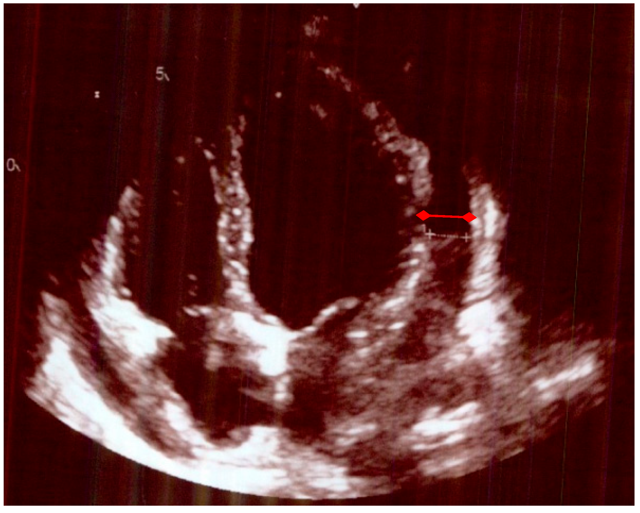
Figure 1. Echocardiogram, with evidence of pericardial effusion (red arrows).
Life 15 01131 g001
Figure 2. CT scan shows evidence of pericardial effusion (red arrows).
Life 15 01131 g002
Figure 3. PRISMA flow diagram of data extraction of the studies included in the systematic review.
Life 15 01131 g003
Table 1. Sequential investigation results at initial presentation and follow-up.
Bloods on each Hospital AdmissionCRP
NORMAL (<4) mg/L
Hb
Normal (115–165) g/L
WCC
Normal (4–11) 10*9/L
Neutrophils
Normal (1.8–7.5) 10 × 9/L
Lymphocytes
Normal (1–4) 10 × 9/L
Alkaline Phosphatase
Norm (30–130) u/L
GGT
Normal (0–39) u/L
Blood Cultures
1st visit (week 0)1711206.04.90.614266Not taken
2nd visit (week 1)191974.53.50.6210115Negative
3rd visit (week 2)1061095.64.80.6214181Negative
4th visit (week 4)741135.94.70.8170159Not taken
At discharge<41233.62.11.178Not takenNot taken
CRP—C-reactive protein; Hb—hemoglobin; ALP—alkaline phosphatase; GGT—gamma-glutamyl transferase.
Table 2. The modified Jones diagnostic criteria of rheumatic fever.
Major Criteria
Low-risk PopulationHigh-risk Population
Carditis (clinical or subclinical)
Arthritis—only polyarthritis
Chorea
Erythema marginatum
Subcutaneous nodules
Carditis (clinical or subclinical)
Arthritis—monoarthritis or polyarthritis
Polyarthralgia
Chorea
Erythema marginatum
Subcutaneous nodules
Minor Criteria
Low-risk populationHigh-risk population
Polyarthralgia
Hyperpyrexia (≥38.5 °C)
ESR ≥ 60 mm/h and/or CRP ≥ 3.0 mg/dL
Prolonged PR interval (after considering the differences related to age, if there is no carditis as a major criterion)
Monoarthralgia
Hyperpyrexia (≥8.0 °C)
ESR ≥ 30 mm/h and/or CRP ≥ 3.0 mg/dL
Prolonged PR interval (after considering the differences related to age, if there is no carditis as a major criterion)
ESR—erythrocyte sedimentation rate.
Table 3. Echocardiogram findings pre and post-treatment.
Echocardiogram Pre-Treatment Echocardiogram Post-Treatment after 4 Months.
A small/moderate global pericardial effusion
Normal LV structure and function.
Valves appeared structurally and functionally normal
Mobile atrial septum, no shunt seen.
No evidence of subacute bacterial endocarditis
No obvious residual pericardial effusion
Normal LV structure and function
No evidence of valve thickening but mild dilation of the non-coronary sinus of Valsalva and trivial AR and MR
Normal RV structure and function
Table 5. Genetic and acquired risk factors and clinical characteristics of acute rheumatic fever (ARF) and its mimickers [21,39,40,41,42,43].
Acute
Rheumatic Fever in
Children
Acute
Rheumatic Fever in Adults
Adults’ Onset Still Disease Post-Streptococcal Reactive Arthritis Rheumatoid Arthritis
Peak age of onset (years)5–15 yrs (rare < 3 yrs)Rare in adults Two peaks: 15–25 and 36–46Bimodal peaks: 8–14 and 21–37 yrsTypically, 40–60
GeneticsHLA-DR7, DR2/DR4; D8/17 B-cells; twin heritability ~60%(HLA-DR7, D8/17)HLA-DRB1No strong HLA links; genetic link unclearHLA-DRB1 shared epitope (DR4); twin concordance low (~12–15%)
Most common clinical features in descending orderFever
Migratory arthritis of large joints
Carditis
Subcutaneous nodules
chorea,
Erythema marginatum
Polyarthralgia (typically large joint)
Fever
Carditis
Quotidian fevers
Salmon rash
Arthritis
Lymphadenopathy
Hepatosplenomegaly
Non-migratory arthritis; small, axial, large joints
Tenosynovitis
Carditis (Rare)
Symmetric polyarthritis of small joints of hands and feet
chronic erosive disease
Environmental triggers Poor socioeconomic status. Usually, recurrence form childhoodTriggered by viral infections, not GASGAS infection, but
closer timing; less
related to poverty
Smoking, silica, periodontal disease, obesity
Sex Female preponderanceFemale preponderanceFemale predominanceEqualFemale preponderance
Most prevalent (region)Asia, Aboriginals in AustraliaN/ANot restricted to any geographical location.Not restricted to any geographical location.Northern Europe and North America
Treatment NSAIDs
Penicillin
Aspirin
NSAIDs
Penicillin
Aspirin
NSAIDs
DMARDS
Corticosteroids
NSAIDs
Corticosteroids
NSAIDs
DMARDS
Corticosteroids
ComplicationsRheumatic heart disease Rheumatic heart disease Macrophage activation syndromeglomerulonephritisJoint deformities
Interstitial lung disease
serositis
GAS—group A streptococcal infection; N/A—not applicable; NSAIDS—non-steroid anti-inflammatory drugs; DMARDS—disease-modifying antirheumatic drugs; HLA—human leucocyte antigen.
Disclaimer/Publisher’s Note: The statements, opinions and data contained in all publications are solely those of the individual author(s) and contributor(s) and not of MDPI and/or the editor(s). MDPI and/or the editor(s) disclaim responsibility for any injury to people or property resulting from any ideas, methods, instructions or products referred to in the content.