Article

Assessing Potential Reservoir of Multidrug-Resistant Bacteria in the Oral Microbiota of Captive Burmese and Royal Pythons †

1
Department of Veterinary Sciences, University of Trás-os-Montes e Alto Douro (UTAD), 5000-801 Vila Real, Portugal
2
Antimicrobials, Biocides and Biofilms Unit (AB2Unit), University of Trás-os-Montes e Alto Douro (UTAD), 5000-801 Vila Real, Portugal
3
Center for the Research and Technology of Agro-Environmental and Biological Sciences (CITAB)-Inov4Agro, University of Trás-os-Montes e Alto Douro (UTAD), 5000-801 Vila Real, Portugal
4
Laboratory of Physiology, Department of Animal Science, University of Trás-os-Montes e Alto Douro (UTAD), 5000-801 Vila Real, Portugal
5
Animal and Veterinary Research Center (CECAV)-AL4AnimalS, University of Trás-os-Montes e Alto Douro (UTAD), 5000-801 Vila Real, Portugal
6
Interdisciplinary Centre of Marine and Environmental Research (CIIMAR), University of Porto, 4450-208 Matosinhos, Portugal
7
Zoo da Maia, 4470-184 Maia, Portugal
*
Authors to whom correspondence should be addressed.
This article is a revised and expanded version of the abstract entitled “Oral microbiota in captive snakes: A study in Burmese and royal pythons” published in the Proceedings of the 31st European Veterinary Dental Forum, Nantes, France, 30 May–1 June 2024.
Life 2025, 15(3), 442; https://doi.org/10.3390/life15030442
Submission received: 30 November 2024 / Revised: 12 February 2025 / Accepted: 24 February 2025 / Published: 12 March 2025
(This article belongs to the Special Issue Trends in Microbiology 2025)

Abstract

:
The oral microbiota has a diversity of microorganisms that together maintain the homeostasis of the oral cavity. Disruptions in the balance of these microbial populations can lead to the development of oral and dental diseases. To characterize the normal oral microbiota of captive ophidians, samples were obtained from the oral cavity of eight Burmese pythons (Python bivittatus) and 11 royal pythons (Python regius), using oral swab, during routine evaluations. In the Laboratory-Antimicrobials, Biocides and Biofilms Unit at University of Trás-os-Montes e Alto Douro, the identification and antimicrobial susceptibility profile was determined using the Vitek® 2 Compact automated device (bioMérieux, Marcy-l’Étoile, France). Of the 106 bacterial isolates obtained, 69% were Gram-negative species and 31% were Gram-positive bacteria. The genus Pseudomonas was the most frequently isolated. Chryseobacterium indologenes, Escherichia coli and Pseudomonas aeruginosa were the most isolated species. Antimicrobial susceptibility testing revealed that the phenotypic resistance was highest for nitrofurans (47.2%), beta-lactams (45.8%) and sulfonamides (30.6%). Twenty-one multidrug-resistant isolates (58.3%) were identified with Acinetobacter baumannii, Serratia plymuthica, Chryseobacterium indologenes, Providencia rettgeri and Pseudomonas aeruginosa showing the highest resistance frequencies.

1. Introduction

The oral cavity of snakes is colonized by a diverse microbiota of both Gram-negative and Gram-positive bacteria. Alterations in this oral microbiota can lead to the colonization of pathogenic organisms and the development of bacterial stomatitis secondary to episodes of immunodepression caused by poor dietary and environmental management, such as inappropriate temperature or nutritional deficits. In these situations, animals can show a variety of clinical signs, such as anorexia, dysphagia, gingivitis, ecchymoses, petechiae and dental exfoliation. If not treated in good time, stomatitis can develop into osteomyelitis and abscesses or cause respiratory tract disease, septicemia and death [1,2,3,4,5]. Due to the strong impact of oral disease on animal health and welfare, the oral microbiota has been the subject of several studies for the knowledge of the organisms present and because it is a niche with differentiating resistance characteristics [6].
Snakes from different regions of the world show significant changes in the oral microbiota. Factors that explain such differences include animal species, origin, wild or captive habitat, health status, type of prey and predation [1,7]. To date, a variety of pathogenic bacteria have been reported in snakes, including Pseudomonas spp., Aeromonas spp., Morganella spp., Escherichia spp., Staphylococcus spp., Corynebacterium spp., Stenotrophomonas spp., Acinetobacter spp., Klebsiella spp., Shigella spp., Clostridium spp., Salmonella spp., Proteus spp. and Providencia spp. [8,9,10,11,12,13,14]. Many of the bacterial agents mentioned are present in the gastrointestinal tract of prey, leading to colonization of the predator’s oral microbiota. Additionally, the gastrointestinal tract of reptiles is also colonized by bacteria such as Enterobacter spp. and Pseudomonas spp., which can contaminate their own water sources, contributing to colonization of the oral cavity [15].
Bacterial stomatitis often involves an overgrowth of Gram-negative bacteria with Pseudomonas spp., Aeromonas spp., Proteus spp., Escherichia spp., Salmonella spp., Klebsiella spp. and Mycobacterium spp. being the most frequently isolated species [3,12,16,17]. Less commonly, bacteria from the genera Fusobacterium, Clostridium, Bacteroides and Peptostreptococcus have also been isolated [3].
Identifying the bacterial agents in the oral microbiota of snakes is crucial for expanding our understanding of these organisms but also for understanding the etiological agents of secondary infections that can arise from animal management. Information regarding the antimicrobial susceptibilities of snake oral bacteria is essential for supporting empirical antimicrobial therapy.
This study aimed to characterize the commensal oral Gram-negative microbiota of captive snakes and evaluate their potential as reservoirs of multidrug-resistant bacteria.

2. Materials and Methods

2.1. Animals

This study included nineteen snakes from the reptile collection at Zoo da Maia, a zoological park located in the north of Portugal, observed during routine oral examinations performed between March and April of 2023. Eight Burmese pythons (Python bivittatus, PB), five females and three males, with an average age of four years old (1–5 years), and 11 royal pythons (Python regius, PR), six females and five males with an average age of six years old (2–12 years), were selected for this study. All procedures were conducted in accordance with the European Animal Welfare Directives (Directive 98/58/CE) and the animals were handled and samples taken by three Federation of European Laboratory Animal Science Associations accredited staff.

2.2. Sample Processing and Isolation

Using sterile AMIES swabs (VWR, Carnaxide, Portugal), 19 samples from the oral cavity were collected under manual restraint of the animal without the need for sedation. The samples were placed in a closed tube containing a transport medium and transferred to the Antimicrobials, Biocides and Biofilms Unit (AB2Unit), Department of Veterinary Sciences at University of Trás-os-Montes e Alto Douro for bacterial isolation and identification.
Oral swabs were spread on different culture (according with the methodologies of laboratory AB2 Unit-CITAB) media such as Chromocult® Coliform Agar (CCA) (Merck, Darmstadt, Germany), MacConkey Agar (Oxoid, Hampshire, UK), Baird Parker Agar (Oxoid, Hampshire, UK), Glutamate Starch Phenol Red Agar (Oxoid, Hampshire, UK), and Mannitol Salt Agar (Oxoid, Hampshire, UK), according to the manufacturer’s instructions. All plates were incubated at 36 ± 1 °C for 24 h and the shape and color of the colonies were observed.

2.3. Identification and Antimicrobial Susceptibility Testing of Bacterial Isolates

After obtaining pure cultures, bacterial identification and antimicrobial susceptibility testing (ID/TSA test) were performed using the automated Vitek® 2 Compact system (bioMérieux, Marcy-l’Étoile, France) in accordance with the methodologies of laboratory AB2 Unit-CITAB. The identification of the microbial species was performed through a card consisting of a miniaturized system of conventional biochemical tests (Vitek® 2 GP and Vitek® 2 GN, bioMérieux, France). The determination of antimicrobial susceptibility was performed using cards for AST composed of multiple antibiotics. In order to obtain the ID and the AST of the bacterial isolate, the protocol established by the manufacturer and used in veterinary microbiology applications was followed.
Twenty-one antimicrobials from seven different classes were tested on Gram-negative isolates (Vitek® 2 AST-GN97, bioMérieux, Craponne, France): beta-lactams: ampicillin (AMP), amoxicillin–clavulanic acid (AMC), cephalexin (CL), cephalothin (KF), cefpodoxime (CPD), cefovecin (CVN), ceftiofur (EFT), ertapenem (ETP), imipenem (IPM), meropenem (MEM); aminoglycosides: amikacin (AK), gentamicin (CN), neomycin (N); fluoroquinolones: enrofloxacin (ENR), marbofloxacin (MRB), pradofloxacin (PFX); tetracyclines: doxycycline (DO), tetracycline (TE); nitrofurans: nitrofurantoin (F); amphenicols: chloramphenicol (C); sulfonamides: trimethoprim + sulfamethoxazole (SXT).

3. Results

3.1. Bacterial Species Isolated

Following the isolation process, thirty-six Gram-negative isolates were identified, including Chryseobacterium indologenes (n = 6), Escherichia coli (n = 4), Pseudomonas aeruginosa (n = 4), Pseudomonas fluorescens (n = 2), Pseudomonas putida (n = 1), Bordetella hinzii (n = 2), Pandoraea spp. (n = 2), Providencia rettgeri (n = 2), Acinetobacter baumannii (n = 1), Achromobacter denitrificans (n = 1), Achromobacter xylosoxidans (n = 1), Delftia acidovorans (n = 1), Enterobacter cloacae complex (n = 1), Lechercia adecarboxylata (n = 1), Pasteurella pneumotropica (n = 1), Proteus mirabilis (n = 1), Rahnella aquatilis (n = 1), Salmonella group (n = 1), Serratia plymuthica (n = 1), Sphingobacterium thalpophilum (n = 1) and Sphingomonas paucimobilis (n = 1) (Table 1).
The genus Pseudomonas was the most identified (19.4%) with seven isolates of the species P. aeruginosa (n = 4), P. fluorescens (n = 2), and P. putida (n = 1). This genus was identified in 12.5% (n = 2) of the PB samples and in 20% of PR samples (P. aeruginosa, 5%; P. fluorescens, 10% and P. putida, 5%). Overall, 55.6% (n = 20) of the isolates were derived from PR, while 44.4% (n = 16) were isolated from the oral cavities of PB (Table 2 and Table 3).
The majority of animals in the study exhibited polymicrobial cultures except for PB7, PR3, PR6, and PR7. Several bacterial species were isolated exclusively in either PB or PR species, such as the Salmonella genus or Acinetobacter baumannii only isolated in the oral cavities of PB and PR, respectively. Bordetella hinzii was isolated in both python species, unlike Escherichia coli, which was found only in the oral cavity of PB. Bacteria from the genus Chryseobacterium were isolated and identified exclusively in the PR population. The three most common organisms were Chryseobacterium indologenes, Escherichia coli and Pseudomonas aeruginosa.

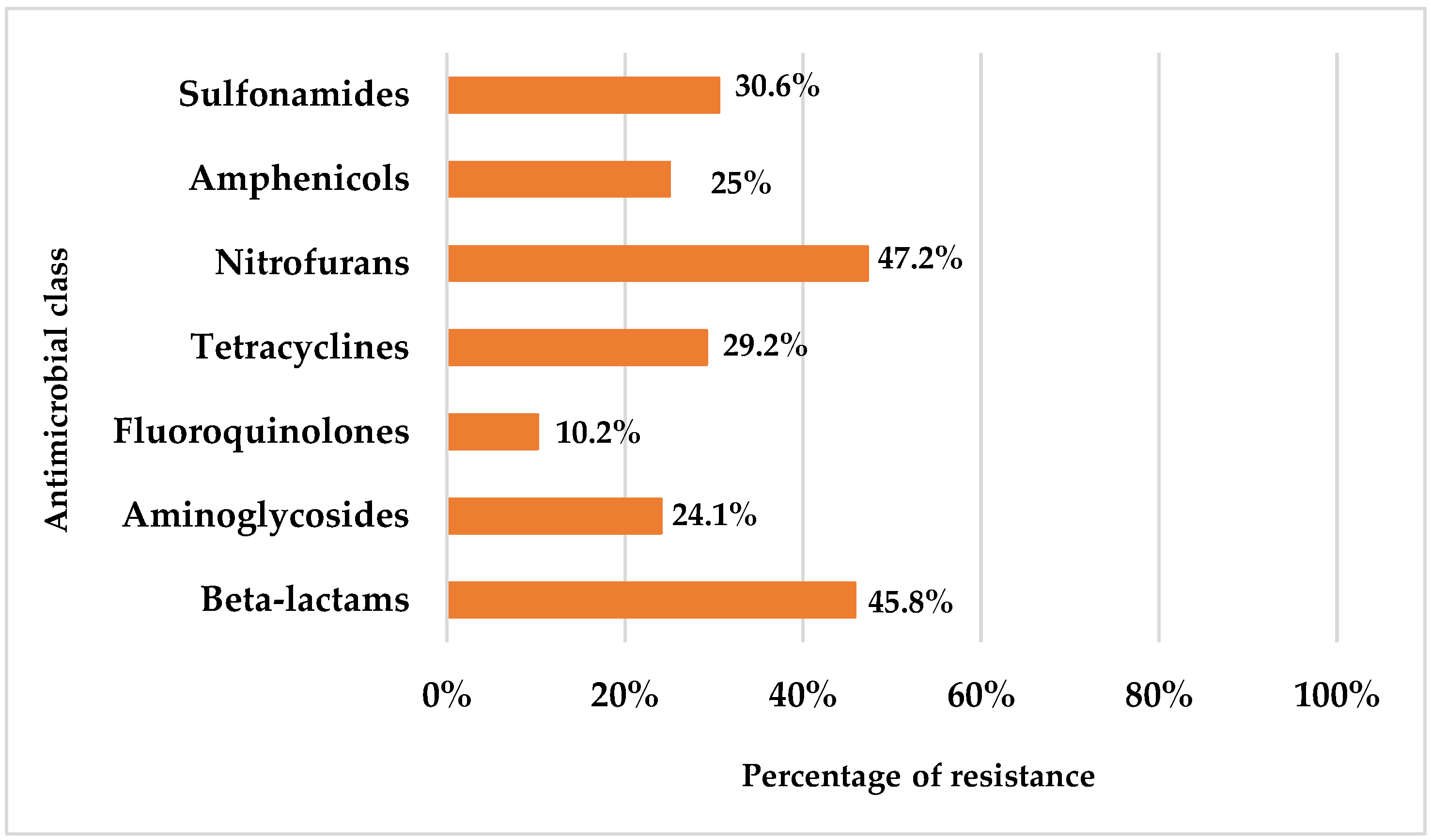
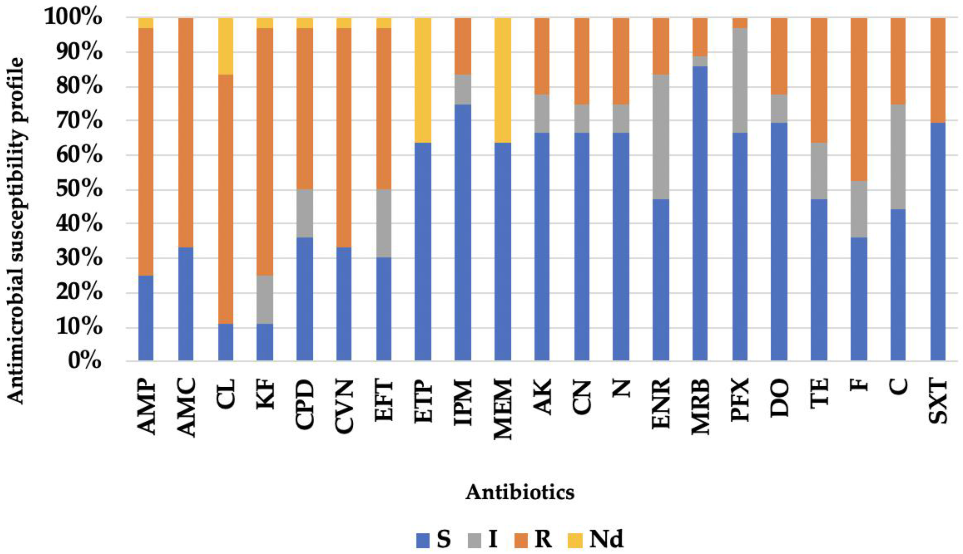
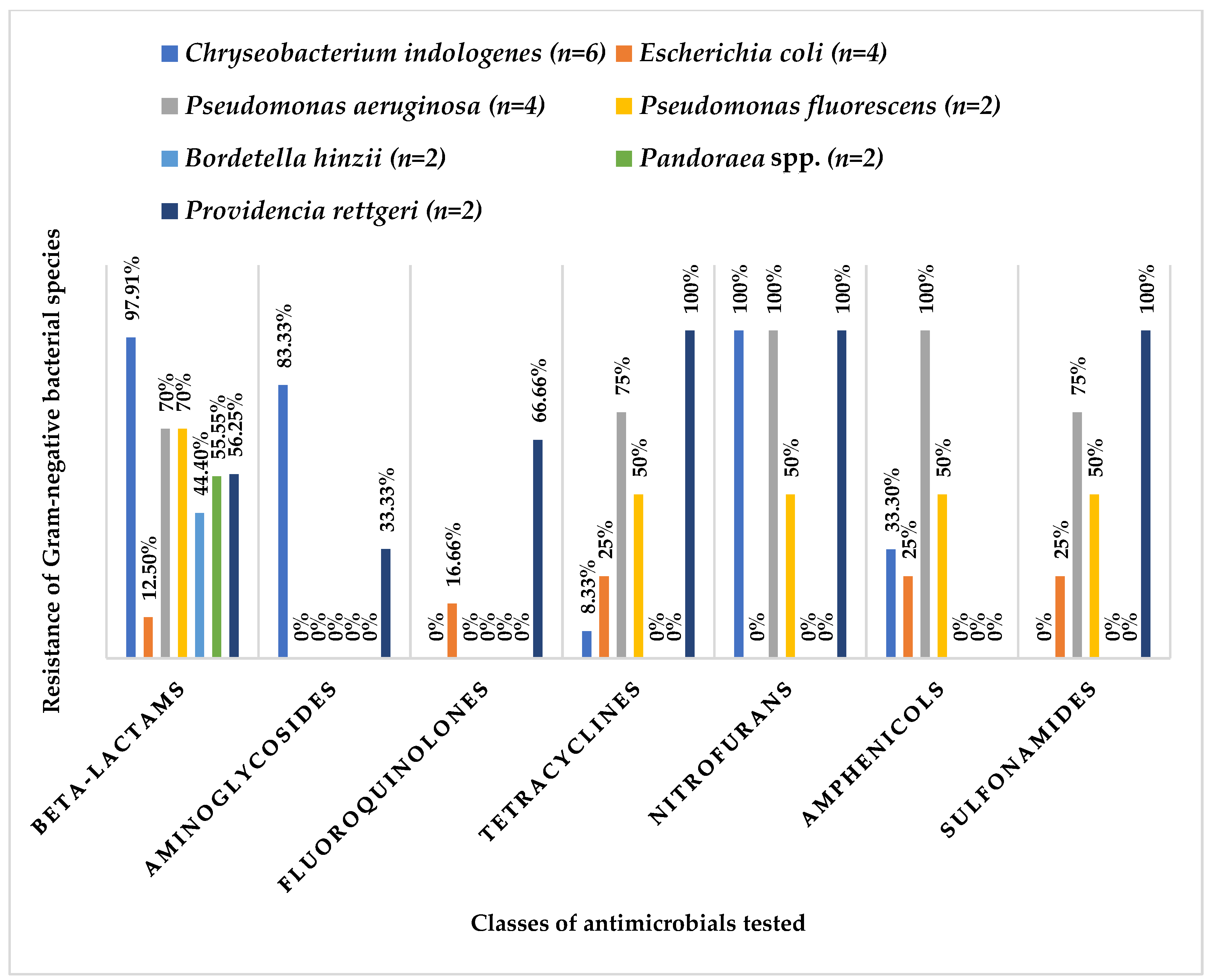
3.2. Antibiotic Resistance Profile

The results showed high levels of phenotypic resistance, particularly to nitrofurans (47.2%), beta-lactams (45.8%) and sulfonamides (30.6%), while resistance to quinolones was comparatively lower (10.2%) (Figure 1).
When assessing individual resistance to each antimicrobial, beta-lactams exhibited the highest overall resistance, with ampicillin, cephalexin, and cephalothin all showing a resistance rate of 72.2%, which were followed by amoxicillin–clavulanic acid (66.7%), and ceftiofur (63.9%). Concerning aminoglycosides, resistance rates were similar for gentamicin and neomycin, with eight isolates (22.2%) resistant to amikacin, and four displaying intermediate behavior (11.1%). Within the fluoroquinolone class, marbofloxacin exhibited the highest susceptibility (86.1%; n = 31), which was followed by pradofloxacin (66.7%; n = 24) and enrofloxacin (47.2%; n = 17). Six isolates (16.7%) demonstrated resistance to enrofloxacin, four demonstrated resistance to marbofloxacin (11.1%), and only one isolate showed resistance to pradofloxacin (2.8%). The tetracycline class also exhibited enhanced antimicrobial activity with doxycycline proving more effective than tetracycline (69.4% and 47.2%, respectively). Additionally, resistance rates to trimethoprim + sulfamethoxazole, nitrofurantoin, and chloramphenicol were 47.2%, 30.6%, and 25%, respectively. Finally, it is noteworthy that 16.7% (n = 6) were identified as resistant to imipenem (Figure 2).
Among the species identified in the microbiota, Chryseobacterium indologenes, Pseudomonas aeruginosa and Providencia rettgeri exhibited the highest resistance to the different classes of antimicrobials tested. Acinetobacter baumannii and Serratia plymuthica were the two species with the highest resistance phenotypes compared to the other species. In this study, A. baumannii was shown to be resistant to cephalexin, cephalothin, cefovecin, and ceftiofur, all of which belong to the beta-lactam class. It also showed a resistant phenotype to tetracycline (tetracyclines), nitrofurantoin (nitrofurans), chloramphenicol (phenicols) and trimethoprim + sulfamethoxazole (sulfonamides). However, resistance to aminoglycosides or fluoroquinolones was not observed. S. plymuthica demonstrated resistance to beta-lactams (ampicillin, cephalexin, cephalothin, cefovecin and imipenem), aminoglycosides (amikacin and gentamicin), fluoroquinolones (enrofloxacin and pradofloxacin), sulfonamides (trimethoprim/sulfamethoxazole) and tetracyclines (doxycycline and tetracycline) (Figure 3).
It is crucial to highlight that multidrug resistance (MDR) was observed in 21 out of 36 isolates (58,3%), indicating simultaneous resistance to three or more antimicrobial families. The MDR pattern was distributed by Chryseobacterium indologenes (6/6), Escherichia coli (1/4), Pseudomonas aeruginosa (4/4), Pseudomonas putida (1/1), Pseudomonas fluorescens (1/2), Providencia rettgeri (2/2), Acinetobacter baumannii (1/1), Delftia acidovorans (1/1), Lechercia adecarboxylata (1/1), Proteus mirabilis (1/1), Serratia plymuthica (1/1), and Sphingomonas paucimobilis (1/1). The MDR profile table shows that resistance to βLC (beta-lactams) in most isolates is often accompanied by resistance to AMN (aminoglycosides) (Table 4).

4. Discussion

The identification of bacteria with an MDR profile in samples from the oral cavitiy and skin of clinically healthy captivity snakes emphasized a public health problem that concerns the emergence of these microorganisms [18,19]. Due to the close contact between humans and snakes, it is highly possible that humans will be infected with multidrug-resistant strains.
This study is clinically relevant since commensal bacteria can act as opportunistic pathogens and produce infections that are difficult to treat [20].
Several bacterial species were isolated only in Burmese pythons (PB) or in royal pythons (PR), such as Escherichia coli, which was only identified in PB. Escherichia coli is a Gram-negative, facultative anaerobic bacterium that is considered a commensal organism in the intestines and present in the environment. It can act as a principal pathogen or as an opportunistic one and can be highly pathogenic. It is associated with bacterial stomatitis in reptiles [3,15,21,22].
One of the bacteria isolated exclusively in the oral cavity of PR was Chryseobacterium indologenes. This bacterial agent is found sporadically in soil, water, waste, food sources, animals and aquatic environments [23]. In snakes, C. indologenes has been reported in an oral abscess in a PR [24].
As mentioned above, the genus Pseudomonas was the most identified. In the study by Jho and colleagues (2011) [12], the genus Pseudomonas was also the most frequently isolated in the oral cavity of snakes (33% of the samples) with the identification of P. aeruginosa and P. putida. In PR, Dipineto and collaborators (2014) [25] isolated Pseudomonas spp. in 85% of the animals with P. putida found in nine animals and P. aeruginosa in 42 animals. Yak and colleagues (2015) [26] confirmed the abundance of the genus Pseudomonas in the oral cavity of reticulated pythons (Python reticulatus) with P. aeruginosa isolated in 20% of the specimens and P. staminae in 10%. The genus Pseudomonas is considered an opportunistic pathogen with a high capacity for developing resistance to antimicrobials and various virulence factors, which is why it is associated with high morbidity and mortality rates. These bacteria can be responsible for pneumonia, septicemia, skin and oral lesions such as ulcerative stomatitis [16,26].
In the present study, Acinetobacter baumannii and Serratia plymuthica were the two species with the highest resistance phenotypes compared to the other species with only one isolated each. A. baumannii is an opportunistic hospital pathogen that causes severe and invasive nosocomial infections associated with high mortality rates, posing a threat to public health [27,28]. The high resistance to antimicrobials shown by this bacterial genus is associated with the presence of multiple resistance mechanisms that make A. baumannii resistant to most beta-lactam antimicrobials, aminoglycosides, fluoroquinolones, tetracyclines, macrolides, phenicols and sulfonamides [29]. The species S. plymuthica, which belongs to the Enterobacteriaceae family, is predominantly found in water, soil, plants, and wild animals. Species from this genus are associated with various infections such as pneumonia, meningitis and urinary tract infections [30].
The bacterial species with at least two isolates that showed the highest resistance to the different classes of antimicrobials tested were Chryseobacterium indologenes, Pseudomonas aeruginosa and Providencia rettgeri. The genus Chryseobacterium is considered an opportunistic and MDR bacterial agent with the capacity to cause serious infections in neonates, pregnant women and immunosuppressed patients [24]. C. indologenes is intrinsically resistant to aminoglycosides, aminopenicillins, first-generation cephalosporins and aztreonam [24,31]. P. aeruginosa is commonly found in intensive care units and is associated with various types of infections in humans and animals, and it can also be found in soil and water. P. aeruginosa is resistant to various antimicrobials, including beta-lactams (3rd generation cephalosporins and carbapenems), fluoroquinolones, and aminoglycosides [32,33]. The genus Providencia, which was identified in two isolates (P. rettgeri) from PB, is also described in the oral cavity of several snakes, namely Protobothrops mucrosquamatus, Bungarus multicinctus and Daboia siamensis [34]. Bacteria of the Providencia genus are commensals of the gastrointestinal tract and have been identified in various animals as well as in water and soil [35,36].
In the treatment of bacterial stomatitis in snakes, fluoroquinolones and aminoglycosides are the common choices of first-line antibiotics while waiting for the results of AST [37,38]. Looking at the percentages of resistance in each class of antimicrobials tested on this study, nitrofurans, beta-lactams and sulfonamides presented the highest phenotypic resistance. These classes of antimicrobials belong to category D according to the EMA (European Medicines Agency) categorization and are used as first-line antimicrobials. The high phenotypic resistance observed in these classes can significantly reduce the effectiveness of antibiotic therapy, demonstrating the importance of clinical laboratory diagnosis in veterinary microbiology associated with animal health. Five isolates of the species Chryseobacterium indologenes and one isolate of Serratia plymuthica showed resistance to imipenem, which is an antibiotic used as a last resort in infections with MDR bacteria and only in hospital settings [39].

5. Conclusions

Wild animals play a critical role in the One Health concept, representing potential natural reservoirs of antimicrobial resistance and infection. They are also actively involved in the spread of bacteria and resistance determinants between different habitats. Emerging infectious diseases often result from the interplay between wildlife, humans, and the environment. These findings highlight the importance of personal protective equipment for a variety of roles, including animal keepers, veterinarians, and healthcare professionals.
This study has shown that snakes can be reservoirs of pathogens with zoonotic potential, carrying genes that confer resistance/multidrug resistance to highly relevant antimicrobials. The distinction between intrinsic and acquired resistance is fundamental in the study of antibiotic resistance, as it influences approaches to the treatment and control of infections. In terms of public health, acquired resistance is of greater concern, as it can spread between different bacterial species and make it difficult to treat infections, so a detailed future study would be essential. Due to the growing increase in antibiotic resistance, it is imperative to conduct studies to monitor and control antibiotic resistance, always considering a holistic approach. Antimicrobial resistance and the increase in multidrug-resistant bacteria emphasize the importance of medicine becoming increasingly aware of the correct use of antimicrobials and of raising awareness among regulatory bodies, veterinarians, animal health professionals, and guardians of the emerging problem and the risk to public health.

Author Contributions

Conceptualization, J.F.R. and M.J.S.; Methodology, I.M., A.R.P., P.T., P.M., M.C., N.A., J.J.M., J.F.R. and M.J.S.; Formal Analysis, I.M., A.R.P., J.F.R. and M.J.S.; Writing—Original Draft Preparation, I.M., J.F.R. and M.J.S.; Writing—Review and Editing, I.M., A.R.P., P.T., P.M., M.C., N.A., J.J.M., J.F.R. and M.J.S.; Supervision, J.F.R. and M.J.S.; Funding Acquisition, J.F.R., J.J.M. and M.J.S. All authors have read and agreed to the published version of the manuscript.

Funding

This research was funded by national funds through the Portuguese Foundation for Science and Technology (FCT) under the projects UIDB/CVT/00772/2020 (CECAV), UIDB/AGR/04033/2020 (CITAB), LA/P/0059/2020 (AL4AnimalS), LA/00037/2022 (Inov4Agro) and under the Scientific Employment Stimulus-Institutional Call—CEECINS/00127/2018 (J.F.R.).

Institutional Review Board Statement

The animal study protocol was developed under the scope the routine clinical examination of the animals and approved by the Board of Maia Zoo (Doc. A/2019 0145; 18 February 2019). The entire procedure was conducted in accordance with the European Animal Welfare Directives (Directive 98/58/CE and Decreto-lei no 64/2000).

Informed Consent Statement

Patient consent was waived, because the animals were part of a zoological collection and were sampled as part of a clinical evaluation by their veterinarian.

Data Availability Statement

All raw data are available from the corresponding authors upon reasonable request.

Acknowledgments

This article is a revised and expanded version of an abstract [40] presented as oral communication at the 31st European Veterinary Dental Forum, 1 June 2024.

Conflicts of Interest

The authors declare no conflicts of interest or relationship, financial or otherwise that might be perceived as influencing their objectivity in the reporting of this study.

References

  1. Padhi, L.; Panda, S.K.; Mohapatra, P.P.; Sahoo, G. Antibiotic susceptibility of cultivable aerobic microbiota from the oral cavity of Echis carinatus from Odisha (India). Microb. Pathog. 2020, 143, 104121. [Google Scholar] [CrossRef] [PubMed]
  2. Mehler, S.J.; Bennett, R.A. Oral, dental, and beak disorders of reptiles. Vet. Clin. North Am. Exot. Anim. Pract. 2003, 6, 477–503. [Google Scholar] [CrossRef]
  3. Hedley, J. Anatomy and disorders of the oral cavity of reptiles and amphibians. Vet. Clin. North Am. Exot. Anim. Pract. 2016, 19, 689–706. [Google Scholar] [CrossRef] [PubMed]
  4. Mustafa, S.; Popova, T. Enterobacter agglomerans—A cause of stomatitis in a snake. Tradit. Mod. Vet. Med. 2017, 1, 39–44. [Google Scholar]
  5. Grego, K.F.; Carvalho, M.P.N.; Cunha, M.P.V.; Knobl, T.; Pogliani, F.C.; Catão-Dias, J.L.; Sant´Anna, S.S.; Ribeiro, M.S.; Sellera, F.P. Antimicrobial photodynamic therapy for infectious stomatitis in snakes: Clinical views and microbiological findings. Photodiagnosis Photodyn. Ther. 2017, 20, 196–200. [Google Scholar] [CrossRef]
  6. Shek, K.; Tsui, K.; Lam, K.; Crow, P.; Ng, K.; Ades, G.; Yip, K.; Grioni, A.; Tan, K.; Lung, D.; et al. Oral bacterial flora of the Chinese cobra (Naja atra) and bamboo pit viper (Trimeresurus albolabris) in Hong Kong SAR, China. Hong Kong Med. J. 2009, 15, 183–190. [Google Scholar]
  7. Artavia-León, A.; Romero-Guerrero, A.; Sancho-Blanco, C.; Rojas, N.; Umaña-Castro, R. Diversity of aerobic bacteria isolated from oral and cloacal cavities from free-living snakes species in Costa Rica rainforest. Int. Sch. Res. Not. 2017, 2017, 8934285. [Google Scholar] [CrossRef]
  8. Babalola, M.O.; Balogun, J.A. The ecology and potential health risk of the oral microflora of Python regius and Clelia scyntalina. Int. J. Microbiol. Res. 2013, 5, 349–356. [Google Scholar] [CrossRef]
  9. Lukač, M.; Horvatek Tomić, D.; Mandac, Z.; Mihoković, S.; Prukner-Radovčić, E.; Horvatek Tomić, D.; ManDac, Z.; Mihoković, S.; Prukner, E. Oral and cloacal aerobic bacterial and fungal flora of free-living four-lined snakes (Elaphe quatuorlineata) from Croatia. Veterinarski Arhiv 2017, 87, 351–361. [Google Scholar] [CrossRef]
  10. Dehghani, R.; Sharif, M.R.; Moniri, R.; Sharif, A.; Kashani, H.H. The identification of bacterial flora in oral cavity of snakes. Comp. Clin. Pathol. 2016, 25, 279–283. [Google Scholar] [CrossRef]
  11. Hedley, J.; Whitehead, M.L.; Munns, C.; Pellett, S.; Abou-Zahr, T.; Calvo Carrasco, D.; Wissink-Argilaga, N. Antibiotic stewardship for reptiles. J. Small Anim. Pract. 2021, 62, 829–839. [Google Scholar] [CrossRef] [PubMed]
  12. Jho, Y.-S.; Park, D.-H.; Lee, J.-H.; Cha, S.-Y.; Han, J.S. Identification of bacteria from the oral cavity and cloaca of snakes imported from Vietnam. Lab. Anim. Res. 2011, 27, 213. [Google Scholar] [CrossRef] [PubMed]
  13. Zancolli, G.; Mahsberg, D.; Sickel, W.; Keller, A. Reptiles as reservoirs of bacterial infections: Real threat or methodological bias. Microb. Ecol. 2015, 70, 579–584. [Google Scholar] [CrossRef]
  14. Gospodinova, I.R.; Mustafa, S.; Popova, T.P. Microbiological analysis of laryngeal and oral swab samples from captive-bred snakes with respiratory tract infections in Bulgaria. Acta Microbiol. Bulg. 2024, 40, 405–411. [Google Scholar] [CrossRef]
  15. Ghosh, T.; Biswas, M.K.; Roy, P.; Guin, C. Short review of different microflora from the oral cavity of snakes. Uttar Pradesh J. Zool. 2017, 37, 30–34. [Google Scholar]
  16. Zhang, F.; Cheng, W. The mechanism of Bbcterial resistance and potential bacteriostatic strategies. Antibiotics 2022, 11, 1215. [Google Scholar] [CrossRef]
  17. Mitchell, M.A.; Diaz-Figueroa, O. Clinical reptile gastroenterology. Vet. Clin. North Am. Exot. Anim. Pract. 2005, 8, 277–298. [Google Scholar] [CrossRef]
  18. Silva, C.; Requicha, J.F.; Martins, J.J.; Duarte, A.; Dias, I.R.; Viegas, C.A.; Saavedra, M.J. Black-and-white ruffed lemur (Varecia variegata) in captivity: Analysis of the oral microbiota in a one health perspective. Animals 2021, 11, 2905. [Google Scholar] [CrossRef] [PubMed]
  19. Marques, I.; Alvura, N.; Martins, J.J.; Requicha, J.F.; Saavedra, M.J. First report of Kocuria kristinae in the skin of a Cuban boa (Epicrates angulifer). Life 2023, 13, 2133. [Google Scholar] [CrossRef]
  20. Pimenta, J.; Pinto, A.R.; Saavedra, M.J.; Cotovio, M. Equine Gram-negative oral microbiota: An antimicrobial aesistances watcher? Antibiotics 2023, 12, 792. [Google Scholar] [CrossRef]
  21. Cheng, L.; Cheng, X.; Deng, M.; Deng, X.; Du, Q.; Ge, Y.; Guo, Q.; He, J.; Jia, W.; Kang, D.; et al. Oral mucosal microbes. In Atlas of Oral Microbiology: From Healthy Microflora to Disease; Zhou, X., Li, Y., Eds.; Academic Press: Cambridge, MA, USA, 2015; pp. 95–107. [Google Scholar]
  22. D’Cruze, N.; Bates, J.; Assou, D.; Ronfot, D.; Coulthard, E.; Segniagbeto, G.H.; Auliya, M.; Megson, D.; Rowntree, J. A preliminary assessment of bacteria in “ranched” ball pythons (Python regius), Togo, West Africa. Nat. Conserv. 2020, 39, 73–86. [Google Scholar] [CrossRef]
  23. Das, P.; Karade, S.; Kaur, K.; Ramamurthy, R.; Ranjan, P. Chryseobacterium indologenes pneumonitis in an infant: A case report. J. Clin. Diagn. Res. 2017, 11, 7–8. [Google Scholar] [CrossRef] [PubMed]
  24. Tamai, I.A.; Pakbin, B.; Kafi, Z.Z.; Brück, W.M. Oral abscess caused by Chryseobacterium indologenes in ball python (Python regius): A case report. Antibiotics 2021, 10, 686. [Google Scholar] [CrossRef]
  25. Dipineto, L.; Russo, T.P.; Calabria, M.; De Rosa, L.; Capasso, M.; Menna, L.F.; Borrelli, L.; Fioretti, A. Oral flora of Python regius kept as pets. Lett. Appl. Microbiol. 2014, 58, 462–465. [Google Scholar] [CrossRef] [PubMed]
  26. Yak, R.; Lundin, A.-C.; Pin, P.Y.; Sebastin, S.J. Oral bacterial microflora of free-living reticulated pythons (Python reticulatus) in Singapore. J. Herpetol. Med. Surg. 2015, 25, 40–44. [Google Scholar] [CrossRef]
  27. Basatian-Tashkan, B.; Niakan, M.; Khaledi, M.; Afkhami, H.; Sameni, F.; Bakhti, S.; Mirnejad, R. Antibiotic resistance assessment of Acinetobacter baumannii isolates from Tehran hospitals due to the presence of efflux pumps encoding genes (adeA and adeS genes) by molecular method. BMC Res. Notes 2020, 13, 2–6. [Google Scholar] [CrossRef]
  28. Kyriakidis, I.; Vasileiou, E.; Pana, Z.D.; Tragiannidis, A. Acinetobacter baumannii antibiotic resistance mechanisms. Pathogens 2021, 10, 373. [Google Scholar] [CrossRef]
  29. Foti, M.; Giacopello, C.; Fisichella, V.; Latella, G. Multidrug-resistant Pseudomonas aeruginosa isolates from captive reptiles. J. Exot. Pet Med. 2013, 22, 270–274. [Google Scholar] [CrossRef]
  30. Van Houdt, R.; Moons, P.; Jansen, A.; Vanoirbeek, K.; Michiels, C.W. Genotypic and phenotypic characterization of a biofilm-forming Serratia plymuthica isolate from a raw vegetable processing line. FEMS Microbiol. Lett. 2005, 246, 265–272. [Google Scholar] [CrossRef]
  31. Izaguirre-Anariba, D.E.; Sivapalan, V. Chryseobacterium indologenes, an emerging bacteria: A case report and review of literature. Cureus. 2020, 12, e6720. [Google Scholar] [CrossRef]
  32. Pachori, P.; Gothalwal, R.; Gandhi, P. Emergence of antibiotic resistance Pseudomonas aeruginosa in intensive care unit; a critical review. Genes Dis. 2019, 6, 109–119. [Google Scholar] [CrossRef]
  33. Roulová, N.; Mot’ková, P.; Brožková, I.; Pejchalová, M. Antibiotic resistance of Pseudomonas aeruginosa isolated from hospital wastewater in the Czech Republic. J. Water Health 2022, 20, 692–701. [Google Scholar] [CrossRef] [PubMed]
  34. Chuang, P.C.; Lin, W.H.; Chen, Y.C.; Chien, C.C.; Chiu IMTsai, T.S. Oral bacteria and their antibiotic susceptibilities in Taiwanese venomous snakes. Microorganisms 2022, 10, 951. [Google Scholar] [CrossRef]
  35. Benedict, S.; Shilton, C.M. Providencia rettgeri septicaemia in farmed crocodiles. Microbiol. Aust. 2016, 37, 114–117. [Google Scholar] [CrossRef]
  36. Lacerda, L.E.; Portela, R.W. Perfil de sensibilidade a antibióticos de um isolado de Providencia rettgeri proveniente de equino. Rev. Ciências Médicas Biológicas 2021, 20, 413–417. [Google Scholar] [CrossRef]
  37. Sharina, O.; Jian, H.S.; Azlan, C. Bacterial stomatitis in wild reticulated pythons (Malayopython reticulatus) in Malaysia. World Vet. J. 2023, 13, 409–419. [Google Scholar] [CrossRef]
  38. Singh, J.; Mallik, S.; Nath, I.; Acharya, A.; Das, S.P.; Sethi, S.; Sahoo, M. Infectious stomatitis in an Indian rock python (Python molurus) and its therapeutic management. J. Entomol. Zool. Stud. 2018, 6, 392–394. [Google Scholar]
  39. European Medicines Agency. Categorisation of Antibiotics Used in Animals Promotes Responsible Use to Protect Public and Animal Health. 2020. Available online: https://www.ema.europa.eu/en/documents/report/categorisation-antibiotics-european-union-answer-request-european-commission-updating-scientific-advice-impact-public-health-and-animal-health-use-antibiotics-animals_en.pdf (accessed on 12 February 2025).
  40. Marques, I.; Pinto, A.R.; Alvura, N.; Telinhos, P.; Mendes, P.; Correia, M.; Martins, J.J.; Requicha, J.F.; Saavedra, M.J. Oral Microbiota in captive snakes: A study in Burmese and royal pythons. In Proceedings of the Book of the 31st European Veterinary Dental Forum, Nantes, France, 30 May–1 June 2024. [Google Scholar]
Figure 1. Percentage of resistance to each antimicrobial class.
Life 15 00442 g001
Figure 2. Antimicrobial susceptibility profile of the set of 36 Gram-negative isolates studied. Legend: beta-lactams: ampicillin (AMP), amoxicillin–clavulanic acid (AMC), cephalexin (CL), cephalothin (KF), cefpodoxime (CPD), cefovecin (CVN), ceftiofur (EFT), ertapenem (ETP), imipenem (IPM), meropenem (MEM); aminoglycosides: amikacin (AK), gentamicin (CN), neomycin (N); fluoroquinolones: enrofloxacin (ENR), marbofloxacin (MRB), pradofloxacin (PFX); tetracyclines: doxycycline (DO), tetracycline (TE); nitrofurans: nitrofurantoin (F); amphenicols: chloramphenicol (C); sulfonamides: trimethoprim + sulfamethoxazole (SXT); S—susceptible; I—intermediate; R—resistant; Nd—not determined.
Life 15 00442 g002
Figure 3. Relative frequency of resistance of Gram-negative bacterial species identified with at least two isolates in relation to the classes of antimicrobials tested.
Life 15 00442 g003
Table 1. List of bacterial species identified in the snake samples studied (n = 19).
IdentificationNo of Isolates (n = 36)
Chryseobacterium indologenes6
Escherichia coli4
Pseudomonas aeruginosa4
Pseudomonas fluorescens2
Pseudomonas putida1
Bordetella hinzii2
Pandoraea spp.2
Providencia rettgeri2
Acinetobacter baumannii1
Achromobacter denitrificans1
Achromobacter xylosoxidans1
Delftia acidovorans1
Enterobacter cloacae complex1
Leclercia adecarboxylata1
Pasteurella pneumotropica1
Proteus mirabilis1
Rahnella aquatilis1
Salmonella group1
Serratia plymuthica1
Sphingobacterium thalpophilum1
Sphingomonas paucimobilis1
Table 2. Bacterial species identified in each Burmese python (n = 8). Legend: F—female, M—male.
AnimalSexAge (Years)No of IsolatesSpecies Identified
PB1M12Achromobacter xylosoxidans
Bordetella hinzii
PB2F42Providencia rettgeri
Providencia rettgeri
PB3M52Pasteurella pneumotropica
Achromobacter denitrificans
PB4F52Escherichia coli
Escherichia coli
PB5F53Escherichia coli
Delftia acidovorans
Salmonella group
PB6F42Proteus mirabilis
Escherichia coli
PB7F41Pseudomonas aeruginosa
PB8M42Pseudomonas aeruginosa
Pseudomonas aeruginosa
Table 3. Bacterial species identified in each royal python (n = 11). Legend: F—female, M—male.
AnimalSexAge (Years)No of IsolatesSpecies Identified
PR1F102Chryseobacterium indologenes
Chryseobacterium indologenes
PR2M22Pseudomonas fluorescens
Pseudomonas aeruginosa
PR3F21Pandoraea spp.
PR4F42Chryseobacterium indologenes
Chryseobacterium indologenes
PR5F123Acinetobacter baumannii
Leclercia adecarboxylata
Enterobacter cloacae complex
PR6M21Sphingobacterium thalpophilum
PR7M101Pseudomonas putida
PR8F102Rahnella aquatilis
Serratia plymuthica
PR9F22Pseudomonas fluorescens
Chryseobacterium indologenes
PR10M122Pandoraea spp.
Sphingomonas paucimobilis
PR11M42Bordetella hinzii
Chryseobacterium indologenes
Table 4. Multidrug-resistant profile of oral microbiota bacteria from Python bivittatus and Python regius. Legend: βLC—beta-lactams, IMP—imipenem, AMN—aminoglycosides, FL—fluoroquinolones, TET—tetracyclines, NIT—nitrofurans, ANF—amphenicols, SUL—sulfonamides, “n”—number of isolates.
Chryseobacterium indologenesnEscherichia colinPseudomonas aeruginosanPseudomonas putidan
βLC-IMP-AMN-NIT4βLC-FL-TET-ANF-SUL1βLC-TET-NIT-ANF-SUL3βLC-NIT-SUL1
βLC-AMN-TET-NIT-ANF1 βLC-TET-NIT-ANF1
βLC-IMP-AMN-NIT-ANF1
Pseudomonas fluorescensnProvidencia rettgerinAcinetobacter baumanniinDelftia acidovoransn
βLC-TET-NIT-ANF-SUL1βLC-AMN-FL-TET-NIT-SUL2βLC-TET-SUL1βLC-AMN-FL1
Lechercia adecarboxylatanProteus mirabilisnSerratia plymuthicanSphingomonas paucimobilisn
AMN-FL-TET-SUL1βLC-TET-NIT1βLC- IMP-AMN-FL-TET-SUL1βLC-AMN-NIT1
Disclaimer/Publisher’s Note: The statements, opinions and data contained in all publications are solely those of the individual author(s) and contributor(s) and not of MDPI and/or the editor(s). MDPI and/or the editor(s) disclaim responsibility for any injury to people or property resulting from any ideas, methods, instructions or products referred to in the content.

