Article

Genetic Profiling of MRSA and MSSA from Food Contact Surfaces: Antibiotic, Heavy Metal and Benzalkonium Chloride Resistance

by
María Guadalupe Avila-Novoa
1,
Oscar Alberto Solis-Velazquez
1,
Jean Pierre González-Gómez
2,
Pedro Javier Guerrero-Medina
1,* and
Melesio Gutiérrez-Lomelí
1,*
1
Centro de Investigación en Biotecnología Microbiana y Alimentaria, Departamento de Ciencias Básicas, División de Desarrollo Biotecnológico, Centro Universitario de la Ciénega, Universidad de Guadalajara, Ocotlan 47820, Jalisco, Mexico
2
Laboratorio Nacional para la Investigación en Inocuidad Alimentaria (LANIIA), Centro de Investigación en Alimentación y Desarrollo, A.C. (CIAD), Culiacan 80110, Sinaloa, Mexico
*
Authors to whom correspondence should be addressed.
Life 2025, 15(12), 1811; https://doi.org/10.3390/life15121811
Submission received: 13 October 2025 / Revised: 23 November 2025 / Accepted: 25 November 2025 / Published: 26 November 2025
(This article belongs to the Special Issue 2nd Edition—Food Microbiological Contamination)

Abstract

Staphylococcus aureus is a foodborne pathogen that affects animals and humans. The persistence of this pathogen in the environment is associated with its ability to form biofilms and/or develop resistance mechanisms to antibiotics and sanitizers. A total of 67 S. aureus strains collected from food contact surfaces (FCSs) made of stainless steel and FCS-polypropylene used in dairy industries in Jalisco, México, were selected for this study. The genetic diversity and genes indicating antibiotic resistance were determined using PCR; antimicrobial susceptibility, resistance to cadmium chloride (CdCl2), and the minimum inhibitory concentration (MIC) of benzalkonium chloride (BC) were determined using the agar diffusion method and broth microdilution. Additionally, the effects of BC treatment on biofilm removal were evaluated. A total of 41.7% of the strains were MRSA [SCCmec Types II (20.8%), V (13.4%), and IV (7.4%)], and 58.2% were MSSA. Genes encoding antibiotic resistance—ermC (2.9%), ermA (2.9%), ermB (10.4%), aacA-aphD (10.4%), tetM (17.9%), and blaZ (88%)—were detected. A phenotypic test showed that 62.6% of the strains were cadmium-resistant S. aureus (>400 µg/mL CdCl2), and the MICs of 97% of isolates lay between 1.56 and 25 µg/mL BC. Treatment with BC + MR (100 µg/mL + 1% milk residues) led to a smaller reduction in biofilm (2.11–2.25 log10 cfu/cm2; p < 0.05) compared to BC (3.75–4.03 log10 cfu/cm2; at 5–10 min). MSSA and MSRA can develop biofilms that harbor mechanism resistance-associated genes, which are a public health hazard and a food safety concern.

1. Introduction

Staphylococcus aureus represents a significant concern for public health due to its ability to infect both humans and animals, causing a wide spectrum of illnesses such as food poisoning, osteomyelitis, endocarditis, pneumonia, toxic shock syndrome, bloodstream infections, and diverse skin lesions, including abscesses and wound infections [1,2,3]. Additionally, one of the main pathologies associated with S. aureus in the agricultural sector is clinical and subclinical mastitis in milk-producing ruminants, generating food safety problems and affecting the commercial value of products such as milk and meat [4,5,6].
S. aureus is one of the primary pathogens associated with both clinical and subclinical mastitis in dairy cattle. Comprehensive information on the prevalence and epidemiological status of S. aureus–related bovine mastitis in México is still limited and appears to differ among regions, production systems, and herd management practices. Reported rates of subclinical and clinical mastitis range from 39.1 to 67% and 9.3 to 54.5%, respectively, in the states of Guanajuato and Jalisco [7,8]. S. aureus can be transmitted to the population by the handling and consumption of contaminated food of animal origin, such as raw milk and dairy products; however, S. aureus exhibits diverse resistance mechanisms to various classes of antimicrobials, such as (i) the efflux pump, (ii) immunity and bypassing, (iii) target modification, and (iv) enzyme inactivation [9].
Methicillin-resistant S. aureus (MRSA) is a worldwide problem in both healthcare institutions and community settings; moreover, this pathogen has high morbidity and mortality rates and hospital costs [10]. In 2017, the Centers for Disease Control and Prevention (CDC) estimated that MRSA caused 323,700 hospitalizations and 10,600 deaths, with an estimated attributable healthcare cost of USD 1.7B in the United States [11]. According to data from the World Health Organization (WHO), there was a worldwide increase in the proportion of bloodstream infections caused by MRSA from 2016 to 2020, ranging from 20.6% to 32.2% [12].
Moreover, projections indicate that antimicrobial resistance (AMR) could lead to approximately 10 million deaths annually by 2025, with an estimated economic loss of around USD 100 trillion worldwide [13,14]. In the agri-food industry, the continued use of antimicrobials in animal production systems to preserve herd health and productivity has contributed to the emergence of resistant strains [15]. Additionally, compounds such as streptomycin, oxytetracycline, and gentamicin have been applied to control bacterial pathogens in fruit crops including apple, pear, and peach [16].
Furthermore, the generation of waste can negatively impact the diversity of the environmental microbiome, as selection pressure promotes the mobilization and transfer of antibiotic resistance genes (AGRs) to various bacterial species, particularly S. aureus, which causes diseases [17,18]. Co-expression of resistance to antibiotics and heavy metals, especially in S. aureus, is a public health problem; in addition, this confers resistance to other antibiotics, complicating treatment and posing significant threats to human health and medical practice [10,19].
In the dairy industry, S. aureus can form biofilms in food processing environments, such as on food contact surfaces of equipment and/or utensils [20,21]. Biofilms are less susceptible to antimicrobial agents than planktonic cells. In fact, the mechanisms of biofilm resistance and tolerance decrease the effectiveness of antibiotics and disinfectants. For example, (i) the antimicrobial recalcitrance mechanism decreases antibiotic penetration through the biofilm matrix; (ii) β-lactamases secreted into the biofilm matrix inactivate β-lactam antibiotics; (iii) extracellular DNA has high-level binding affinity to vancomycin, which limits its entry into cells embedded in the biofilm matrix; (iv) determinant genes, such as qac of Staphylococcus spp., encode Qac efflux pumps that pump biocides and disinfectants out of the cell, as well as fluoroquinolones, β-lactams, cetrimide, quaternary ammonium compounds (benzalkonium chloride), and chlorhexidine; and (v) these genes promote the horizontal spread of determinants of antibiotic resistance and complicate the treatment of S. aureus infections [22,23,24].
Therefore, the objectives of this research were to determine (i) the prevalence of MSSA and MRSA SCCmec Types I–V on food contact surfaces, (ii) the genes associated with the antibiotic and multidrug-resistant (MDR) profiles of MRSA and MSSA on food contact surfaces, (iii) the resistance and/or susceptibility of MRSA and MSSA to benzalkonium chloride (BC) and cadmium chloride (CdCl2), and (iv) to evaluate the effects of treatment with BC on the removal of MRSA SCCmec Types II, IV, V and MSSA biofilms on stainless steel.

2. Materials and Methods

2.1. Bacterial Strains

For this research, 67 isolates of S. aureus were grown at the Center for Research in Microbial and Food Biotechnology, the Department of Basic Sciences, the Division of Biotechnological Development, the University Center of the Ciénega, the University of Guadalajara. Therefore, this study is retrospective and focused on identifying a comprehensive set of genetic and phenotypic determinants associated with S. aureus biofilm formation (Supplementary Table S1). The isolates were obtained from food contact surfaces (FCSs) made of stainless steel and FCS-polypropylene from dairy industries in Jalisco, México [21,25,26].

2.2. Detection of Genes Involved in Antibiotic Resistance and Staphylococcal Chromosomal Cassette mec (SCCmec) Typing

The S. aureus strains were reactivated in tryptic soy broth (TSB; Becton Dickinson Bioxon, Le Pont de Claix, France) for 24 h at 35 °C. Genomic DNA was extracted using a Bacteria DNA Preparation Kit (Jena Bioscience, Jena, Germany) according to the manufacturer’s instructions. All the S. aureus strains were studied for the detection of antibiotic resistance genes (blaZ, mecA, ermA, ermB, ermC, aacA-aphD, and tetM) using the protocols of Gan et al. [27] and Deepak et al. [28].
The PCR amplification conditions were as follows: 5 min at 95 °C, 30 cycles of 30 s at 95 °C, annealing for 45 s at different temperatures for each gene (Supplementary Table S2), and extension for 45 s at 72 °C, followed by a final extension of 15 min at 72 °C.
Subsequently, the SCCmec Types I–V of MRSA were determined by PCR using the protocol described by Shah et al. [29]. The amplification conditions for the SCCmec I–V genes were as follows: 5 min at 94 °C, 30 cycles of 1 min at 94 °C, 1 min at 51 °C, and 1.5 min at 72 °C, followed by a final extension of 10 min at 72 °C (Supplementary Table S2). The products of the amplification were electrophoresed on 1% (w/v) agarose gel (UltraPure agarose, Invitrogen, Carlsbad, USA) using SYBR Green (Sigma-Aldrich, St. Louis, MO, USA). S. aureus ATCC 43300, S. aureus ATCC 33598, and S. aureus ATCC 6538 were used as the controls.

2.3. Antimicrobial Susceptibility Testing

Antimicrobial susceptibility was determined using the agar diffusion method according to the CLSI guidelines [30]. Suspensions of 0.5 McFarland of each S. aureus strain were cultured on Mueller–Hinton agar (MHA; Becton Dickinson Bioxon, Le Pont de Claix, France), incubated at 35 °C for 24 h. The following antimicrobial agents were included: penicillin (PE: 10 U); ciprofloxacin (CPF: 5 µg); clindamycin (CLM: 30 µg); erythromycin (E: 15 µg), tetracycline (TE: 30 µg); gentamicin (GE: 10 μg), and sulfamethoxazole–trimethoprim (SXT: 2.5/23.75 μg) (BBLTM Sensi-DiscTM, Becton Dickinson, Le Pont de Claix, France). After 24 h of incubation, the inhibition zone was measured and interpreted in accordance with the breakpoints for S. aureus [30]. Further, the minimum inhibitory concentration (MIC) of vancomycin (Sigma-Aldrich, St. Louis, MO, USA) and oxacillin (Sigma-Aldrich, St. Louis, MO, USA) was determined for all the isolates, following the broth microdilution method recommended by CLSI guidelines [30].
The criteria for defining multidrug resistance (MDR) in S. aureus are as follows [31] (one or more of these must apply): (i) an MRSA is always considered MDR; (ii) non-susceptibility to ≥1 agent in ≥3 antimicrobial categories [32]. In addition, the multiple antimicrobial resistance (MAR) index of each S. aureus was determined using the methods described by Krumperman [33] and Blasco et al. [34].

2.4. Disinfectant and Heavy Metal Sensitivity

Benzalkonium chloride (BC) (Sigma-Aldrich, St. Louis, MO, USA) was used to determine the sensitivity of the S. aureus strains using the protocol of Ebrahimi et al. [35] with modifications. The standard MIC was determined by the broth microdilution method. Briefly, 100 µL of bacterial suspension (~108 CFU/mL) in Mueller–Hinton broth (MHB; Becton Dickinson Bioxon, Le Pont de Claix, France) was added to 100 µL BC at various concentrations of 100, 50, 25, 12.5, 6.2, 3.1, 1.5, and 0.7 µg/mL. Each strain was tested in triplicate, with the positive (100 µL of MHB + 100 µL of S. aureus ATCC 25923) and negative control wells containing only 200 µL of MHB. Cadmium chloride (CdCl2; Sigma-Aldrich, St. Louis, MO, USA) was used to determine the resistance of S. aureus to heavy metal cadmium. Each S. aureus isolate was adjusted to ~108 CFU/mL and was inoculated onto the MHA supplemented with different concentrations of CdCl2 (400, 200, 100, 70, 50, 25, and 12.5 µg/mL), which were then incubated at 35 °C/24 h in triplicate. An isolate with growth on an agar plate was considered resistant/tolerant to the concentration of heavy metal [10,19].

2.5. Treatment with BC with or Without Milk Residues for Removal of Biofilm Methicillin-Resistant S. aureus (MRSA) and Methicillin-Susceptible S. aureus (MSSA)

Biofilms of MRSA and MSSA were developed on stainless steel (SS) coupons (AISI 316, 0.8 × 2.0 × 0.1 cm; CIMA Inoxidable, Jalisco, Mexico) using a protocol described by Avila-Novoa et al. [36]. The coupons with mono-species biofilms of MRSA and MSSA were treated with (i) BC in distilled H2O at 100 µg/mL or (ii) BC in distilled H2O at 100 µg/mL + 1% of milk residues (BC + MR). BC and BC + MR were applied at 22 °C with two exposure times: 5 and 10 min. After the exposure period, each coupon was transferred to Modified Letheen broth (MLB; Becton Dickinson Bioxon, Le Pont de Claix, France) for 30 min. Bacterial enumeration was estimated by standard plate counting on tryptic soy agar (TSA; Becton Dickinson Bioxon, Le Pont de Claix, France) at 35 °C for 24 h. Each assay was performed in triplicate, and the controls with distilled water and 1% milk residue were included.

2.6. Evaluation of Cell Viability and Scanning Electron Microscopy (SEM)

The cell viability of the MRSA and MSSA biofilms on the SS coupons was determined before and after the treatments mentioned in Section 2.5 using the LIVE/DEAD™ BacLight™ Bacterial Viability Kit (Thermo Fisher Scientific, Eugene, OR, USA) examined under a Nikon Eclipse E400 epifluorescence microscope. Simultaneously, SEM analysis of the SS coupons was performed (Section 2.5) according to the protocols described by Borucki et al. [37] and Fratesi et al. [38]. The biofilms were observed using a TESCAN Mira3 LMU scanning electron microscope (Tezcan, Czech Republic).

2.7. Statistical Analysis

Each experiment was carried out three times independently, and the data were subjected to analysis of variance (ANOVA) with post hoc comparisons using the least significant difference (LSD) test in Statgraphics Centurion XVI (StatPoint Technologies, Inc., Warrenton, VA, USA).

3. Results

3.1. Antimicrobial Resistance and Sensitivity to Disinfectants and Heavy Metals of MRSA and MSSA

A total of 41.7% (28/67; MIC of ≥4 μg/mL oxacillin) of the S. aureus strains harbored the mecA gene, indicating that they were MRSA; moreover, 20.8% (14/67) belonged to SCCmec Type II, 13.4% (9/67) to SCCmec Type V, and 7.4% (5/67) to SCCmec Type IV. In 58.2% (39/67) of the isolates, the mecA gene was not detected, and the isolate was considered MSSA.
Subsequently, all the isolates were screened for antibiotic resistance genes: erythromycin ribosome methylase ermC (2.9%; 2/67), ermA (2.9%; 2/67), ermB (10.4%; 7/67); and the bifunctional aminoglycoside N-acetyltransferase and aminoglycoside phosphotransferase with aacA-aphD (10.4%; 7/67), which showed resistance to gentamicin. Tetracycline (tetM) and penicillin resistance (blaZ) were detected in 17.9% (12/67) and 88% (59/67), respectively (Table 1).
Additionally, the antimicrobial sensitivity and resistance profiles of the MRSA and MSSA isolates against the 8 antibiotics tested are shown in Table 1. Overall, the S. aureus isolates (both MRSA and MSSA) showed susceptibility to trimethoprim–sulfamethoxazole (82%; 55/67), tetracycline (83.5; 56/67), erythromycin (86.5%; 58/67), gentamicin (88%; 59/67), and vancomycin (92.5%; 62/67).
Table 2 shows the genetic characteristics and the antibiotic resistance profiles of the MRSA isolates.
In addition, 71.64% (48/67) of MRSA and MSSA were MDR (Table 3), and nineteen S. aureus isolates were not considered to be MDR (Table 3). The MAR index for MRSA and MSSA isolates ranged from 0.12 to 0.75, with 34.3% presenting an MAR index of 0.25. In addition, the MICs of 97% (65/67) of isolates lay between 1.56 and 25 µg/mL BC, while 2.9% (2/67) had an MIC of 50 µg/mL.
Overall, 100% of S. aureus isolates were cadmium-resistant; however, 62.6% (42/67) were resistant to cadmium at >400 µg/mL CdCl2 [MRSA (34.3%; 23/67) and MSSA (28.3%; 19/67)], and 38.5% (25/67) were resistant to cadmium at 25–200 µg/mL of CdCl2 (Table 4).

3.2. Reduction in Biofilms of MSSA and MRSA with BC with or Without Milk Residues

Six S. aureus strains (MSRA-7, MSRA-18, MSRA-21, MSSA-16, MSSA-35, and MSSA-54) were selected based on their genotypic and phenotypic characteristics associated with SCCmec and antimicrobial resistance (Table 2). In addition to the ability to produce biofilm, it is essential to highlight that these strains have already been characterized as biofilm formers by detecting the responsible genes in their genomes [21,25]. Subsequently, it was determined that there was a significant difference in the treatments with BC (100 µg/mL) and BC + MR (100 µg/mL+ 1% milk residues), which was applied at 22 °C (p < 0.05). The treatment with BC + MR led to a smaller reduction in biofilm (2.11–2.25 log10 cfu/cm2; p < 0.05) with exposure times of 5 and 10 min compared to BC (3.75–4.03 log10 cfu/cm2 at 5–10 min) (Figure 1).
MSSA-35 had a larger biofilm reduction (4.59 ± 0.06 log10 cfu/cm2; p < 0.05 at 5 min) with the BC treatment compared to MRSA-7 SCCmec II (4.22 ± 0.01 log10 cfu/cm2), MSSA-54 (3.59 ± 0.03 log10 cfu/cm2), MRSA-18 SCCmec V (3.55 ± 0.01 log10 cfu/cm2), MRSA-21 SCCmec IV (3.30 ± 0.05 log10 cfu/cm2), and MSSA-16 (3.27 ± 0.08 log10 cfu/cm2).
However, in the 10 min exposure BC treatment, there was a greater reduction in biofilm (4.22–4.59 log10 cfu/cm2; p < 0.05) formed by the strains MRSA-7 SCCmec II, MSSA-35, and MRSA-18 SCCmec V compared to MSSA-16, MRSA-21 SCCmec IV, and MSSA-54 (3.46–3.79 log10 cfu/cm2).
In addition, MSSA-16 and MSSA-54 (1.26–1.49 log10 cfu/cm2; p < 0.05) exhibited a smaller reduction in biofilm in the BC + MR treatment with an exposure time of 5 min compared to MRSA-21 SCCmec IV, MRSA-7 SCCmec II, MSSA-35, and MRSA-18 SCCmec V. MSSA-35 had a biofilm reduction of 3 log10 cfu/cm2 (p < 0.05) at 10 min exposure time with BC + MR compared to MSSA-16, MRSA-7 SCCmec II, MRSA-21 SCCmec IV, MRSA-18 SCCmec V, and MSSA-54.
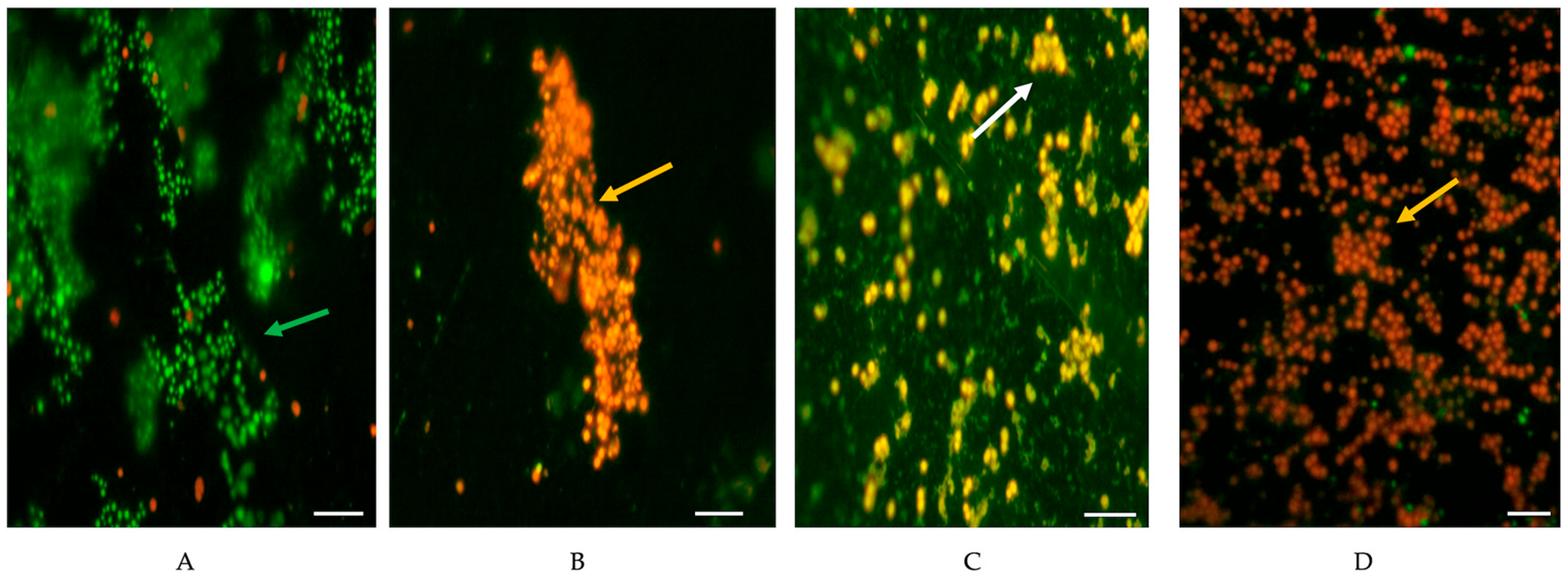
Figure 2 shows the live and dead cells of the MRSA and MSSA biofilms before and after the treatment with BC and BC + MR.
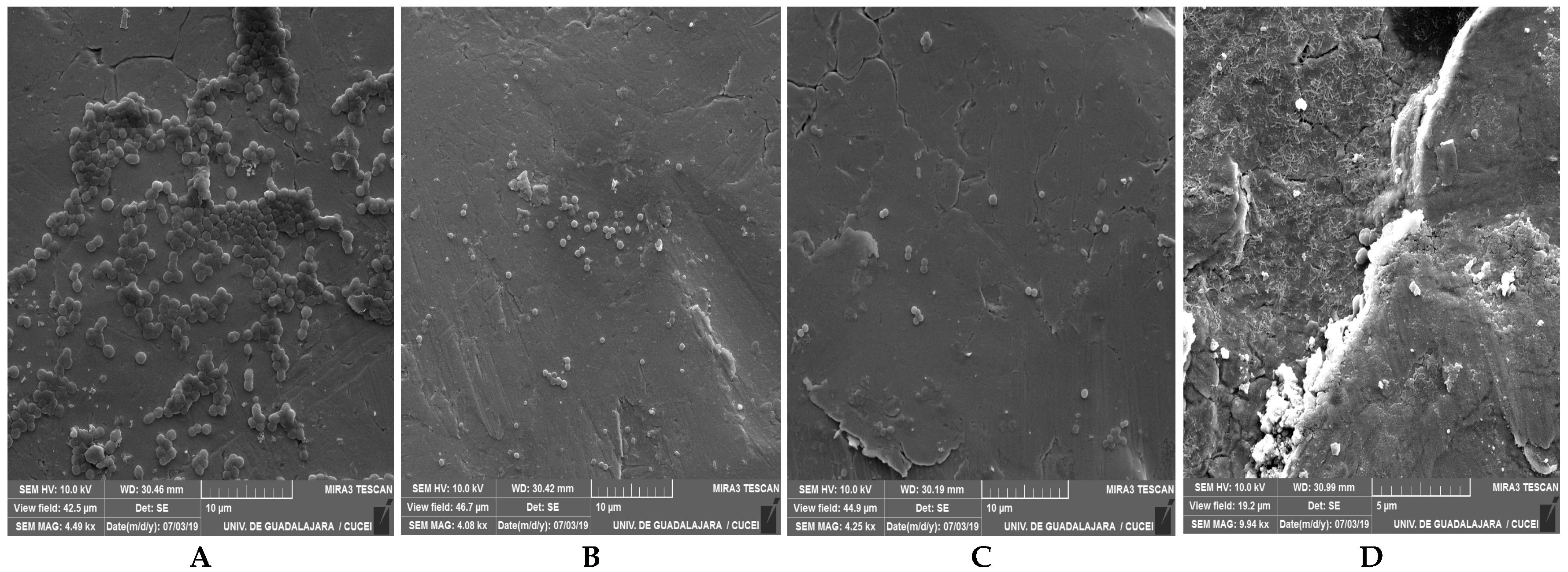
SEM analysis of the biofilms formed by the representative mono-species MRSA and MSSA revealed that the cells were linked to each other and embedded in dense EPS (Figure 3).

4. Discussion

S. aureus and MRSA are considered to be a public health problem due to their ability to contaminate various foods, such as milk and dairy products, as well as colonize and infect humans and animals. In 2024, MRSA was ranked 14th on the Bacterial Priority Pathogens List (BPPL). They are considered high-priority-group pathogens and are therefore a global health problem [12].
In this study, we analyzed a total of 67 S. aureus samples collected from food contact surfaces in the dairy industry, 41.7% of which were MRSA. Some authors have reported a similar prevalence for MRSA (51.6–95%) in raw milk samples (including cow, clinical mastitis, camel, and horse milk) [39,40]; however, several authors identified a lower prevalence (1.23–14%) for MRSA collected from sheep and goat bulk tanks, animal (rectal and nasal swabs) mastitis milk samples, dairy products, and ready-to-eat (RTE) foods [31,36,41,42,43,44]. Likewise, Kotzamanidis et al. [5] did not detect MRSA among the S. aureus strains recovered from goats, sheep, and bovines with clinical and subclinical mastitis. This investigation suggests that the prevalence of S. aureus and MRSA is associated with various factors, including (i) a diversity of sources and mechanisms of contamination during the obtaining of milk and the finished product, where food contact surfaces and food handlers are involved [20,21,45]; (ii) the sample size, type of food, and methods used for the detection and characterization of pathogens; (iii) geographical distribution of the prevalence of S. aureus in developed or developing countries [31]; and (iv) implementation and verification of control measures within the industry, such as sanitary prerequisites, good agricultural practices, and standard operating procedures for sanitation.
Regarding genetic characterization, SCCmec Types II (20.8%), V (13.4%), and IV (7.4%) were detected in this study. Similarly, other studies have reported SCCmec Types II (28.4–94%), IV (6–22.2%), and V (0.9%) in mastitis milk (clinical and subclinical cow), clinical strains of MRSA, clinical samples, and surgical materials [42,46,47]. Likewise, Annamanedi et al. [6] reported that the prevalence of SCCmec IVb, SCCmec IVd, and SCCmec V was 16.2%, 1.8% and 1.2% among the clinical and subclinical mastitis cases. SCCmec IVa has been associated with serious human infections and is spread primarily in community settings in humans and livestock populations [31,48,49]. Notably, the characterization of the pathogens and risk associated with food categories would aid in the prevention and treatment of infections with these pathogens in humans and animals.
Overall, the MSSA and MRSA isolates detected in this study exhibited antibiotic resistance (Table 1). Previous investigations revealed similar wide-ranging prevalences of antimicrobial resistance to penicillin (58.5–92.5%), ciprofloxacin (4.6–22.2%), clindamycin (17.5–97.5), erythromycin (8.3–87.5%), vancomycin (3.2%), tetracycline (11.3–77.8%), gentamicin (12.3–30.6%), and sulfamethoxazole–trimethoprim (19.4%) in S. aureus and MRSA isolates from raw milk samples of different varieties (goat bulk tank milk, mastitis milk samples, dairy products, and ready-to-eat (RTE) foods) [31,39,40,42,50,51]. The use of antibiotics in animal production, overprescription, and inappropriate use of antimicrobials in animal and human medicine may have contributed to the prevalence of heterogeneity of resistance to various antimicrobial classes [36,51,52].
Additionally, 71.64% of the MRSA (41.79%; 28/67) and MSSA (29.85%; 20/67) strains was MDR (Table 3), with an MAR index of >0.25 (86.56%; ranged 0.25–0.75), indicating a higher risk of contamination (MAR index of >0.2) associated with the continuous use of antibiotics [33]. Other investigators have also reported similar observations [10,19,40] that demonstrated the prevalence of MDR S. aureus (9.4–90.8%) with an MAR index of >0.2; however, the variation in the prevalence of MDR and MAR indexes is associated with the geographical area, seasons, sample size [53], the amount of antimicrobial classes, and the phenotypic and genotypic characteristics of S. aureus.
The detection of mecA, blaZ, tetM, aacA-aphD, ermA, ermB, and ermC in the S. aureus isolates in our study may be related to various resistance mechanisms, such as penicillin-binding protein 2a (PBP2a) activity and hyperproduction of β-lactamases, which confers resistance to β-lactam antibiotics [9,31], and the efflux pump and the bifunctional enzyme with acetyltransferase and phosphotransferase activity that confers resistance to tetracycline and gentamicin and methylases that modify A20258/A2059 in rRNA 23S and inhibit macrolide, lycosamide, and streptogramin B MLSB [54]. This is in accordance with the detection of mecA, blaZ, tetM, aacA-aphD, ermA, ermB, and ermC in S. aureus and MRSA isolates from milk samples and clinical specimens [28,39,40,55].
This research suggests that there is a high prevalence of resistance to β-lactams used in the treatment of bovine mastitis, such as penicillin and ampicillin [56,57], indicating the necessity for regulatory adjustments and optimized treatment protocols to strengthen control and regulation of bovine mastitis [40,42]. Furthermore, resistance to MLSB, which is a second-line drug used by patients with an allergy to β-lactams [43], can prolong and worsen the disease, increase healthcare costs, and increase the risk of death [58].
Additionally, the presence of heavy metal residues and antibacterial biocides can co-select antibiotic-resistant bacteria (cross-resistance) or cause co-resistance via interaction and genetic exchange through plasmids [18]. Resistance to cadmium in S. aureus is caused by the gene cadA that encodes efflux mechanisms consisting of Zn(II)/Cd(II)/Pb(II)-translocating ATPase [59].
In this study, 62.6% of the S. aureus isolates were cadmium-resistant (≥400 µg/mL). Other researchers [10,19] have reported similar findings regarding the progressive cadmium resistance (from 50 to 1500 µg/mL) [56.3% and 91.2% (100 µg/mL) or 39.4% and 88.9% (1500 µg/mL)] in S. aureus collected from livestock animals and the environment. Environmental cadmium contamination in different ecological settings, including agricultural soil and ground-, drinking, and wastewater, can be attributed to anthropogenic sources, including the industrial sector, such as the use of phosphate fertilizers and detergents within the sanitary or agricultural prerequisites [60,61,62]. Notably, S. aureus exhibits co-expression of antibiotic and heavy metal resistance, which complicates the prevention or treatment of pathologies caused by S. aureus and MRSA in the host [19,61] or in disinfection processes for the control of foodborne pathogens in the food industry.
Quaternary ammonium compounds (QACs), such as BC, are used in the food industry as sanitizers to control foodborne pathogens [63]. In this study, 97% of S. aureus showed MICs = 1.56 ≤ 25 µg/mL and had a 2.9% MIC = 50 µg/mL (Table 4); moreover, the BC [4MIC (MIC = 6.25 µg/mL)] more effectively removed the MRSA-7 SCCmec II, MRSA-18 SCCmec V, and MSSA-35 strain biofilms (p < 0.05) than the MSSA-16, MRSA-21 SCCmec IV and, MSSA-54 types (Figure 1). Therefore, BC is an effective disinfectant for preventing environmental niches and forming a biofilm in industry to control MRSA and MSSA on food contact surfaces. Indeed, other studies have revealed the antibacterial activity of BC against S. aureus and MRSA at conventional in-use concentrations for sanitizers [63,64,65]. In fact, the MIC or a serial increase in this (from 2MIC to 8MIC) for BC inhibited planktonic growth and biofilm formation with S. aureus and S. epidermidis CIP54124 [35,66].
The co-occurrence of heavy metal and antibiotic resistance is highly relevant, as the former can enhance the latter or lead to cross-resistance between these compounds, as previously reported [67]. Komijani et al. [68] demonstrated a strong correlation between the presence of antimicrobial resistance genes and heavy metal concentrations in aquatic environments, an association even stronger than that observed with the antibiotics themselves. Similarly, Zhou et al. [69] found that in dairy farm environments, ARGs and metal resistance genes are positively correlated, suggesting that heavy metals not only promote the emergence of metal resistance but also contribute to the co-selection of antibiotic resistance. This phenomenon is particularly significant in dairy facilities, where MRSA and MSSA strains are prevalent.
Finally, the anti-biofilm effect of MRSA and MSSA of BC is affected by the presence of organic matter (p < 0.05); in addition, MRSA-18 SCCmec V, MSSA-54, and MRSA-7 SCCmec II showed the lowest biofilm reduction in BC + MR with 10 min of exposure (p < 0.05) compared to MSSA-16, MRSA-21 SCCmec IV, and MSSA-35 (Figure 1) in this study. This result suggests that the decrease in BC efficiency is related to (i) the BC resistance mechanism or S. aureus biofilm matrix components decreasing the diffusion of sanitizers or antibiotics [39,66], (ii) sub-MIC exposure to BC inducing biofilm formation of S. aureus [35], (iii) the decrease in biofilm shrinkage associated with the slow growth rate of S. aureus embedded within the biofilm matrix [70], and (iv) the presence of organic matter favors pre-conditioning on the surface and bacterial adhesion or the low pH limiting the production of extracellular proteases within the biofilm matrix, favoring its formation [71,72,73]. Consequently, an S. aureus biofilm is a potential source of contamination among food products, generating major food safety problems and economic losses for the food industry. In addition, hazard characterization based on the determination of antimicrobial resistance factors or co-resistance to heavy metals and disinfectants is helpful for constant improvement of treatments and cleaning and disinfection procedures used in the food industry.

5. Conclusions

This study demonstrated that S. aureus strains isolated from FCSs in the dairy industry exhibit a high prevalence of methicillin and multidrug resistance, with SCCmec Types II, IV, and V being the most common. The co-occurrence of resistance to antibiotics, benzalkonium chloride, and cadmium highlights the capacity of these strains to persist in industrial environments, even under disinfection conditions. These findings underscore the urgent need to strengthen hygiene monitoring and antimicrobial stewardship within food production facilities. More importantly, this work provides molecular and phenotypic evidence that food contact surfaces can act as long-term reservoirs for MRSA and MSSA, representing a critical point for intervention to reduce the risk of foodborne transmission and improve food safety.

Supplementary Materials

The following supporting information can be downloaded at https://www.mdpi.com/article/10.3390/life15121811/s1, Table S1. Distribution and genetic analysis of S. aureus (n = 67) isolates collected from food contact surfaces (FCSs) in the dairy industry; Table S2: Primers for amplification of resistance genes in S. aureus and the conditions of annealing for PCR.

Author Contributions

Conceptualization; formal analysis; methodology; validation; writing—original draft: M.G.A.-N. Formal analysis; methodology; writing—review and editing: O.A.S.-V. Formal analysis; methodology; writing—review and editing: J.P.G.-G. Conceptualization; validation; writing—review and editing: P.J.G.-M. Conceptualization; funding acquisition; validation; writing—review and editing: M.G.-L. All authors have read and agreed to the published version of the manuscript.

Funding

This research received no external funding.

Institutional Review Board Statement

Not applicable.

Informed Consent Statement

Not applicable.

Data Availability Statement

The original contributions presented in this study are included in the article/Supplementary Materials. Further inquiries can be directed to the corresponding authors.

Acknowledgments

We also acknowledge the Secretaría de Ciencia, Humanidades, Tecnología e Innovación (SECIHTI) of Mexico, for its support through the Estancias Posdoctorales por México 2023 (1) program granted to Jean Pierre González-Gómez [818472].

Conflicts of Interest

The authors declare no conflicts of interest.

References

  1. Lister, J.L.; Horswill, A.R. Staphylococcus aureus biofilms: Recent developments in biofilm dispersal. Front. Cell. Infect. Microbiol. 2014, 4, 178. [Google Scholar] [CrossRef]
  2. Cheung, G.Y.C.; Bae, J.S.; Otto, M. Pathogenicity and virulence of Staphylococcus aureus. Virulence 2021, 12, 547–569. [Google Scholar] [CrossRef] [PubMed]
  3. Chapman, J.E.; George, S.E.; Wolz, C.; Olson, M.E. Biofilms: A developmental niche for vancomycin-intermediate Staphylococcus aureus. Infect. Genet. Evol. 2024, 117, 105545. [Google Scholar] [CrossRef] [PubMed]
  4. Abril, A.G.; Villa, T.G.; Barros-Velázquez, J.; Cañas, B.; Sánchez-Pérez, A.; Calo-Mata, P.; Carrera, M. Staphylococcus aureus Exotoxins and Their Detection in the Dairy Industry and Mastitis. Toxins 2020, 12, 537. [Google Scholar] [CrossRef] [PubMed]
  5. Kotzamanidis, C.; Vafeas, G.; Giantzi, V.; Anastasiadou, S.; Mygdalias, S.; Malousi, A.; Loukia, E.; Daniel, S.; Zdragas, A. Staphylococcus aureus Isolated from Ruminants with Mastitis in Northern Greece Dairy Herds: Genetic Relatedness and Phenotypic and Genotypic Characterization. Toxins 2021, 13, 176. [Google Scholar] [CrossRef]
  6. Annamanedi, M.; Sheela, P.; Sundareshan, S.; Isloor, S.; Gupta, P.; Jasmeen, P.; Gargi, M.; Mallick, S.; Hegde, N.R. Molecular fingerprinting of bovine mastitis-associated Staphylococcus aureus isolates from India. Sci. Rep. 2021, 11, 15228. [Google Scholar] [CrossRef]
  7. León-Galván, M.F.; Barboza-Corona, J.E.; Lechuga-Arana, A.A.; Valencia-Posadas, M.; Aguayo, D.D.; Cedillo-Pelaez, C.; Martínez-Ortega, E.A.; Gutierrez-Chavez, A.J. Molecular detection and sensitivity to antibiotics and bacteriocins of pathogens isolated from bovine mastitis in family dairy herds of central Mexico. Biomed. Res. Int. 2015, 2015, 615153. [Google Scholar] [CrossRef]
  8. Vázquez, H.C.; Jäger, S.; Wolter, W.; Zschöck, M.; Vazquez, M.A.C.; El-Sayed, A. Isolation and identification of main mastitis pathogens in Mexico. Arq. Bras. Med. Vet. Zootec. 2013, 65, 377–382. [Google Scholar] [CrossRef]
  9. Ahmad, S.; Raqeeb, A.; Ali, F.; Anwar, M. Characterization of novel antibiotic resistance genes in Staphylococcal aureus. J. Bacteriol. Mycol. 2018, 6, 8–10. [Google Scholar] [CrossRef]
  10. Nwobi, O.C.; Anyanwu, M.U.; Jaja, I.F.; Nwankwo, I.O.; Okolo, C.C.; Nwobi, C.A.; Ezenduka, E.V.; Oguttu, J.W. Staphylococcus aureus in Horses in Nigeria: Occurrence, Antimicrobial, Methicillin and Heavy Metal Resistance and Virulence Potentials. Antibiotics 2023, 12, 242. [Google Scholar] [CrossRef]
  11. Centers for Disease Control and Prevention (CDC). Methicillin-Resistant Staphylococcus aureus. Available online: https://www.cdc.gov/antimicrobial-resistance/media/pdfs/MRSA-508.pdf (accessed on 2 August 2025).
  12. World Health Organization (WHO). WHO Bacterial Priority Pathogens List, 2024. Bacterial Pathogens of Public Health Importance to Guide Research, Development and Strategies to Prevent and Control Antimicrobial Resistance. Available online: https://data.who.int/indicators/i/918081E/5DD9606 (accessed on 21 September 2025).
  13. O’Neill, J. Tackling Drug-Resistant Infections Globally: Final Report and Recommendations. The Review on Antimicrobial Resistance. Available online: https://amr-review.org/sites/default/files/160518_Final%20paper_with%20cover.pdf (accessed on 5 August 2025).
  14. Guo, H.; Tong, Y.; Cheng, J.; Abbas, Z.; Li, Z.; Wang, J.; Zhou, Y.; Si, D.; Zhang, R. Biofilm and Small Colony Variants—An Update on Staphylococcus aureus Strategies toward Drug Resistance. Int. J. Mol. Sci. 2022, 23, 1241. [Google Scholar] [CrossRef]
  15. Van Boeckel, T.P.; Brower, C.; Gilbert, M.; Grenfell, B.T.; Levin, S.A.; Robinson, T.P.; Teillant, A.; Laxminarayan, R. Global trends in antimicrobial use in food animals. Proc. Natl. Acad. Sci. USA 2015, 112, 5649–5654. [Google Scholar] [CrossRef]
  16. Amábile-Cuevas, C.F. Antibiotic usage and resistance in Mexico: An update after a decade of change. J. Infect. Dev. Ctries. 2021, 15, 442–449. [Google Scholar] [CrossRef] [PubMed]
  17. Alcock, B.P.; Raphenya, A.R.; Lau, T.T.Y.; Tsang, K.K.; Bouchard, M.; Edalatmand, A.; Huynh, W.; Nguyen, A.V.; Cheng, A.A.; Liu, S.; et al. CARD 2020: Antibiotic resistome surveillance with the comprehensive antibiotic resistance database. Nucleic Acids Res. 2020, 48, D517–D525. [Google Scholar] [CrossRef] [PubMed]
  18. Larsson, D.G.J.; Flach, C.F. Antibiotic resistance in the environment. Nat. Rev. Microbiol. 2022, 20, 257–269. [Google Scholar] [CrossRef]
  19. Dweba, C.C.; Zishiri, O.T.; El Zowalaty, M.E. Isolation and molecular identification of virulence, antimicrobial and heavy metal resistance genes in livestock-associated methicillin-resistant Staphylococcus aureus. Pathogens 2019, 8, 79. [Google Scholar] [CrossRef]
  20. Di Ciccio, P.; Vergara, A.; Festino, A.R.; Paludi, D.; Zanardi, E.; Ghidini, S.; Ianieri, A. Biofilm formation by Staphylococcus aureus on food contact surfaces: Relationship with temperature and cell surface hydrophobicity. Food Control 2015, 50, 930–936. [Google Scholar] [CrossRef]
  21. Avila-Novoa, M.G.; Iñíguez-Moreno, M.; Solís-Velázquez, O.A.; González-Gómez, J.P.; Guerrero-Medina, P.J.; Gutiérrez-Lomelí, M. Biofilm Formation by Staphylococcus aureus Isolated from Food Contact Surfaces in the Dairy Industry of Jalisco, Mexico. J. Food Qual. 2018, 2018, 72–78. [Google Scholar] [CrossRef]
  22. Bridier, A.; Sanchez-Vizuete, P.; Guilbaud, M.; Piard, J.C.; Naïtali, M.; Briandet, R. Biofilm-associated persistence of food-borne pathogens. Food Microbiol. 2015, 45, 167–178. [Google Scholar] [CrossRef]
  23. Hall, C.W.; Mah, T.F. Molecular mechanisms of biofilm-based antibiotic resistance and tolerance in pathogenic bacteria. FEMS Microbiol. Rev. 2017, 41, 276–301. [Google Scholar] [CrossRef]
  24. Wassenaar, T.; Ussery, D.; Nielsen, L.; Ingmer, H. Review and phylogenetic analysis of qac genes that reduce susceptibility to quaternary ammonium compounds in Staphylococcus species. Eur. J. Microbiol. Immunol. 2015, 5, 44–61. [Google Scholar] [CrossRef]
  25. Avila-Novoa, M.G.; Solis-Velazquez, O.A.; Guerrero-Medina, P.J.; González-Gómez, J.P.; González-Torres, B.; Velázquez-Suárez, N.Y.; Martínez-Chávez, L.; Martínez-Gonzáles, N.E.; De la Cruz-Color, L.; Ibarra-Velázquez, L.M.; et al. Genetic and compositional analysis of biofilm formed by Staphylococcus aureus isolated from food contact surfaces. Front. Microbiol. 2022, 13, 1001700. [Google Scholar] [CrossRef] [PubMed]
  26. Avila-Novoa, M.G.; Iñiguez-Moreno, M.; González-Gómez, J.P.; Zacarías-Castillo, E.; Guerrero-Medina, P.J.; Padilla-Frausto, J.J.; Navarro-Villarruel, C.L.; Gutiérrez-Lomelí, M. Detection of enterotoxin genes of Staphylococcus aureus isolates from food contact surfaces in the dairy industry of Jalisco, Mexico. Biotecnia 2018, 20, 72–78. [Google Scholar] [CrossRef]
  27. Gan, T.; Shu, G.; Fu, H.; Yan, Q.; Zhang, W.; Tang, H.; Yin, L.; Zhao, L.; Lin, J. Antimicrobial resistance and genotyping of Staphylococcus aureus obtained from food animals in Sichuan Province, China. BMC Vet. Res. 2021, 17, 177. [Google Scholar] [CrossRef] [PubMed]
  28. Deepak, S.J.; Kannan, P.; Savariraj, W.R.; Ayyasamy, E.; Tuticorin Maragatham Alagesan, S.K.; Ravindran, N.B.; Sundaram, S.; Mohanadasse, N.Q.; Kang, Q.; Cull, C.A.; et al. Characterization of Staphylococcus aureus isolated from milk samples for their virulence, biofilm, and antimicrobial resistance. Sci. Rep. 2024, 14, 25635. [Google Scholar] [CrossRef]
  29. Shah, M.S.; Qureshi, S.; Kashoo, Z.; Farooq, S.; Wani, S.A.; Hussain, M.I.; Banday, M.S.; Khan, A.A.; Gull, B.; Habib, A.; et al. Methicillin resistance genes and in vitro biofilm formation among Staphylococcus aureus isolates from bovine mastitis in India. Comp. Immunol. Microbiol. Infect. Dis. 2019, 64, 117–124. [Google Scholar] [CrossRef]
  30. CLSI M100-ED35:2025; Performance Standards for Antimicrobial Susceptibility Testing. 35th ed. CLSI Supplement M100S. Clinical and Laboratory Standards Institute: Wayne, PA, USA, 2025.
  31. Mekhloufi, O.A.; Chieffi, D.; Hammoudi, A.; Bensefia, S.A.; Fanelli, F.; Fusco, V. Prevalence, enterotoxigenic potential and antimicrobial resistance of Staphylococcus aureus and methicillin-resistant Staphylococcus aureus (MRSA) isolated from Algerian ready to eat foods. Toxins 2021, 13, 835. [Google Scholar] [CrossRef]
  32. Magiorakos, A.P.; Srinivasan, A.; Carey, R.B.; Carmeli, Y.; Falagas, M.E.; Giske, C.G.; Harbarth, S.; Hindler, J.F.; Kahlmeter, G.; Olsson-Liljequist, B.; et al. Multidrug-resistant, extensively drug-resistant and pandrug-resistant bacteria: An international expert proposal for interim standard definitions for acquired resistance. Clin. Microbiol. Infect. 2012, 18, 268–281. [Google Scholar] [CrossRef]
  33. Krumperman, P.H. Multiple antibiotic resistance indexing of Escherichia coli to identify high-risk sources of fecal contamination of foods. Appl. Environ. Microbiol. 1983, 46, 165–170. [Google Scholar] [CrossRef]
  34. Blasco, M.D.; Esteve, C.; Alcaide, E. Multiresistant waterborne pathogens isolated from water reservoirs and cooling systems. J. Appl. Microbiol. 2008, 105, 469–475. [Google Scholar] [CrossRef]
  35. Ebrahimi, A.; Hemati, M.; Shabanpour, Z.; Dehkordi, S.H.; Bahadoran, S.; Lotalian, S.; Khoibani, S. Effects of benzalkonium chloride on planktonic growth and biofilm formation by animal bacterial pathogens. Jundishapur J. Microbiol. 2015, 8, e16058. [Google Scholar] [CrossRef]
  36. Avila-Novoa, M.G.; González-Gómez, J.P.; Guerrero-Medina, P.J.; Cardona-López, M.A.; Ibarra-Velazquez, L.M.; Velazquez-Suarez, N.Y.; Morales-del Río, J.A.; Gutiérrez-Lomelí, M. Staphylococcus aureus and methicillin-resistant S. aureus (MRSA) strains isolated from dairy products: Relationship of ica-dependent/independent and components of biofilms produced in vitro. Int. Dairy J. 2021, 119, 105066. [Google Scholar] [CrossRef]
  37. Borucki, M.K.; Peppin, J.D.; White, D.; Loge, F.; Call, D.R. Variation in biofilm formation among strains of Listeria monocytogenes. Appl. Environ. Microbiol. 2003, 69, 7336–7342. [Google Scholar] [CrossRef] [PubMed]
  38. Fratesi, S.E.; Lynch, F.L.; Kirkland, B.L.; Brown, L.R. Effects of SEM preparation techniques on the appearance of bacteria and biofilms in the Carter Sandstone. J. Sediment. Res. 2004, 74, 858–867. [Google Scholar] [CrossRef]
  39. Kou, X.; Cai, H.; Huang, S.; Ni, Y.; Luo, B.; Qian, H.; Ji, H.; Wang, X. Prevalence and Characteristics of Staphylococcus aureus Isolated from Retail Raw Milk in Northern Xinjiang, China. Front. Microbiol. 2021, 12, 705947. [Google Scholar] [CrossRef] [PubMed]
  40. Eidaroos, N.H.; Algammal, A.M.; Mohamaden, W.I.; Alenzi, A.M.; Alghamdi, S.; Kabrah, A.; El-Mahallawy, H.S.; Eid, H.M.; Algwad, A.A.; Asfor, S.A.; et al. Virulence traits, agr typing, multidrug resistance patterns, and biofilm ability of MDR Staphylococcus aureus recovered from clinical and subclinical mastitis in dairy cows. BMC Microbiol. 2025, 25, 155. [Google Scholar] [CrossRef]
  41. Caruso, M.; Latorre, L.; Santagada, G.; Fraccalvieri, R.; Miccolupo, A.; Sottili, R.; Palazzo, L.; Parisi, A. Methicillin-resistant Staphylococcus aureus (MRSA) in sheep and goat bulk tank milk from Southern Italy. Small Rumin. Res. 2016, 135, 26–31. [Google Scholar] [CrossRef]
  42. Chen, C.; Sun, C.; Li, J.; Ji, X.; Wang, Y.; Song, C.; Wang, G. Characterisation of Staphylococcus aureus isolates from bovine mastitis in Ningxia, Western China. J. Glob. Antimicrob. Resist. 2021, 25, 232–237. [Google Scholar] [CrossRef]
  43. Szczuka, E.; Porada, K.; Wesołowska, M.; Łęska, B. Occurrence and characteristics of Staphylococcus aureus isolated from dairy products. Molecules 2022, 27, 4649. [Google Scholar] [CrossRef]
  44. Biswas, S.; Islam, M.A.; Islam, J.; Khatun, M.M.; Rahman, M.Z. Detection of multidrug-resistant methicillin-resistant Staphylococcus aureus from healthy black Bengal goat in Bangladesh. J. Infect. Dev. Ctries. 2024, 18, 1891–1898. [Google Scholar] [CrossRef]
  45. Gutiérrez, D.; Delgado, S.; Vázquez-Sánchez, D.; Martínez, B.; Cabo, M.L.; Rodríguez, A.; Herrera, J.J.; García, P. Incidence of Staphylococcus aureus and analysis of associated bacterial communities on food industry surfaces. Appl. Environ. Microbiol. 2012, 78, 8547–8554. [Google Scholar] [CrossRef]
  46. Takata, T.; Shirakawa, T.; Ito, J.; Okamoto, A.; Massi, M.N.; Kinoshita, S.; Hatta, M.; Kawabata, M. SCCmec typing and detection of VISA-related genes in methicillin-resistant Staphylococcus aureus clinical strains from Kobe University Hospital, Japan. Southeast Asian J. Trop. Med. Public Health 2006, 37, 1149–1155. [Google Scholar] [PubMed]
  47. Sağlam, M.; Kılıç, İ.H.; Zer, Y. Investigation of SCCmec types using the real time PCR method in cefoxitin-resistant Staphylococcus aureus isolates. Indian J. Med. Microbiol. 2024, 50, 100649. [Google Scholar] [CrossRef] [PubMed]
  48. Cuny, C.; Strommenger, B.; Witte, W.; Stanek, C. Clusters of infections in horses with MRSA ST1, ST254, and ST398 in a veterinary hospital. Microb. Drug Resist. 2008, 14, 307–310. [Google Scholar] [CrossRef] [PubMed]
  49. Otter, J.A.; Havill, N.L.; Boyce, J.M.; French, G.L. Comparison of community-associated methicillin-resistant Staphylococcus aureus from teaching hospitals in London and the USA, 2004–2006: Where is USA300 in the UK? Eur. J. Clin. Microbiol. Infect. Dis. 2009, 28, 835–839. [Google Scholar] [CrossRef]
  50. Dastmalchi Saei, H.; Panahi, M. Genotyping and antimicrobial resistance of Staphylococcus aureus isolates from dairy ruminants: Differences in the distribution of clonal types between cattle and small ruminants. Arch. Microbiol. 2020, 202, 115–125. [Google Scholar] [CrossRef]
  51. Ren, Q.; Liao, G.; Wu, Z.; Lv, J.; Chen, W. Prevalence and characterization of Staphylococcus aureus isolates from subclinical bovine mastitis in southern Xinjiang, China. J. Dairy Sci. 2020, 103, 3368–3380. [Google Scholar] [CrossRef]
  52. Oniciuc, E.A.; Nicolau, A.I.; Hernández, M.; Rodríguez-Lázaro, D. Presence of methicillin-resistant Staphylococcus aureus in the food chain. Trends Food Sci. Technol. 2017, 61, 49–59. [Google Scholar] [CrossRef]
  53. Tartor, Y.H.; Enany, M.E.; Ismail, N.I.; El-Demerdash, A.S.; Eidaroos, N.H.; Algendy, R.M.; Mahmmod, Y.; Elsohaby, I. Vancomycin-resistant Staphylococcus aureus endangers Egyptian dairy herds. Sci. Rep. 2024, 14, 30606. [Google Scholar] [CrossRef]
  54. Wendlandt, S.; Feßler, A.T.; Monecke, S.; Ehricht, R.; Schwarz, S.; Kadlec, K. The diversity of antimicrobial resistance genes among staphylococci of animal origin. Int. J. Med. Microbiol. 2013, 303, 338–349. [Google Scholar] [CrossRef]
  55. El-Banna, T.E.S.; Sonbol, F.I.; Kamer, A.M.A.; Badr, S.A.M.M. Genetic diversity of macrolides resistant Staphylococcus aureus clinical isolates and the potential synergistic effect of vitamins, C and K3. BMC Microbiol. 2024, 24, 30. [Google Scholar] [CrossRef]
  56. Page, S.W.; Gautier, P. Use of antimicrobial agents in livestock. Rev. Sci. Tech. 2012, 31, 145–188. [Google Scholar] [CrossRef] [PubMed]
  57. Sharifi, A.; Sobhani, K.; Mahmoudi, P. A systematic review and meta-analysis revealed a high-level antibiotic resistance of bovine mastitis Staphylococcus aureus in Iran. Res. Vet. Sci. 2023, 161, 23–30. [Google Scholar] [CrossRef] [PubMed]
  58. Painuli, S.; Semwal, P.; Sharma, R.; Akash, S. Superbugs or multidrug resistant microbes: A new threat to the society. Health Sci. Rep. 2023, 6, e1480. [Google Scholar] [CrossRef] [PubMed]
  59. Chudobova, D.; Dostalova, S.; Blazkova, I.; Michalek, P.; Ruttkay-Nedecky, B.; Sklenar, M.; Nejdl, L.; Kudr, J.; Gumulec, J.; Tmejova, K.; et al. Effect of ampicillin, streptomycin, penicillin and tetracycline on metal resistant and non-resistant Staphylococcus aureus. Int. J. Environ. Res. Public Health 2014, 11, 3233–3255. [Google Scholar] [CrossRef]
  60. Aonghusa, C.N.; Gray, N.F. Laundry detergents as a source of heavy metals in Irish domestic wastewater. J. Environ. Sci. Health A Toxic Hazard. Subst. Environ. Eng. 2002, 37, 1–6. [Google Scholar] [CrossRef]
  61. Eggers, S.; Safdar, N.; Malecki, K.M. Heavy metal exposure and nasal Staphylococcus aureus colonization: Analysis of the National Health and Nutrition Examination Survey (NHANES). Environ. Health 2018, 17, 2. [Google Scholar] [CrossRef]
  62. Agency for Toxic Substances and Disease Registry (ATSDR). Cadmium ToxGuide. (Listeriosis). Available online: https://www.atsdr.cdc.gov/toxguides/toxguide-5.pdf (accessed on 8 September 2025).
  63. Bondurant, S.; Mckinney, T.; Bondurant, L.; Fitzpatrick, L. Evaluation of a benzalkonium chloride hand sanitizer in reducing transient Staphylococcus aureus bacterial skin contamination in health care workers. Am. J. Infect. Control 2020, 48, 522–526. [Google Scholar] [CrossRef]
  64. Chojnacki, M.; Dobrotka, C.; Osborn, R.; Johnson, W.; Young, M.; Meyer, B.; Laskey, E.; Wozniak, R.A.F.; Dewhurst, S.; Dunman, P.M. Evaluating the Antimicrobial Properties of Commercial Hand Sanitizers. mSphere 2021, 6, e00062-21. [Google Scholar] [CrossRef]
  65. Tahir, S.; Emanuel, S.; Inglis, D.W.; Vickery, K.; Deva, A.K.; Hu, H. Mild Positive Pressure Improves the Efficacy of Benzalkonium Chloride against Staphylococcus aureus Biofilm. Bioengineering 2022, 9, 461. [Google Scholar] [CrossRef]
  66. Houari, A.; Di Martino, P. Effect of chlorhexidine and benzalkonium chloride on bacterial biofilm formation. Lett. Appl. Microbiol. 2007, 45, 652–656. [Google Scholar] [CrossRef] [PubMed]
  67. Nguyen, C.C.; Hugie, C.N.; Kile, M.L.; Navab-Daneshmand, T. Association between heavy metals and antibiotic-resistant human pathogens in environmental reservoirs: A review. Front. Environ. Sci. Eng. 2019, 13, 46. [Google Scholar] [CrossRef]
  68. Komijani, M.; Shamabadi, N.S.; Shahin, K.; Eghbalpour, F.; Tahsili, M.R.; Bahram, M. Heavy metal pollution promotes antibiotic resistance potential in the aquatic environment. Environ. Pollut. 2021, 274, 116569. [Google Scholar] [CrossRef] [PubMed]
  69. Zhou, B.; Wang, C.; Zhao, Q.; Wang, Y.; Huo, M.; Wang, J.; Wang, S. Prevalence and dissemination of antibiotic resistance genes and coselection of heavy metals in Chinese dairy farms. J. Hazard. Mater. 2016, 320, 10–17. [Google Scholar] [CrossRef]
  70. Azmi, K.; Qrei, W.; Abdeen, Z. Screening of genes encoding adhesion factors and biofilm production in methicillin resistant strains of Staphylococcus aureus isolated from Palestinian patients. BMC Genom. 2019, 20, 578. [Google Scholar] [CrossRef]
  71. Chmielewski, R.A.N.; Frank, J.F. Biofilm Formation and Control in Food Processing Facilities. Compr. Rev. Food Sci. Food Saf. 2003, 2, 22–32. [Google Scholar] [CrossRef]
  72. Lorite, G.S.; Rodrigues, C.M.; Souza, A.A.; De Kranz, C.; Mizaikoff, B.; Cotta, M.A. The role of conditioning film formation and surface chemical changes on Xylella fastidiosa adhesion and biofilm evolution. J. Colloid Interface Sci. 2011, 359, 289–295. [Google Scholar] [CrossRef]
  73. Sugimoto, S.; Sato, F.; Miyakawa, R.; Chiba, A.; Onodera, S.; Hori, S.; Mizunoe, Y. Broad impact of extracellular DNA on biofilm formation by clinically isolated Methicillin-resistant and -sensitive strains of Staphylococcus aureus. Sci. Rep. 2018, 8, 2254. [Google Scholar] [CrossRef]
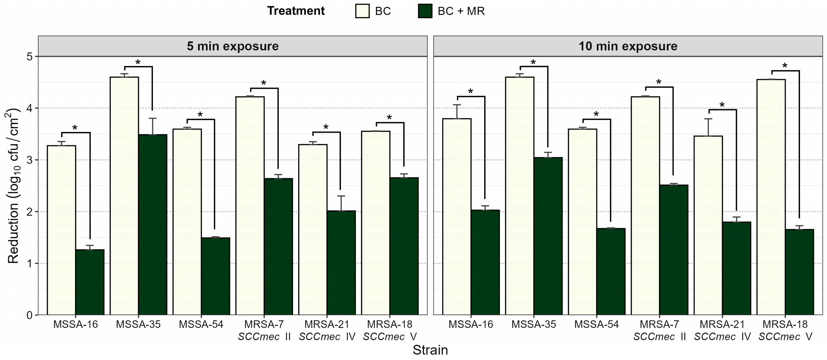
Figure 1. Effect of BC alone and in the presence of milk residues (MRs) on biofilm reduction in MSSA and MRSA strains. Biofilm reduction was assessed after 5 and 10 min of exposure. Data represent mean ± SD of three independent experiments. Asterisks (*) indicate statistically significant differences between treatments (p < 0.05).
Life 15 01811 g001
Figure 2. Micrographs of biofilms before and after removal treatments. Biofilms of MRSA and MSSA were developed on SS during 192 h of incubation at 35 °C. Micrographs were obtained by epifluorescence microscopy (100×), stained with LIVE/DEAD™ BacLight™ before and after removal treatments. (A) MRSA-7 SCCmec II before applied treatment; (B) MRSA-7 SCCmec II after applied treatment with BC (100 µg/mL, 5 min, 22 °C); (C) MSSA-54 after applied treatment with BC + MR (100 µg/mL + 1% of milk residues, 10 min, 22 °C); (D) MRSA-18 SCCmec V after applied treatment with BC (100 µg/mL, 10 min, 22 °C). White bar scale indicates 5 μm. Distinctive fluorescence and patterns can be linked to different cellular states. As for LIVE/DEAD™ BacLight™ stain, SYTO9 penetrates all bacterial membranes and stains cells green (green arrows). Propidium iodide only penetrates cells with damaged membranes. Fluorescing cells turn red (orange arrows), and bacterial cells in an intermediate state between alive and dead or damaged are stained yellow-green (white arrows).
Life 15 01811 g002
Figure 3. Micrographs of biofilms before and after removal treatments. Biofilms of MRSA and MSSA were developed on SS during 192 h of incubation at 35 °C. Micrographs were obtained by scanning electron microscopy (SEM) before and after removal treatments. (A) MRSA-7 SCCmec II before applied treatment; (B) MRSA-7 SCCmec II after applied treatment with BC (100 µg/mL, 5 min, 22 °C); (C) MSSA-35 after applied treatment with BC (100 µg/mL, 10 min, 22 °C); (D) MRSA-18 SCCmec V after applied treatment with BC (100 µg/mL + 1% of milk residues, 10 min, 22 °C).
Life 15 01811 g003
Table 1. Antimicrobial susceptibility test results for MRSA and MSSA.
Antimicrobial Class According to the WHOAntibioticNo. (%) of MRSA (n = 28) and MSSA (n = 39) Strains
ResistantSusceptibleIntermediate
MRSAMSSAMRSAMSSAMRSAMSSA
Highly importantLincosamidesCLM19 (28.3)25 (37.3)9 (13.4)14 (20.8)--
SulfonamidesSTX5 (7.4)7 (10.4)23 (34.3)32 (47.7)--
Cyclic peptidesTE8 (11.9)3 (4.4)20 (29.8)36 (53.7)--
Critically importantMacrolidesE5 (7.4)4 (5.9)23 (34.3)35 (52.2)--
AminoglycosidesGE6 (8.9)2 (2.9)22 (32.8)37 (55.2)--
FluoroquinolonesCPF11 (11.9)22 (32.8)12 (17.9)10 (14.9)5 (7.4)7 (10.4)
β-LactamsPE28 (41.7)39 (58.2)----
GlycopeptidesVA *-5 (7.4)28 (41.7)34 (50.7)--
Abbreviations for antibiotics were defined in Section 2.3. * MIC Test of vancomycin by CLSI guidelines.
Table 2. Genetic analysis and antibiotic resistance results of MRSA (n = 28) isolated from food contact surfaces.
Strain No.SCCmecAntimicrobial Resistance GenesAntibiotic ResistanceMAR Index
ermBermCermAtetMblaZaacA-aphD
MSRA-2SCCmec II----++PE + GE0.25
MSRA-6SCCmec II----+-PE + CLM0.25
MSRA-7SCCmec II++-++-PE + CPF + CLM + E + TE0.62
MSRA-8SCCmec II---++-PE + CLM + TE0.37
MSRA-9SCCmec II----+-PE + CLM0.25
MSRA-10SCCmec II----+-PE0.12
MSRA-11SCCmec II----+-PE0.12
MSRA-23SCCmec II+--+++PE + CPF + CLM + E + TE + GE0.75
MSRA-27SCCmec II+--++-PE + CPF + CLM + E + TE + GE0.75
MSRA-28SCCmec II----+-PE0.12
MSRA-37SCCmec II---++-PE + CLM + TE0.37
MSRA-40SCCmec II------PE + CLM0.25
MSRA-43SCCmec II---++-PE + CLM + TE + STX0.50
MSRA-44SCCmec II----+-PE + CLM0.25
MSRA-3SCCmec IV--+-++PE + CPF + CLM + E + GE0.62
MSRA-13SCCmec IV+----+PE + CLM + E + GE0.50
MSRA-17SCCmec IV----+-PE + CPF + CLM0.37
MSRA-21SCCmec IV---++-PE + CPF + CLM + TE + STX0.62
MSRA-56SCCmec IV--- +-PE0.12
MSRA-4SCCmec V---++-PE + TE0.25
MSRA-5SCCmec V----+-PE + CPF + CLM + STX0.50
MSRA-12SCCmec V---++-PE + CPF + CLM + TE + STX0.62
MSRA-15SCCmec V----+-PE + CLM + STX0.37
MSRA-18SCCmec V------PE + CPF0.25
MSRA-24SCCmec V----+-PE0.12
MSRA-34SCCmec V----++PE + GE0.25
MSRA-39SCCmec V-+--+-PE + CPF + CLM + E0.50
MSRA-62SCCmec V------PE + CPF + CLM0.37
SCCmec, staphylococcal chromosomal cassette mec. Abbreviations for antibiotics were defined in Section 2.3.
Table 3. Antimicrobial resistance patterns and multiple antimicrobial resistance indices of S. aureus (n = 67) from food contact surfaces.
No. of AntimicrobialResistance ProfileStaphylococcus aureusMAR Index
No. (%) of MSSA
(n = 39)
No. (%) of MRSA
(n = 28)
6PE + CPF + CLM + E + TE + GE-2 (2.98)0.75
PE + CPF + CLM + VA + GE + STX1 (1.49)-
PE + CPF + CLM + E + TE + STX1 (1.49)-
5PE + CPF + CLM + TE + STX-2 (2.98)0.62
PE + CPF + CLM + E + TE-1 (1.49)
PE + CPF + CLM + E + GE-1 (1.49)
4PE + CLM + TE + STX-1 (1.49)0.50
PE + CLM + E + GE-1 (1.49)
PE + CPF + CLM + STX-1 (1.49)
PE + CPF + CLM + E-1 (1.49)
PE + CPF + CLM + STX3 (4.47)-
PE + CPF + CLM + VA1 (1.49)-
PE + CPF + CLM + TE1 (1.49)-
PE + CLM + E + VA1 (1.49)-
PE + CLM + VA + STX1 (1.49)-
3PE + CPF + CLM9 (13.43)2 (2.98)0.37
PE + CLM + TE-2 (2.98)
PE + CLM + STX-1 (1.49)
PE + E + TE1 (1.49)-
PE + CLM + VA1 (1.49)-
2PE + TE-1 (1.49)0.25
PE + GE1 (1.49) *2 (2.98)
PE + CPF6 (8.95) *1 (1.49)
PE + CLM6 (8.95) *4 (5.97)
PE + STX1 (1.49) *-
PE + E1 (1.49) *-
1PE1 (1.49) *3 (4.47)0.12
PE2 (2.98) *2 (2.98)
PE1 (1.49) *-
* S. aureus is not MDR. Abbreviations for antibiotics were defined in Section 2.3.
Table 4. MIC of BC and resistance to CdCl2 in MRSA and MSSA.
µg/mLNo. (%) of MRSA (n = 28)No. (%) of MSSA (n = 39)Total
BC
(MIC)
502 (2.9)-2 (2.9)
258 (11.9)12 (17.9)20 (29.8)
12.52 (2.9)2 (2.9)4 (5.9)
6.256 (8.9)1 (1.4)7 (10.4)
3.122 (2.9)7 (10.4)9 (13.4)
1.568 (11.9)17 (25.3)25 (37.3)
ClCd2Higher 40023 (34.3)19 (28.3)42 (62.6)
200-1 (1.4)1 (1.4)
100-1 (1.4)1 (1.4)
701 (1.4)7 (10.4)8 (11.9)
502 (2.9)3 (4.4)5 (7.4)
252 (2.9)8 (11.9)10 (14.9)
Disclaimer/Publisher’s Note: The statements, opinions and data contained in all publications are solely those of the individual author(s) and contributor(s) and not of MDPI and/or the editor(s). MDPI and/or the editor(s) disclaim responsibility for any injury to people or property resulting from any ideas, methods, instructions or products referred to in the content.