Article

Increased Susceptibility to Salmonella Infection in Systemic Lupus Erythematosus Compared with Other Systemic Autoimmune Diseases: Insights from a Retrospective Cohort Study from the Largest Health Care System in Taiwan

1
Division of Chinese Internal Medicine, Center for Traditional Chinese Medicine, Chang Gung Memorial Hospital, Taoyuan 333, Taiwan
2
Division of Rheumatology, Allergy and Immunology, Linkou Chang Gung Memorial Hospital, Taoyuan 333, Taiwan
3
College of Medicine, Chang Gung University, Taoyuan 333, Taiwan
4
Center for Big Data Analytics and Statistics, Linkou Chang Gung Memorial Hospital, Taoyuan 333, Taiwan
*
Author to whom correspondence should be addressed.
Life 2025, 15(10), 1522; https://doi.org/10.3390/life15101522
Submission received: 8 August 2025 / Revised: 18 September 2025 / Accepted: 25 September 2025 / Published: 26 September 2025
(This article belongs to the Section Physiology and Pathology)

Abstract

Systemic lupus erythematosus (SLE) and other systemic autoimmune rheumatic diseases (SARDs) require long-term immunosuppressive therapy, placing patients at increased risk of infection. Salmonella species are particularly concerning due to their invasiveness and potential link to autoimmune activation, notably in SLE. This study aimed to compare the risk of culture-confirmed Salmonella infection between SLE and other SARDs, based on data from the Chang Gung Research Database between 2005 and 2020. After propensity score matching, 3537 patients per group were analyzed. Patients with SLE had a higher incidence of Salmonella infection compared with those with other SARDs (0.54 vs. 0.17 per 1000 person-years), with a significantly greater cumulative incidence (log-rank p < 0.01). The adjusted hazard ratio (HR) for Salmonella infection in SLE was 2.47 (95% confidence interval (CI): 0.95–6.38), and the competing risk model confirmed a significant association (sub-distribution HR 2.58, 95% CI: 1.06–6.29, p = 0.04). Among SLE patients, lymphopenia was the only independent predictor of Salmonella infection (adjusted HR 3.98, 95% CI: 1.83–8.68, p < 0.001). Bloodstream infections were most common (70%), and serogroup D was the predominant strain (80%). These results suggest patients with SLE face higher Salmonella risk than other SARDs, especially those with lymphopenia, underscoring the need for targeted surveillance and preventive strategies.

1. Introduction

Systemic lupus erythematosus (SLE) and other systemic autoimmune rheumatic diseases (SARDs)—including Sjögren’s syndrome, rheumatoid arthritis, systemic sclerosis, dermatomyositis, and polymyositis—are chronic, immune dysregulation disorders with multi-organ involvements. These conditions often necessitate long-term immunosuppressive or immunomodulatory therapy to control disease activity and prevent irreversible damages [1,2].
While advances in immunosuppressive treatment have improved disease control and survival, infection remain a leading cause of morbidity and mortality in these populations [3,4]. For example, among various immunosuppressive strategies, tumor necrosis factor-alpha inhibitors have been identified as important risk factors for opportunistic infections caused by intracellular pathogens such as Salmonella and Mycobacteria, as well as certain fungi [5].
Among these opportunistic infections, Salmonella species are of particular concern in immunocompromised adults due to their heightened risk of invasive disease manifestations such as primary bacteremia and recurrent infections, often associated with increased mortality [6]. Notably, emerging evidence also suggests a possible bidirectional relationship between Salmonella infection and autoimmunity. A nationwide, population-based case–control study in Taiwan reported that individuals with a history of nontyphoidal Salmonella infection had a significantly higher risk of developing SLE, with an adjusted odds ratio exceeding 7 in both conventional and propensity score-matched analyses [7]. This suggests that, in addition to being a common opportunistic pathogen in patients with established SLE, nontyphoidal Salmonella infection may also act as a potential environmental trigger for autoimmune activation through dysregulated inflammatory pathways.
Although Salmonella infection has long been recognized as a clinically important opportunistic infection in patients with SLE [8,9,10]. A single-center study by Abramson et al. identified SLE as the most common underlying condition among patients with Salmonella bacteremia, accounting for 20% of cases, a striking overrepresentation compared to non-Salmonella bacteremia [11]. These observations suggest that SLE patients may be particularly susceptible due to both disease-related immune dysregulation and immunosuppressive therapy. However, it remains unclear whether this heightened vulnerability is unique to SLE or reflective of a broader risk pattern across other SARDs, many of which similarly require prolonged immunomodulatory treatment.
Importantly, a large epidemiological analysis from the Chinese Rheumatism Data Center revealed that SLE is associated with a standardized mortality ratio (SMR) of 3.5—higher than rheumatoid arthritis (SMR 0.96) and Sjogren’s syndrome (SMR 1.53) [12]. Adding to the complexity, nationwide studies in Taiwan have demonstrated that SLE not only imposes a substantial clinical burden, with highest mortality concentrated in newly diagnosed young patients [13], but also represents a significant economic burden, with per-patient societal costs reaching over 33,000 USD annually and infections constituting a major driver of health care utilization [14].
To date, no large-scale cohort study has compared the risk of Salmonella infection across SLE and other SARDs. Therefore, the present study aimed to fill this knowledge gap by evaluating the hazard ratio of Salmonella infection in SLE versus other SARDs utilizing the Chang Gung Research Database (CGRD)—a database from the largest health care system in Taiwan—and identify key clinical predictors, including immune parameters and medication exposures.

2. Materials and Methods

2.1. Data Source

We utilized the CGRD in Taiwan to compare the risk of Salmonella infection between the SLE patients and other SARDs patients. The definition of SLE was determined according to catastrophic illness record with code 710.0 of the International Classification of Disease, Ninth Revision, Clinical Modification (ICD-9-CM) or code M32 of the International Classification of Disease, Tenth Revision, Clinical Modification (ICD-10-CM). Other SARDs included Sjogren’s syndrome, rheumatoid arthritis, dermatomyositis/polymyositis, and systemic sclerosis, which were also defined according to catastrophic illness record. The ICD-9-CM and ICD-10-CM codes for Sjogren’s syndrome are 710.2 and M35.0, respectively. The ICD-9-CM codes for rheumatoid arthritis are 714.0 and 714.30–714.33, and the ICD-10-CM codes are M05.70–M06.09, M06.20–M06.39, M06.8, M06.9, and M08. The ICD-9-CM codes for dermatomyositis/polymyositis are 710.3 and 710.4, and the ICD-10-CM codes are M33.2, M33.00-M33.19, M33.9, and M36.0. The ICD-9-CM and ICD-10-CM codes for systemic sclerosis are 710.1 and M34, respectively.

2.2. Ethical Statement

This study was approved by the Institutional Review Board of Chang Gung Medical Foundation (IRB No. 202202074B0C6004). The IRB approved the waiver of informed consent since de-identified data was used.

2.3. Study Design

We initially included 31,217 patients diagnosed with SLE or other SARDs between 2005 and 2020. After excluding patients diagnosed between 2001 and 2004, those under 20 years of age at diagnosis, and those diagnosed with both SLE and other SARDs during the study period, a total of 19,148 adult patients remained. These were categorized into the newly diagnosed SLE group (n = 3745) and the newly diagnosed other SARDs group (n = 15,403). The index date for each patient was defined as the date of their initial diagnosis of SLE or other SARDs. To ensure comparability between groups, we conducted a 1:1 propensity score matching based on sex, age, year of diagnosis, and comorbidities. By leaving only patients new diagnosed between 2005 and 2020 and matching with the year of first diagnosis, the effect of patients with different lengths of the course of SLE or other SARDs might be mitigated. This yielded a final matched cohort of 7074 patients (3537 in each group), who were followed up until the end of 2022. Figure 1 presents the study flowchart.

2.4. Study Variables

Our main outcome was a Salmonella infection event during the 18 years follow-up period, which was defined as a bacterial culture result with Salmonella in the blood, urine, sputum, ascites, or stool. Comorbidities included chronic diseases with a potential effect on our outcome, such as diabetes mellitus, chronic kidney disease, liver cirrhosis, and lymphoproliferative disorder/cancer. One inpatient hospitalization or two outpatient visits due to any of the chronic diseases mentioned above within 1 year before the index date was considered comorbidity. The criterion for the use of corticosteroids, conventional synthesized disease-modifying antirheumatic drugs (DMARDs), and JAK inhibitors was a medication prescription period of no less than 90 days in the year preceding the index date. The criterion for the use of biological DMARDs was used at least once in the year preceding the index date.

2.5. Statistical Analysis

Age was presented as mean and standard deviation, and Student’s t-test was used for group comparisons. Categorical data were presented as numbers and percentages, and the chi-squared test was used for group comparisons. A standardized difference was also used for group comparisons to demonstrate the effect size. To compare the risk of Salmonella infection between the SLE and other SARDs groups, we executed the Cox proportional hazard regression model adjusted for factors including age, sex, comorbidities, and medications. In addition, the Cox proportional hazard regression model was also used to further analyze the risk factors of Salmonella infection among the SLE patients. We included risk factors with p-value < 0.2 in the stepwise selection into the multivariate analysis.
All statistical analyses were utilized by SAS 9.4 (SAS Institute, Cary, NC, USA). All statistical tests were 2-sided, and a p-value < 0.05 being considered statistically significant.

3. Results

3.1. Baseline Demographic Characteristics

Between 1 January 2005, and 31 December 2020, 19,148 patients with either SLE or other SARDs were included. After matching by propensity score, this cohort comprised 3537 SLE patients and an equal number of patients with other SARDs, ensuring a balanced comparison between the two groups. We analyzed baseline demographic and clinical characteristics between the SLE and other SARDs groups using standardized differences, as shown in Table 1. The effect sizes of age, sex and comorbidities for all characteristics were below 0.2. According to prior studies on standardized difference, the differences in these baseline characteristics between the two groups in this study were negligible [15,16]. However, in terms of drug use, several DMARDs showed notable imbalances in usage between groups: azathioprine (6.73% in SLE vs. 2.21%, effect size = 0.22), methotrexate (0.48% vs. 16.03%, effect size = 0.59), and sulfasalazine (0.40% vs. 11.17%, effect size = 0.50).

3.2. Incidence and Risk Comparison of Salmonella Infection in SLE and Other SARDs

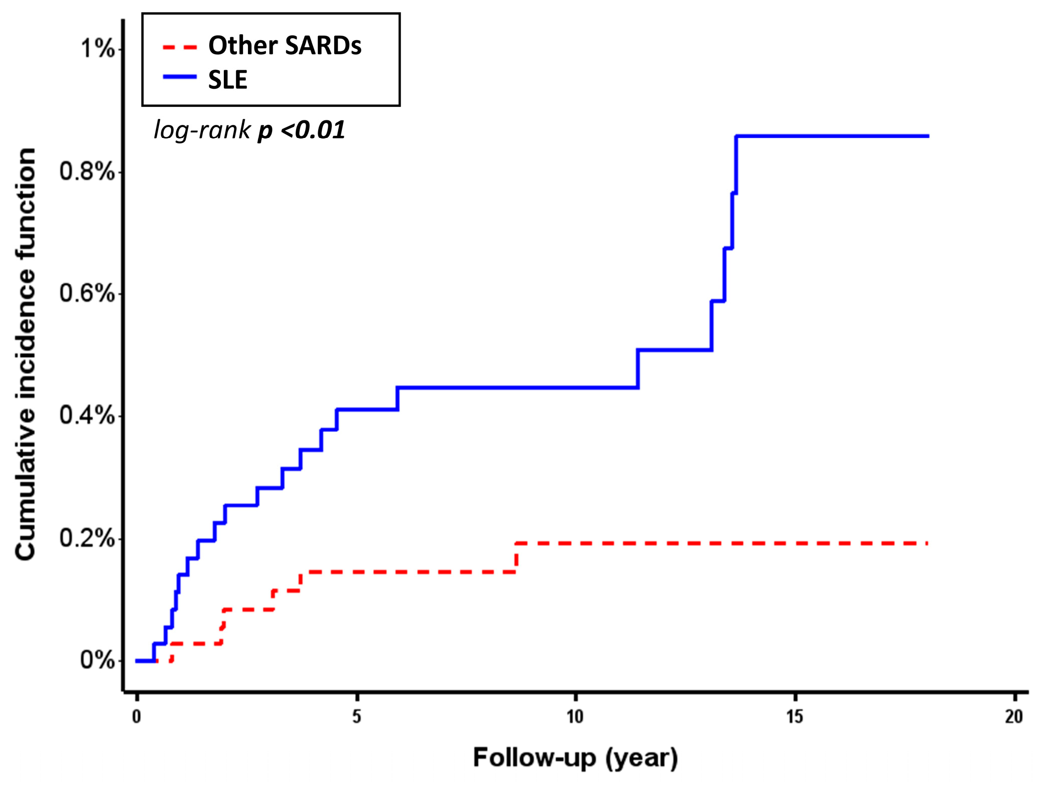
Patients with SLE had a higher incidence of Salmonella infection compared to those with other SARDs. The incidence rate of Salmonella infection in the SLE group was 0.54 per 1000 person-years (95% confidence interval (CI): 0.31–0.78), whereas it was 0.17 per 1000 person-years (95% CI: 0.03–0.30) in the comparison group. Kaplan–Meier analysis showed a significantly higher cumulative incidence of Salmonella infection in the SLE group (log-rank p < 0.01) (Figure 2.)
At the end of the 18-year-follow-up, the Cox proportional hazards model showed a crude hazard ratio (HR) of 3.28 (95% CI: 1.32–8.18) and an adjusted HR 2.47 (95% CI: 0.95–6.38, p = 0.06) for Salmonella infection in comparison of the SLE group with the other SARDs group. Furthermore, a competing risk analysis was performed with death as a competing event, yielding a sub-distribution hazard ratio of 2.58 (95% CI: 1.06–6.29, p = 0.04), confirming an independently increased risk of Salmonella infection in patients with SLE. These findings are summarized in Table 2.

3.3. Risk Factors for Salmonella Infection in the Patients with SLE

In the univariate Cox proportional hazards analysis, several factors were significantly associated with an increased risk of Salmonella infection among patients with SLE, including male sex (HR 2.55, 95% CI: 1.07–6.07, p = 0.03), lymphoproliferative disorder or cancer (HR 3.08, 95% CI: 1.06–8.94, p = 0.04), leukopenia (HR 2.67, 95% CI: 1.19–6.02, p = 0.02), lymphopenia (HR 3.98, 95% CI: 1.83–8.68, p < 0.001), decreased C3 (HR 2.48, 95% CI: 1.13–5.47, p = 0.02), proteinuria (HR 4.06, 95% CI: 1.39–11.92, p = 0.01), and hepatitis (HR 3.17, 95% CI: 1.20–8.41, p = 0.02) (Table 3).
Among DMARDs, azathioprine (HR 3.03, 95% CI: 0.91–10.12, p = 0.07), mycophenolic acid (HR 3.91, 95% CI: 0.53–28.99, p = 0.19), and cyclosporin (HR 4.22, 95% CI: 0.57–31.15, p = 0.16) demonstrated non-significant but suggestive trends toward increased risk.
Variables with a p-value < 0.2 in the univariate analysis were subsequently included in a stepwise multivariate Cox proportional hazards regression model. In the final model, only lymphopenia remained independently associated with a significantly higher risk of Salmonella infection (HR 3.98, 95% CI: 1.83–8.68, p < 0.001).

3.4. Serogroup and Sample Source of Salmonella in SLE

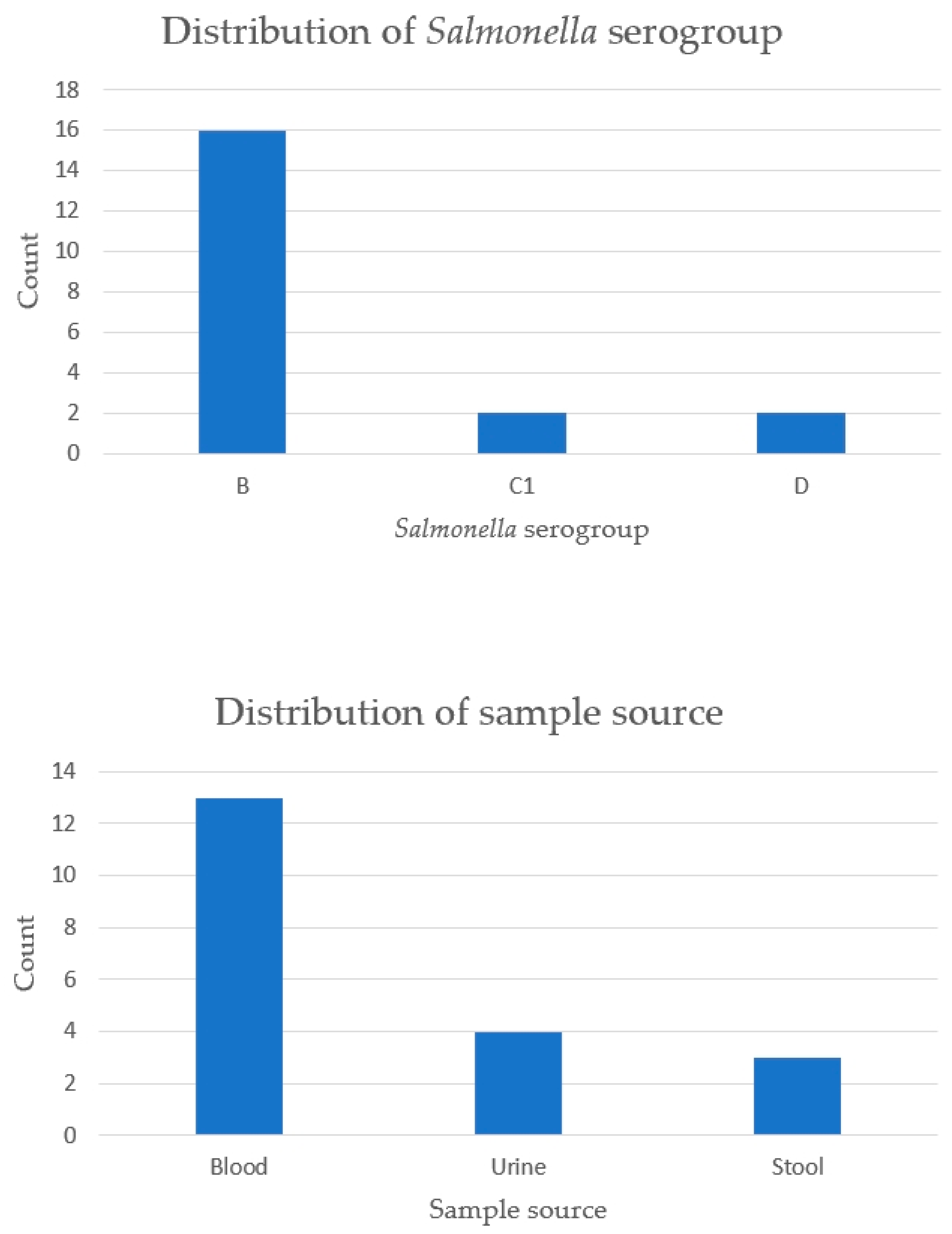
Among the 20 SLE patients with Salmonella infection, serogroup D was the most frequently identified strain (80%), followed by B (15%) and C1 (10%). The most common specimen source was blood (70%), followed by urine (20%) and stool (15%). This distribution suggested that bloodstream infections with serogroup D are the predominant clinical presentation in this population (Figure 3.)

4. Discussion

This large-scale retrospective cohort study demonstrated a significant higher risk of Salmonella infection in SLE patients compared to other SARDs. While several clinical factors were associated with increased risk among the SLE patients in the univariate analysis, lymphopenia emerged as the most robust independent predictor after adjusting for other covariates.
Infection is a major contributor to both morbidity and mortality in patients with SLE, accounting for an estimated 30–50% of overall disease-related complications worldwide [17,18]. An analysis from the United Kingdom revealed a striking rise in hospitalizations for severe infections among SLE patients, with the rate surging more than 12 times between 1996 and 2001 [3].
Among the causative pathogens of infection, Salmonella infection has long been recognized as a clinically important opportunistic infection in patients with SLE [9,11]. Previous studies have reported that patients with SLE are disproportionately vulnerable to Salmonella infection compared to general population. In a retrospective study of adult patients at Bellevue Hospital from 1975 to 1982, Abramson et al. found that SLE was the most frequent underlying disease among those with Salmonella bacteremia, accounting for 20% of cases, compared to only 0.54% in non-Salmonella Gram-negative bacteremia [11]. The disproportionately high rate of Salmonella bacteremia among SLE patients observed in this study may be attributed to their widespread use of immunosuppressive agents and underlying humoral immune defects, such as hypocomplementemia, both of which impair host defense mechanisms against invasive pathogens [11].
Exposure to Salmonella is associated with the development of SLE, which is possibly caused by releasing bacterial amyloid curli/DNA complexes [7,19,20]. Previous study also indicated that nontyphoidal Salmonella enterica infections, particularly those caused by S. typhimurium and S. enteritidis, may contribute to elevated autoantibody levels. Patients demonstrated significantly increased titers of autoantibodies during both the acute and convalescent phases of infection [21]. On the other hand, there is a high association between non-typhoidal salmonellosis and SLE, especially among patients with active disease [22]. As a result, development of the autoantibodies due to exposure to Salmonella among SLE patients could possibly increase disease activity and then result in severe Salmonella infection. As one of the opportunistic infection pathogens, Salmonella in SLE patients might progress to clinically significant infection by the vicious cycle of this bidirectional relationship.
While previous studies have primarily focused on the heightened risk of Salmonella infection in SLE patients compared to the general population, less is known about how this risk compares to that in patients with other SARDs, who also commonly receive immunosuppressive treatment. By directly comparing SLE with other SARDs, our study helps clarify whether this vulnerability is disease-specific or merely reflective of generalized immune dysfunction across autoimmune diseases.
To further characterize disease-specific immune vulnerability, we investigated potential predictors of Salmonella infection among SLE patients. In the univariate Cox proportional hazards analysis, male sex was significantly associated with an increased risk of Salmonella infection. Compared with female SLE patients, male SLE patients have higher disease activity and more severe organ damage and may require more aggressive treatment [23,24], which might explain the greater susceptible to infection. In addition, previous studies showed hematologic involvement was associated with serious infections in SLE patients and antimalarial use was protective [25,26]. Although our study did not reveal significant effects in hydroxychloroquine use, lymphopenia stood out as the only independent risk factor in our multivariate analysis—a finding that aligns with prior observations.
In a retrospective study conducted by Tsao et al. involving 31 hospitalized SLE patients with Salmonella infection, lymphopenia was observed in 74.2% of cases, suggesting a high prevalence of T-cell immune dysfunction in this population [27]. Further supporting the predictive role of lymphopenia, a retrospective case–control study by Merayo-Chalico et al. investigated 167 SLE patients and found that lymphopenia was independently associated with a significantly higher risk of severe infections, with an odds ratio of 5.2 (95% CI: 2.39–11.3), even after adjusting for other variables such as disease activity and immunosuppressive therapy [28].
Lymphopenia is a frequent hematologic abnormality in SLE, affecting T lymphocytes—particularly CD4+ cells—more profoundly than B cells. Its mechanisms are multifactorial, involving increased apoptosis, complement-mediated cytolysis, and impaired lymphopoiesis [29]. Clinically, lymphopenia has been associated with disease activity, renal involvement, and immunosuppressive therapy. In a cross-sectional study of 124 SLE patients, lymphopenia was significantly associated with lupus nephritis, complement consumption, higher steroid dose, and cyclophosphamide use [30]. These findings suggest that lymphopenia may serve as a potential clinical marker of immunologic severity and renal involvement in patients with SLE.
Given that therapeutic regimens may influence infection susceptibility, we first compared immunosuppressive drug use between groups. Standardized difference analysis revealed notable imbalances in the use of DMARDs—particularly methotrexate, sulfasalazine, and azathioprine—between the SLE and other SARDs groups. These discrepancies likely reflect divergent therapeutic strategies tailored to the underlying disease pathology, disease activity, or organ involvement, and may in turn contribute to differential infection risk profiles across groups. To account for these potential confounding effects, we incorporated medication use, including specific DMARDs and corticosteroids, into our multivariate Cox proportional hazards model. This adjustment allowed us to isolate the association between disease groups (SLE vs. other SARDs) and Salmonella infection risk, independent of variations in immunosuppressive treatment regimens.
Beyond assessing overall infection risk, we further analyzed the distribution of serogroups and specimen sources of Salmonella isolates to better understand the microbiological profiles and infection sites in SLE patients. Compared to real-world evidence, the previous study by Yu et al. consistently identified bacteremia as the predominant clinical manifestation of Salmonella infection in SLE patients, with blood cultures yielding the highest proportion of positive isolates [31]. However, the distribution of Salmonella serogroups differed between the two cohorts. In our analysis, serogroup D accounted for the majority of infections (80%), followed by groups B (15%) and C1 (10%). In contrast, Yu et al. reported serogroup B as the most frequently isolated strain (57.2%), with no mention of serogroup D [31]. This discrepancy may reflect temporal changes in circulating Salmonella strains, regional epidemiological differences, or distinct patient immunologic backgrounds and treatment exposures.
Our study offers several strengths that enhance the validity of its findings. First, previous studies had largely concentrated on case-based descriptions of Salmonella infections in SLE, while our study directly compared incidence and risk with other SARDs. Second, outcome analyses were performed using both Cox proportional hazards regression and competing risk models, which accounted for death as a competing event. This dual modeling strategy strengthened the reliability of our risk estimates, particularly in the presence of differential mortality risks. Third, we applied a stepwise variable selection approach in our multivariate modeling, allowing for dynamic inclusion of predictors based on statistical significance. This method helped address multicollinearity and interaction among variables—an important consideration in observational cohorts involving complex immune-mediated diseases and overlapping treatment exposures. Finally, a key strength of this study lies in the use of the CGRD, which provided access to detailed laboratory test results. This enabled us to incorporate objective immune and inflammatory biomarkers into the risk models, offering greater specificity than many administrative databases that lack laboratory data.
Several limitations should be considered in this study. First, medication exposure was assessed at baseline only, and potential time-varying effects throughout the follow-up period could not be fully captured. Second, despite multivariate adjustments, residual confounding may persist. Unmeasured behavioral or environmental factors—such as dietary habits, hygiene practices, and recent travel history—might have influenced individual infection risk but were not available in the database.
Building upon these strengths, and acknowledging the limitations of the study, our findings may carry important clinical implications. Clinicians may consider maintaining a heightened awareness of the potential for Salmonella infection in patients with SLE, particularly those with lymphopenia. Future studies could explore whether monitoring lymphocyte subsets might benefit high-risk subgroups. Moreover, prospective research incorporating dynamic immune profiling and microbiological data may provide further insights into host–pathogen interactions and help refine infection risk assessment in this population.

Author Contributions

Conceptualization, H.-H.Y. and Y.-F.C.; methodology, Y.-F.C.; software, P.-Y.C.; validation, C.-Y.W., H.-H.Y. and Y.-F.C.; formal analysis, P.-Y.C.; investigation, P.-Y.C.; resources, P.-Y.C.; data curation, C.-Y.W. and H.-H.Y.; writing—original draft preparation, C.-Y.W. and H.-H.Y.; writing—review and editing, Y.-F.C.; visualization, C.-Y.W.; supervision, Y.-F.C.; project administration, Y.-F.C.; funding acquisition, C.-Y.W. All authors have read and agreed to the published version of the manuscript.

Funding

This research was funded by Chang Gung Medical Foundation, grant number CGRPG5N0021.

Institutional Review Board Statement

The study was conducted in accordance with the Declaration of Helsinki, and approved by the Institutional Review Board of Chang Gung Medical Foundation (IRB No. 202202074B0C6004 and 1 March 2023).

Informed Consent Statement

Patient consent was waived because de-identified data were used.

Data Availability Statement

The study’s original contributions are detailed within the article. Requests for clarification or additional information can be directed to the corresponding author.

Acknowledgments

The authors wish to acknowledge the Center for Big Data Analytics and Statistics, Chang Gung Memorial Hospital, Linkou for statistical and data analysis assistance and interpretation support. This study is based in part on data from the Chang Gung Research Database provided by Chang Gung Memorial Hospital. The interpretation and conclusions contained herein do not represent the position of Chang Gung Memorial Hospital.

Conflicts of Interest

The authors declare no conflicts of interest.

Abbreviations

The following abbreviations are used in this manuscript:
Anti-dsDNAAntibody to double-stranded deoxyribonucleic acid
C3Complement 3
C4Complement 4
CIConfidence interval
CGRDChang Gung Research Database
DMARDsDisease-modifying antirheumatic drugs
ESRErythrocyte sedimentation rate
HRHazard ratio
ICDInternational Classification of Diseases
IgMImmunoglobulin M
SARDsSystemic autoimmune rheumatic diseases
SLESystemic lupus erythematosus

References

  1. Burmester, G.R.; Bijlsma, J.W.J.; Cutolo, M.; McInnes, I.B. Managing rheumatic and musculoskeletal diseases—Past, present and future. Nat. Rev. Rheumatol. 2017, 13, 443–448. [Google Scholar] [CrossRef]
  2. Fanouriakis, A.; Tziolos, N.; Bertsias, G.; Boumpas, D.T. Update οn the diagnosis and management of systemic lupus erythematosus. Ann. Rheum. Dis. 2021, 80, 14–25. [Google Scholar] [CrossRef] [PubMed]
  3. Tektonidou, M.G.; Wang, Z.; Dasgupta, A.; Ward, M.M. Burden of Serious Infections in Adults With Systemic Lupus Erythematosus: A National Population-Based Study, 1996–2011. Arthritis Care Res. 2015, 67, 1078–1085. [Google Scholar] [CrossRef]
  4. Consani Fernández, S.A.; Díaz Cuña, C.L.; Fernández Rey, L.; Rostán Sellanes, S.; Maciel Oleggini, G.; Facal Castro, J.A. Infections in systemic autoimmune diseases. Reumatol. Clínica (Engl. Ed.) 2021, 17, 582–587. [Google Scholar] [CrossRef]
  5. Crum, N.F.; Lederman, E.R.; Wallace, M.R. Infections associated with tumor necrosis factor-alpha antagonists. Medicine 2005, 84, 291–302. [Google Scholar] [CrossRef]
  6. Gordon, M.A. Salmonella infections in immunocompromised adults. J. Infect. 2008, 56, 413–422. [Google Scholar] [CrossRef]
  7. Tu, T.Y.; Yeh, C.Y.; Hung, Y.M.; Chang, R.; Chen, H.H.; Wei, J.C. Association Between a History of Nontyphoidal Salmonella and the Risk of Systemic Lupus Erythematosus: A Population-Based, Case-Control Study. Front. Immunol. 2021, 12, 725996. [Google Scholar] [CrossRef]
  8. Chen, J.Y.; Luo, S.F.; Wu, Y.J.; Wang, C.M.; Ho, H.H. Salmonella septic arthritis in systemic lupus erythematosus and other systemic diseases. Clin. Rheumatol. 1998, 17, 282–287. [Google Scholar] [CrossRef] [PubMed]
  9. Pablos, J.L.; Aragon, A.; Gomez-Reino, J.J. Salmonellosis and systemic lupus erythematosus. Report of ten cases. Br. J. Rheumatol. 1994, 33, 129–132. [Google Scholar] [CrossRef]
  10. Shahram, F.; Akbarian, M.; Davatchi, F. Salmonella infection in systemic lupus erythematosus. Lupus 1993, 2, 55–59. [Google Scholar] [CrossRef]
  11. Abramson, S.; Kramer, S.B.; Radin, A.; Holzman, R. Salmonella bacteremia in systemic lupus erythematosus. Eight-year experience at a municipal hospital. Arthritis Rheum. 1985, 28, 75–79. [Google Scholar] [CrossRef]
  12. Li, M.; Wu, C.; Yin, P.; Qian, J.; Zhao, J.; Wang, Q.; Xu, D.; Su, J.; Leng, X.; Zheng, W.; et al. Mortality-related health metrics in systemic autoimmune diseases: An epidemiological analysis of a nationwide register-based cohort. Sci. Bull. 2025, 70, 492–495. [Google Scholar] [CrossRef] [PubMed]
  13. Yeh, K.W.; Yu, C.H.; Chan, P.C.; Horng, J.T.; Huang, J.L. Burden of systemic lupus erythematosus in Taiwan: A population-based survey. Rheumatol. Int. 2013, 33, 1805–1811. [Google Scholar] [CrossRef] [PubMed]
  14. Elsisi, G.H.; Hsieh, S.-C.; Chen, D.-Y. The economic burden of systemic lupus erythematosus in Taiwan. J. Med. Econ. 2024, 27, 56–66. [Google Scholar] [CrossRef]
  15. Cummings, P. Arguments for and against standardized mean differences (effect sizes). Arch. Pediatr. Adolesc. Med. 2011, 165, 592–596. [Google Scholar] [CrossRef]
  16. Wu, M.Y.; Huang, M.C.; Liao, H.H.; Chiang, J.H.; Lee, Y.C.; Hsu, C.Y.; Sun, M.F.; Yen, H.R. Acupuncture decreased the risk of coronary heart disease in patients with rheumatoid arthritis in Taiwan: A Nationwide propensity score-matched study. BMC Complement. Altern. Med. 2018, 18, 341. [Google Scholar] [CrossRef] [PubMed]
  17. Zandman-Goddard, G.; Shoenfeld, Y. Infections and SLE. Autoimmunity 2005, 38, 473–485. [Google Scholar] [CrossRef]
  18. Alarcón, G.S. Infections in systemic connective tissue diseases: Systemic lupus erythematosus, scleroderma, and polymyositis/dermatomyositis. Infect. Dis. Clin. North. Am. 2006, 20, 849–875. [Google Scholar] [CrossRef]
  19. Gallo, P.M.; Rapsinski, G.J.; Wilson, R.P.; Oppong, G.O.; Sriram, U.; Goulian, M.; Buttaro, B.; Caricchio, R.; Gallucci, S.; Tükel, Ç. Amyloid-DNA Composites of Bacterial Biofilms Stimulate Autoimmunity. Immunity 2015, 42, 1171–1184. [Google Scholar] [CrossRef]
  20. Qiu, C.C.; Caricchio, R.; Gallucci, S. Triggers of Autoimmunity: The Role of Bacterial Infections in the Extracellular Exposure of Lupus Nuclear Autoantigens. Front. Immunol. 2019, 10, 2608. [Google Scholar] [CrossRef]
  21. Ktsoyan, Z.; Budaghyan, L.; Agababova, M.; Mnatsakanyan, A.; Arakelova, K.; Gevorgyan, Z.; Sedrakyan, A.; Hovhannisyan, A.; Mkrtchyan, M.; Zakharyan, M.; et al. Potential Involvement of Salmonella Infection in Autoimmunity. Pathogens 2019, 8, 96. [Google Scholar] [CrossRef] [PubMed]
  22. Lim, E.; Koh, W.H.; Loh, S.F.; Lam, M.S.; Howe, H.S. Non-thyphoidal salmonellosis in patients with systemic lupus erythematosus. A study of fifty patients and a review of the literature. Lupus 2001, 10, 87–92. [Google Scholar] [CrossRef]
  23. Albrecht, K.; Troll, W.; Callhoff, J.; Strangfeld, A.; Ohrndorf, S.; Mucke, J. Sex- and gender-related differences in systemic lupus erythematosus: A scoping review. Rheumatol. Int. 2025, 45, 160. [Google Scholar] [CrossRef]
  24. Gui, Y.; Bai, W.; Xu, J.; Duan, X.; Zhan, F.; Zhao, C.; Jiang, Z.; Li, Z.; Wu, L.; Liu, S.; et al. Sex differences in systemic lupus erythematosus (SLE): An inception cohort of the Chinese SLE Treatment and Research Group (CSTAR) registry XVII. Chin. Med. J. 2022, 135, 2191–2199. [Google Scholar] [CrossRef]
  25. Jung, J.Y.; Yoon, D.; Choi, Y.; Kim, H.A.; Suh, C.H. Associated clinical factors for serious infections in patients with systemic lupus erythematosus. Sci. Rep. 2019, 9, 9704. [Google Scholar] [CrossRef] [PubMed]
  26. Pimentel-Quiroz, V.R.; Ugarte-Gil, M.F.; Harvey, G.B.; Wojdyla, D.; Pons-Estel, G.J.; Quintana, R.; Esposto, A.; García, M.A.; Catoggio, L.J.; Cardiel, M.H.; et al. Factors predictive of serious infections over time in systemic lupus erythematosus patients: Data from a multi-ethnic, multi-national, Latin American lupus cohort. Lupus 2019, 28, 1101–1110. [Google Scholar] [CrossRef] [PubMed]
  27. Tsao, C.H.; Chen, C.Y.; Ou, L.S.; Huang, J.L. Risk factors of mortality for Salmonella infection in systemic lupus erythematosus. J. Rheumatol. 2002, 29, 1214–1218. [Google Scholar]
  28. Merayo-Chalico, J.; Gómez-Martín, D.; Piñeirúa-Menéndez, A.; Santana-De Anda, K.; Alcocer-Varela, J. Lymphopenia as risk factor for development of severe infections in patients with systemic lupus erythematosus: A case-control study. Qjm 2013, 106, 451–457. [Google Scholar] [CrossRef]
  29. Martin, M.; Guffroy, A.; Argemi, X.; Martin, T. Systemic lupus erythematosus and lymphopenia: Clinical and pathophysiological features. Rev. Med. Interne 2017, 38, 603–613. [Google Scholar] [CrossRef]
  30. Sobhy, N.; Niazy, M.H.; Kamal, A. Lymphopenia in systemic lupus erythematosus patients: Is it more than a laboratory finding? Egypt. Rheumatol. 2020, 42, 23–26. [Google Scholar] [CrossRef]
  31. Yu, S.-F.; Cheng, T.-T.; Chen, Y.-C.; Wu, C.-H.; Chiu, C.-K.; Lai, H.-M.; Lee, C.-H. Salmonella infection in systemic lupus erythematosus: Seventeen-year experience at a medical center in southern Taiwan. Formos. J. Rheumatol. 2004, 18, 35–46. (In Chinese) [Google Scholar] [CrossRef]
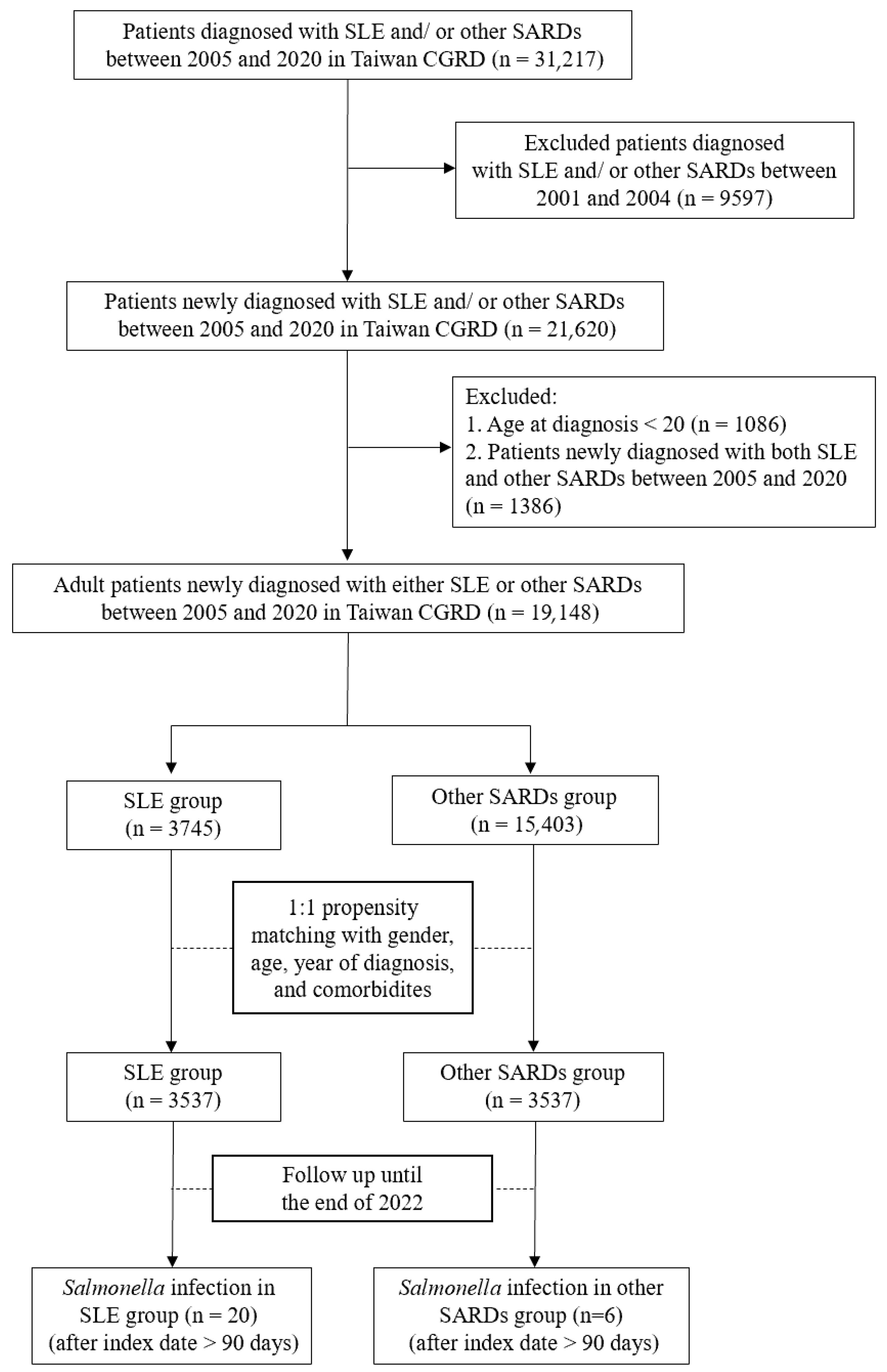
Figure 1. Flow chart of the study. Abbreviations: SLE, systemic lupus erythematosus; SARDs, systemic autoimmune rheumatic diseases.
Life 15 01522 g001
Figure 2. Cumulative incidence of Salmonella infection over 18 years in SLE and other SARDs. Abbreviations: SARDs, systemic autoimmune rheumatic diseases; SLE, systemic lupus erythematosus.
Life 15 01522 g002
Figure 3. Serogroup and sample source of Salmonella infection among SLE patients. Abbreviation: SLE, systemic lupus erythematosus.
Life 15 01522 g003
Table 1. Baseline patient characteristics in the SLE and other SARDs groups.
SLE
(n = 3537)
Other SARDs
(n = 3537)
p-Value aStandardized Difference b
Age42.93 ± 15.0244.00 ± 14.31<0.010.07
Male445 (12.58)445 (12.58)10
Comorbidity
    Diabetes mellitus166 (4.69)216 (6.11)0.010.06
    Chronic kidney disease522 (14.76)444 (12.55)0.010.06
    Cirrhosis64 (1.81)63 (1.78)0.93<0.01
    Lymphoproliferative disorder/cancer187 (5.29)219 (6.19)0.100.04
Drug
    Steroids1263 (35.71)1047 (29.60)<0.0010.13
DMARDS
    Hydroxychloroquine1040 (29.40)1041 (29.43)0.98<0.001
    Azathioprine238 (6.73)78 (2.21)<0.0010.22
    Methotrexate17 (0.48)567 (16.03)<0.0010.59
    Leflunomide2 (0.06)61 (1.72)<0.0010.18
    Mycophenolic acid78 (2.21)12 (0.34)<0.0010.17
    Cyclosporin27 (0.76)49 (1.39)0.110.09
    Cyclophosphamide17 (0.48)9 (0.25)0.120.03
    Sulfasalazine14 (0.40)395 (11.17)<0.0010.50
    Tofacitinib0 (0.00)0 (0.00)--
    Baricitinib0 (0.00)0 (0.00)--
    Upadacitinib0 (0.00)0 (0.00)--
    Rituximab7 (0.20)8 (0.23)0.800.01
    Etanercept1 (0.03)13 (0.37)<0.010.08
    Adalimumab0 (0.00)7 (0.20)0.020.06
    Golimumab0 (0.00)2 (0.06)0.500.03
    Certolizumab0 (0.00)7 (0.20)0.020.06
    Infliximab0 (0.00)0 (0.00)--
    Tocilizumab0 (0.00)14 (0.40)<0.0010.09
    Abatacept0 (0.00)3 (0.08)0.250.04
    Belimumab0 (0.00)0 (0.00)--
a Chi-squared test for categorical data and unpaired Student t-test for continuous data. b Calculation of effect size: mean for continuous data and percentage for categorical data. Abbreviations: DMARDs, disease-modifying antirheumatic drugs; SARDs, systemic autoimmune rheumatic diseases; SLE, systemic lupus erythematosus.
Table 2. Incidence and risk comparison of Salmonella infection in SLE and other SARDs.
NumberFollow-Up
(Years)
Person-Years of Follow-UpIncidence Rate,
Per 1000 Person-Years
Crude HR
(95% CI)
Adjusted HR a
(95% CI)
Competing Risk Regression Model b
Adjusted HR a
(95% CI)
SLE20/353710.38 ± 4.7536,703.710.54
(0.31–0.78)
3.28
(1.32–8.18)
2.47
(0.95–6.38)
2.58
(1.06–6.29)
Other SARDs6/353710.19 ± 4.5736,059.530.17
(0.03–0.30)
---
p-value<0.01---0.010.060.04
a Adjusted for age, sex, baseline comorbidities, and medications; b death as a competing event. Abbreviations: SARDs, systemic autoimmune rheumatic diseases; SLE, systemic lupus erythematosus; HR, hazard ratio; CI, confidence interval.
Table 3. Risk factors of Salmonella infection in patients with SLE.
VariableUnivariate AnalysisMultivariate Analysis
HR (95% CI)p-ValueHR (95% CI)p-Value
Age ≥ 500.86 (0.36–2.05)0.74-
Male2.55 (1.07–6.07)0.03-
Comorbidity
 Diabetes mellitus2.38 (0.72–7.94)0.16-
 Chronic kidney disease1.08 (0.37–3.13)0.89-
 Cirrhosis2.33 (0.32–17.16)0.41-
 Lymphoproliferative disorder/cancer3.08 (1.06–8.94)0.04-
DMARDS
 Hydroxychloroquine1.30 (0.58–2.92)0.53-
 Azathioprine3.03 (0.91–10.12)0.07-
 Mycophenolic acid3.91 (0.53–28.90)0.18-
 Cyclosporin4.22 (0.57–31.15)0.16-
Laboratory data
 Leukopenia2.67 (1.19–6.02)0.02-
 Lymphopenia3.98 (1.83–8.68)<0.0013.98 (1.83–8.68)<0.001
 Decreased IgM5.63 (0.76–41.56)0.09-
 Decreased C32.48 (1.13–5.47)0.02-
 Decreased C42.41 (1.02–5.74)0.05-
 Positivity of anti-dsDNA1.02 (0.45–2.28)0.97-
 Increased ferritin2.66 (0.63–11.28)0.18-
 Proteinuria4.06 (1.39–11.92)0.01-
 Hepatitis3.17 (1.20–8.41)0.02-
 Renal impairment1.87 (0.71–4.96)0.21-
 Hypoalbuminemia1.68 (0.68–4.19)0.26-
 Elevated ESR0.37 (0.13–1.06)0.06-
Abbreviations: SLE, systemic lupus erythematosus; IgM, Immunoglobulin M; C3, complement 3; C4, complement 4; anti-dsDNA, antibody to double-stranded deoxyribonucleic acid; ESR, erythrocyte sedimentation rate; CI, confidence interval; HR, hazard ratio.
Disclaimer/Publisher’s Note: The statements, opinions and data contained in all publications are solely those of the individual author(s) and contributor(s) and not of MDPI and/or the editor(s). MDPI and/or the editor(s) disclaim responsibility for any injury to people or property resulting from any ideas, methods, instructions or products referred to in the content.