

Spontaneous Intracranial Hypotension and Subdural Hematomas Treatment Management Using MMA Embolization and Target Blood Patch: A Case Report

 , , ,
, , ,  and
and 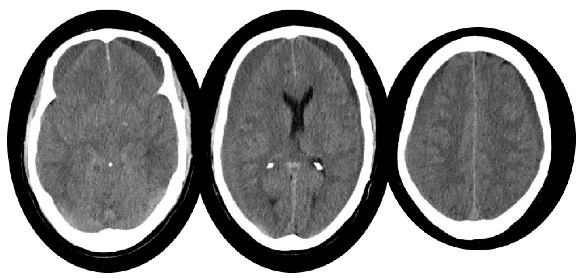
Abstract
1. Introduction
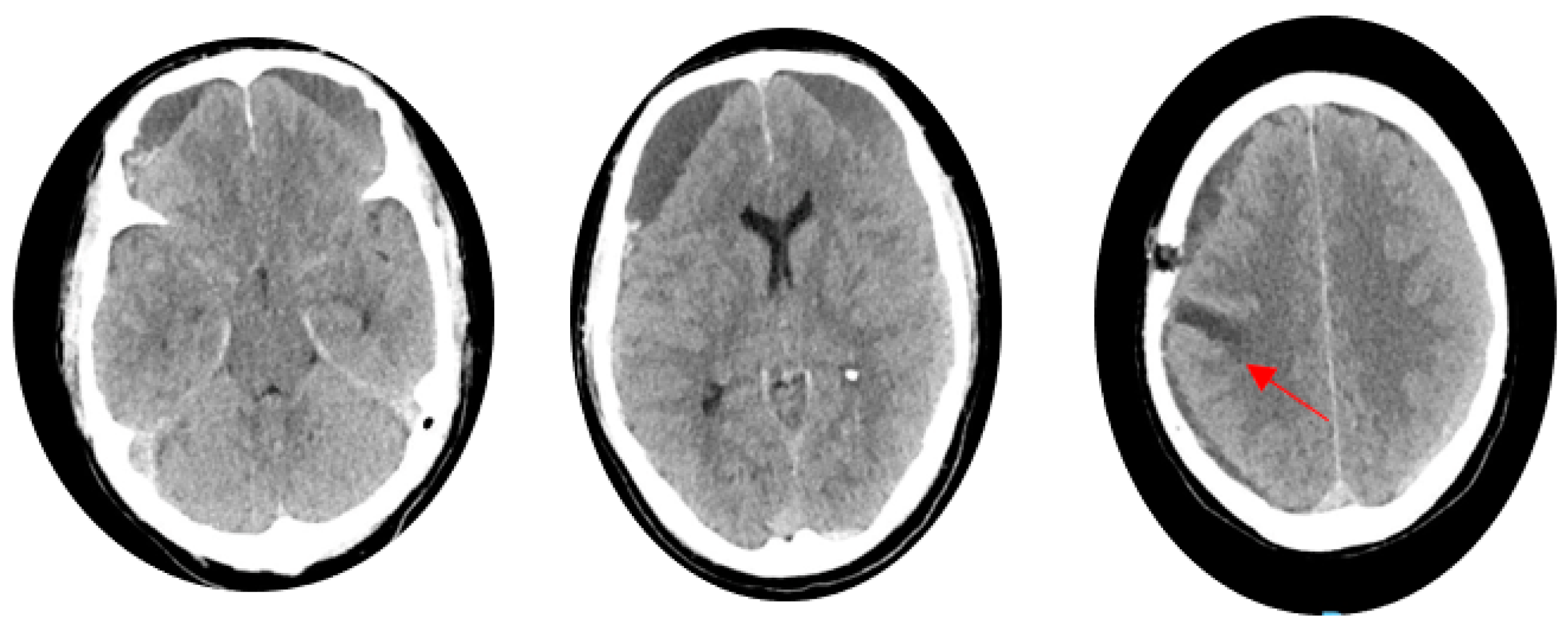
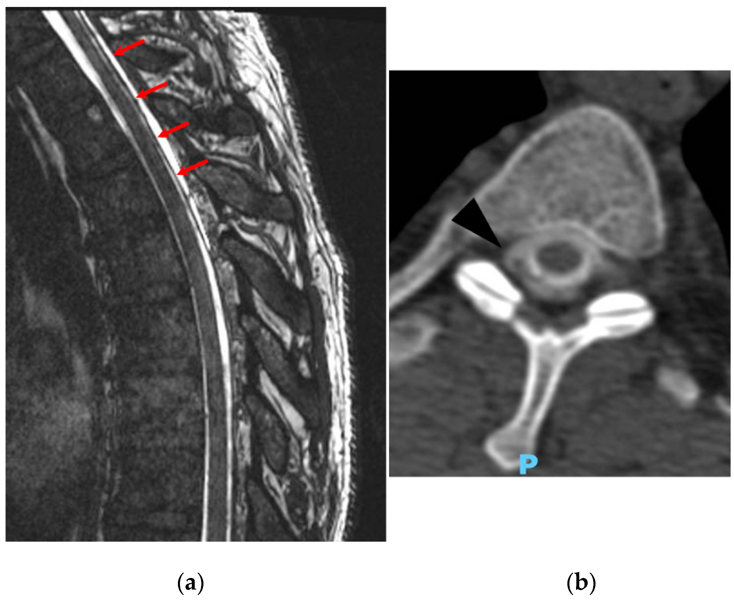
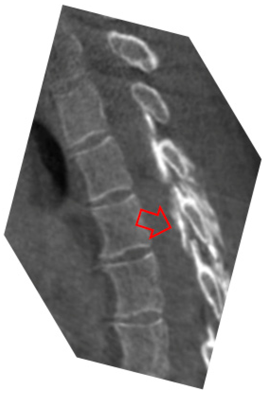
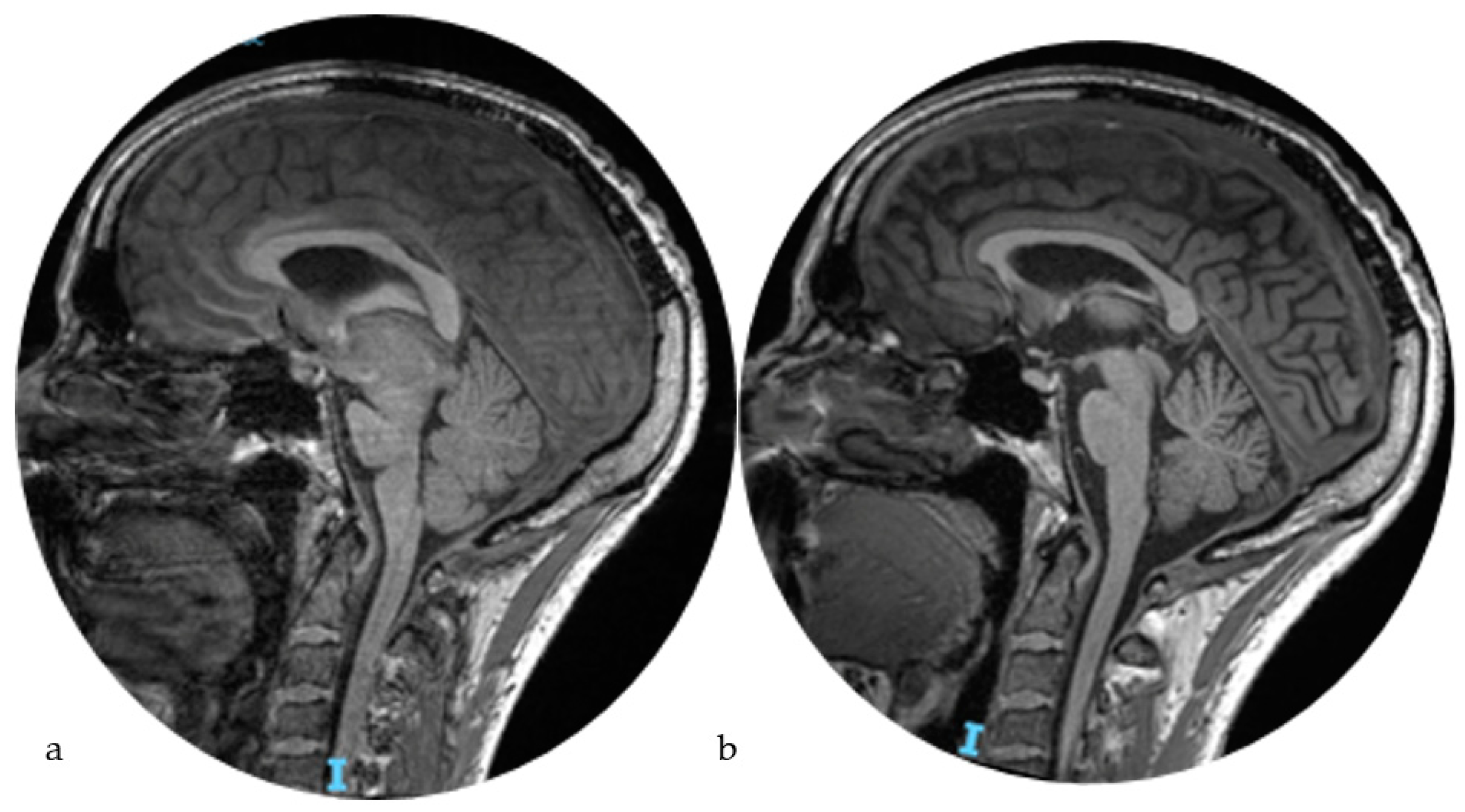
2. Case Report
- T0:
- It is time zero, the first measurement of the test, with the patient in a supine position.
- T1:
- It is the second measurement of the test after the patient has been in an upright position for 5 min.
- T2:
- It is the third measurement of the test after the patient has been in an upright position for 20 min.
- T0:
- Headache NRS 2–3/10, with only slight occipital discomfort. FAB score 17/18, losing one point in the grip test. T1: Stable headache. FAB score 16/17, losing one point in the grip test and one point in verbal fluency. T2: Stable headache. FAB score 15/17, losing one point in the grip test, one point in verbal fluency, and one point in the Go-no-go test.
3. Discussion
4. Conclusions
Author Contributions
Funding
Institutional Review Board Statement
Informed Consent Statement
Data Availability Statement
Conflicts of Interest
References
- Schievink, W.I. Spontaneous Spinal Cerebrospinal Fluid Leaks and Intracranial Hypotension. JAMA 2006, 295, 2286–2296. [Google Scholar] [CrossRef]
- Shah, L.M.; McLean, L.A.; Heilbrun, M.E.; Salzman, K.L. Intracranial Hypotension: Improved MRI Detection with Diagnostic Intracranial Angles. Am. J. Roentgenol. 2013, 200, 400–407. [Google Scholar] [CrossRef]
- Kranz, P.G.; Gray, L.; Malinzak, M.D.; Amrhein, T.J. Spontaneous Intracranial Hypotension: Pathogenesis, Diagnosis, and Treatment. Neuroimaging Clin. N. Am. 2019, 29, 581–594. [Google Scholar] [CrossRef] [PubMed]
- Luetzen, N.; Dovi-Akue, P.; Fung, C.; Beck, J.; Urbach, H. Spontaneous Intracranial Hypotension: Diagnostic and Therapeutic Workup. Neuroradiology 2021, 63, 1765–1772. [Google Scholar] [CrossRef] [PubMed]
- Dobrocky, T.; Grunder, L.; Breiding, P.S.; Branca, M.; Limacher, A.; Mosimann, P.J.; Mordasini, P.; Zibold, F.; Haeni, L.; Jesse, C.M.; et al. Assessing Spinal Cerebrospinal Fluid Leaks in Spontaneous Intracranial Hypotension with a Scoring System Based on Brain Magnetic Resonance Imaging Findings. JAMA Neurol. 2019, 76, 580–587. [Google Scholar] [CrossRef] [PubMed]
- Watanabe, A.; Horikoshi, T.; Uchida, M.; Koizumi, H.; Yagishita, T.; Kinouchi, H. Diagnostic Value of Spinal MR Imaging in Spontaneous Intracranial Hypotension Syndrome. Am. J. Neuroradiol. 2009, 30, 147–151. [Google Scholar] [CrossRef] [PubMed]
- Lee, G.H.; Kim, J.; Kim, H.W.; Cho, J.W. Comparisons of Clinical Characteristics, Brain MRI Findings, and Responses to Epidural Blood Patch between Spontaneous Intracranial Hypotension and Post-Dural Puncture Headache: Retrospective Study. BMC Neurol. 2021, 21, 253. [Google Scholar] [CrossRef] [PubMed]
- Cheema, S.; Anderson, J.; Angus-Leppan, H.; Armstrong, P.; Butteriss, D.; Carlton Jones, L.; Choi, D.; Chotai, A.; D’Antona, L.; Davagnanam, I.; et al. Multidisciplinary Consensus Guideline for the Diagnosis and Management of Spontaneous Intracranial Hypotension. J. Neurol. Neurosurg. Psychiatry 2023, 94, 835–843. [Google Scholar] [CrossRef]
- Kranz, P.G.; Luetmer, P.H.; Diehn, F.E.; Amrhein, T.J.; Tanpitukpongse, T.P.; Gray, L. Myelographic Techniques for the Detection of Spinal CSF Leaks in Spontaneous Intracranial Hypotension. Am. J. Roentgenol. 2016, 206, 8–19. [Google Scholar] [CrossRef]
- Schievink, W.I.; Maya, M.M.; Moser, F.G.; Tourje, J. Spectrum of Subdural Fluid Collections in Spontaneous Intracranial Hypotension. J. Neurosurg. 2005, 103, 608–613. [Google Scholar] [CrossRef]
- Rudy, R.F.; Catapano, J.S.; Jadhav, A.P.; Albuquerque, F.C.; Ducruet, A.F. Middle Meningeal Artery Embolization to Treat Chronic Subdural Hematoma. Stroke Vasc. Interv. Neurol. 2023, 3, e000490. [Google Scholar] [CrossRef]
- Iyer, A.M.; Venkataraman, S.S.; Kittel, C.A.; Fargen, K.M. Coil Embolization Alone Appears Sufficient for Middle Meningeal Artery Embolization. Interv. Neuroradiol. 2023. online first. [Google Scholar] [CrossRef] [PubMed]
- Dubois, B.; Slachevsky, A.; Litvan, I.; Pillon, B.F.A.B. The FAB: A Frontal Assessment Battery at Bedside. Neurology 2000, 55, 1621–1626. [Google Scholar] [CrossRef]
- Kim, Y.J.; Cho, H.Y.; Seo, D.W.; Sohn, C.H.; Ahn, S.; Lee, Y.S.; Kim, W.Y.; Lim, K.S. Misdiagnosis of Spontaneous Intracranial Hypotension as a Risk Factor for Subdural Hematoma. Headache 2017, 57, 1593–1600. [Google Scholar] [CrossRef] [PubMed]
- Kim, B.W.; Jung, Y.J.; Kim, M.S.; Choi, B.Y. Chronic Subdural Hematoma after Spontaneous Intracranial Hypotension: A Case Treated with Epidural Blood Patch on C1-2. J. Korean Neurosurg. Soc. 2011, 50, 274–276. [Google Scholar] [CrossRef] [PubMed]
- Choi, S.H.; Lee, Y.Y.; Kim, W.J. Epidural Blood Patch for Spontaneous Intracranial Hypotension with Subdural Hematoma: A Case Report and Review of Literature. World J. Clin. Cases 2022, 10, 388–396. [Google Scholar] [CrossRef]
- Armstrong, N.; Williamson, C.; Williamson, N.; Fortes, M.; Tjauw, I.; Vij, V.; Trojan, R. Dural Diverticulum with a Symptomatic Cerebrospinal Fluid Leak. Radiol. Case Rep. 2016, 11, 16–20. [Google Scholar] [CrossRef][Green Version]
- Enriquez-Marulanda, A.; Gomez-Paz, S.; Salem, M.M.; Mallick, A.; Motiei-Langroudi, R.; Arle, J.E.; Stippler, M.; Papavassiliou, E.; Alterman, R.L.; Ogilvy, C.S.; et al. Middle Meningeal Artery Embolization versus Conventional Treatment of Chronic Subdural Hematomas. Neurosurgery 2021, 89, 486–495. [Google Scholar] [CrossRef]
- Markwalder, T.-M. Chronic Subdural Hematomas: A Review. J. Neurosurg. 1981, 54, 637–645. [Google Scholar] [CrossRef]
- Hashimoto, T.; Ohashi, T.; Watanabe, D.; Koyama, S.; Namatame, H.; Izawa, H.; Haraoka, R.; Okada, H.; Ichimasu, N.; Akimoto, J.; et al. Usefulness of Embolization of the Middle Meningeal Artery for Refractory Chronic Subdural Hematomas. Surg. Neurol. Int. 2013, 4, 104. [Google Scholar] [CrossRef]
- Tanaka, T.; Kaimori, M. Histological Study of Vascular Structure between the Dura Mater and the Outer Membrane in Chronic Subdural Hematoma in an Adult. Neurol. Surg. 1999, 27, 431–436. [Google Scholar]
- Désir, L.L.; D’Amico, R.; Link, T.; Silva, D.; Ellis, J.A.; Doron, O.; Langer, D.J.; Ortiz, R.; Serulle, Y. Middle Meningeal Artery Embolization and the Treatment of a Chronic Subdural Hematoma. Cureus 2021, 13, e18868. [Google Scholar] [CrossRef] [PubMed]
- Sattari, S.A.; Yang, W.; Shahbandi, A.; Feghali, J.; Lee, R.P.; Xu, R.; Jackson, C.; Gonzalez, L.F.; Tamargo, R.J.; Huang, J.; et al. Middle Meningeal Artery Embolization versus Conventional Management for Patients with Chronic Subdural Hematoma: A Systematic Review and Meta-Analysis. Neurosurgery 2023, 92, 1142–1154. [Google Scholar] [CrossRef] [PubMed]
- Catapano, J.S.; Nguyen, C.L.; Wakim, A.A.; Albuquerque, F.C.; Ducruet, A.F. Middle Meningeal Artery Embolization for Chronic Subdural Hematoma. Front. Neurol. 2020, 11, 557233. [Google Scholar] [CrossRef]


Disclaimer/Publisher’s Note: The statements, opinions and data contained in all publications are solely those of the individual author(s) and contributor(s) and not of MDPI and/or the editor(s). MDPI and/or the editor(s) disclaim responsibility for any injury to people or property resulting from any ideas, methods, instructions or products referred to in the content. |
© 2024 by the authors. Licensee MDPI, Basel, Switzerland. This article is an open access article distributed under the terms and conditions of the Creative Commons Attribution (CC BY) license (https://creativecommons.org/licenses/by/4.0/).
Share and Cite
Cirillo, L.; Verna, F.; Princiotta, C.; Dall’Olio, M.; Rustici, A.; Bortolotti, C.; Badaloni, F.; Mascarella, D.; Cortelli, P.; Cevoli, S. Spontaneous Intracranial Hypotension and Subdural Hematomas Treatment Management Using MMA Embolization and Target Blood Patch: A Case Report. Life 2024, 14, 250. https://doi.org/10.3390/life14020250
Cirillo L, Verna F, Princiotta C, Dall’Olio M, Rustici A, Bortolotti C, Badaloni F, Mascarella D, Cortelli P, Cevoli S. Spontaneous Intracranial Hypotension and Subdural Hematomas Treatment Management Using MMA Embolization and Target Blood Patch: A Case Report. Life. 2024; 14(2):250. https://doi.org/10.3390/life14020250
Chicago/Turabian StyleCirillo, Luigi, Francesca Verna, Ciro Princiotta, Massimo Dall’Olio, Arianna Rustici, Carlo Bortolotti, Filippo Badaloni, Davide Mascarella, Pietro Cortelli, and Sabina Cevoli. 2024. "Spontaneous Intracranial Hypotension and Subdural Hematomas Treatment Management Using MMA Embolization and Target Blood Patch: A Case Report" Life 14, no. 2: 250. https://doi.org/10.3390/life14020250
APA StyleCirillo, L., Verna, F., Princiotta, C., Dall’Olio, M., Rustici, A., Bortolotti, C., Badaloni, F., Mascarella, D., Cortelli, P., & Cevoli, S. (2024). Spontaneous Intracranial Hypotension and Subdural Hematomas Treatment Management Using MMA Embolization and Target Blood Patch: A Case Report. Life, 14(2), 250. https://doi.org/10.3390/life14020250