Article

Diversity of Endophytes of Actinidia arguta in Different Seasons

1
Institute of Special Animal and Plant Sciences, Chinese Academy of Agricultural Sciences, Changchun 130112, China
2
College of Animal Science and Veterinary Medicine, Heilongjiang Bayi Agricultural University, Daqing 163319, China
3
College of Horticulture, Jilin Agricultural University, Changchun 130112, China
*
Author to whom correspondence should be addressed.
Life 2024, 14(1), 149; https://doi.org/10.3390/life14010149
Submission received: 12 December 2023 / Revised: 10 January 2024 / Accepted: 13 January 2024 / Published: 19 January 2024
(This article belongs to the Special Issue Rhizosphere Microbes and Plant Stress)

Abstract

:
The seasonal changes in environmental conditions can alter the growth states of host plants, thereby affecting the living environment of endophytes and forming different endophytic communities. This study employs Illumina MiSeq next-generation sequencing to analyze the 16SrRNA and ITS rDNA of endophytes in 24 samples of Actinidia arguta stem tissues across different seasons. The results revealed a high richness and diversity of endophytes in Actinidia arguta, with significant seasonal variations in microbial community richness. This study identified 897 genera across 36 phyla for bacteria and 251 genera across 8 phyla for fungi. Notably, 69 bacterial genera and 19 fungal genera significantly contributed to the differences in community structure across seasons. A distinctive feature of coexistence in the endophytic community, both specific and conservative across different seasons, was observed. The bacterial community in winter demonstrated significantly higher richness and diversity compared to the other seasons. Environmental factors likely influence the optimal timing for endophyte colonization. Solar radiation, temperature, precipitation, and relative humidity significantly impact the diversity of endophytic bacteria and fungi. In addition, seasonal variations show significant differences in the nutritional modes of fungal endophytes and the degradation, ligninolysis, and ureolysis functions of bacterial endophytes. This study elucidates the potential role of endophytes in assisting Actinidia arguta in adapting to seasonal changes and provides a theoretical basis for further exploration of functional microbial strains.

1. Introduction

Endophytes are microorganisms that reside in plant tissues without harming the host and are present in almost every plant species [1]. Most endophytes beneficially influence plant growth, and without them plants would struggle to combat biotic and abiotic stresses [2]. Endophytes play critical roles in plant ecology, evolution, and development, including in nitrogen fixation [3], growth promotion [4], and the production of antimicrobial and antitumor substances [5]. They are also pivotal in enhancing plant stress resistance [6] and in environmental pollution remediation [7].
Recent studies confirmed the crucial role of various plant endophytes in helping hosts adapt to biotic stresses and adverse environmental conditions [8]. Wang et al. isolated three species of endophytic fungi, Trichoderma spp., from the roots of Gentiana, which, when inoculated, increased the susceptibility of Gentiana to leaf spot disease [9]. Adhikari and Pandey isolated endophytic bacteria Burkholderia GBPI_TWL and Enterobacter GBPI_TWr from the roots of Taxus wallichiana Zucc. Both bacteria could grow at a wide range of temperatures (5–40 °C, opt = 25 °C) and pHs (1.5–11.0, opt = 6–7), and could tolerate salt concentrations up to 12% [10]. Li et al. used the cold-sensitive banana variety Musa acuminate as the material to study the impact of the endophytic fungus Piriformospora indica on the cold resistance of bananas. It was concluded that Piriformospora indica confers bananas with enhanced cold resistance by stimulating antioxidant capacity, SS accumulation, and the expression of cold-responsive genes in leaves [11]. Jaiswal and others isolated the endophytic bacteria Enterobacter cloacae subsp. cloacae strain CPR5B and the endophytic fungus Penicillium citrinum strain CPL1F from the drought-resistant plant Calotropis procera. Both were found to produce indole acetic acid (IAA) and 1-aminocyclopropane-1-carboxylic acid (ACC) deaminase, and were capable of solubilizing phosphate, thus promoting plant growth [12]. Zhang et al. isolated the endophytic fungus Aspergillus ochraceus XZC-1 from Lagopsis supina, which showed significant antibacterial activity against Staphylococcus and Fusarium graminearum. The secondary metabolites produced by this fungus, particularly 2-methoxy-6-methyl-1,4-benzoquinone and penicillic acid, were found to exhibit selective cytotoxicity towards cancer cells [13]. Endophytes assist plants in coping with abiotic stress while also being constrained by environmental factors such as intense UV radiation, drought and extreme cold, which can limit the vitality of some endophytes [14]. Hence, environmental conditions not only affect plant species and growth status but also define microbial community structure. In extreme environments, through dual selection by plants and the environment, specialized endophyte resources can become enriched, forming an adaptive capability to adverse conditions within plant tissues [15].
Many studies have shown that seasonal changes are determining factors in the composition of endophytic microbial communities in plants. For instance, different seasons dictate the composition of the endophytic microbial communities in Kalidium schrenkianum. The highest diversity of endophytic bacteria was observed during summer and autumn, while the diversity of endophytic fungal communities peaked in spring [16]. The endophytic bacteria in the bark and leaves of Eucommia ulmoides exhibit pronounced seasonal and tissue-specific characteristics. The seasonal characteristics are associated with the accumulation of active components of Delftia and Sphingomonas paucimobilis in medicinal tissues [17]. The diversity of endophytic fungi in the roots, stems, and leaves of Huperzia serrata varies significantly across different seasons, with lower diversity observed during summer compared to other seasons [18]. Furthermore, research indicates that the season is a key factor in determining the endophytic bacterial community in mulberry trees, followed by host varieties [19].
Actinidia arguta, a large deciduous vine in the Actinidia genus of the Actinidiaceae family, is a valuable cold-resistant fruit tree native to China. Its peach-like fruit is uniquely flavored and rich in proteins, amino acids, dietary fibers, and active ingredients like polyphenols, volatile oils, and proanthocyanidins [20] offering anti-cancer, antioxidant and laxative properties [21]. The plant has a well-developed root system and vigorous growth, living up to a century. It withstands temperatures as low as −40 °C [22], making it the most cold-resistant species in the Actinidia genus. Compared to Actinidia chinensis and Actinidia deliciosa, it exhibits greater resistance to ulcer disease [23]. Based on the plant characteristics of Actinidia arguta, we hypothesize the presence of microbial strains within Actinidia arguta that exhibit growth-promoting, disease-resistant, and cold-tolerant functionalities. By employing high-throughput sequencing methods, we aim to delineate the community structure of endophytic microbes residing within Actinidia arguta. This effort will serve as a foundational step towards further exploration and identification of microbial resources with functional properties within Actinidia arguta.
Traditional cultivation methods can only recover 0.1–10% of beneficial plant endophytes [24]. In recent years, the development of the Illumina MiSeq second-generation gene sequencing technology has not only allowed for in situ analysis of microbial population structures in their natural state, providing a comprehensive and objective determination of microbial community composition in target environments [25], but has also assisted in studying the diversity of symbiotic microbial ecological functions [26]. To explore the community structure and dynamic changes of endophytes in Actinidia arguta, as well as the relationships between endophytes and environmental factors and their functions, this study employed Illumina MiSeq sequencing technology to analyze the endophytic community structure and diversity in Actinidia arguta across different seasons. The results will guide our understanding of the microecosystem of Actinidia arguta and aid in the exploration of potential functional microbes in disease prevention.

2. Materials and Methods

2.1. Sample Collection and Processing

This study was conducted from January to December 2021, with samples collected from the Actinidia arguta orchard of the Institute of Special Animal and Plant Sciences, Chinese Academy of Agricultural Sciences, Changchun of Jilin province (N 44°00′; E 126°01′). Healthy, vigorously growing plants, free from diseases and pests, were chosen for sampling. Due to the low temperatures in winter, the plants had shed their leaves and the soil was frozen, making it impossible to collect leaf and root samples. Therefore, we selected biennial stem tissues in spring, summer, autumn, and winter as the study materials (Table 1). In each season, stems were collected from 18 plants, with 3 plant stems combined to create one sample, totaling 24 samples overall. After collection, the stems were temporarily stored in a foam box with ice packs. They were transported to the laboratory within an approximately one-hour drive and were processed immediately. Stems were rinsed under running water for 30 min and cut into 2–3 cm segments in a laminar flow hood (ZHJH-C1112C, Shanghai, China). The segments were first disinfected with 75% ethanol for 90 s, washed 3 times with sterile water and then continuously shaken in 2.5% sodium hypochlorite (NaOCl) for 8 min. Finally, sterilized stems were washed 3 times with sterile water and excess water was removed using sterile filter paper [27]. The sterile material was cut into 0.2 cm thick stem segments and stored in new sterile tubes at −80 °C in a cryogenic refrigerator (700 Series, Thermo, Waltham, MA, USA) for total endophytic DNA extraction. All samples were sequenced in a single batch to avoid machine-related errors.

2.2. Genomic DNA Extraction, PCR Amplification, and Sequencing

Total microbial genomic DNA was extracted from Actinidia arguta samples using the FastDNA™ SPIN kit(MP Biomedicals, Eschwege, Germany) according to the manufacturer’s instructions. The quality and concentration of DNA samples were determined by 1.0% agarose gel electrophoresis and a NanoDrop® ND-2000 spectrophotometer (Thermo Scientific Inc., USA) and they were kept at −80 °C for further use. The hypervariable region V5–V7 of the bacterial 16S rRNA gene was amplified with the primer pairs 799F (5′-AACMGGATTAGATACCCKG-3′) and 1193R (5′-ACGTCATCCCCACCTTCC-3′) [28] using an ABI GeneAmp 9700 PCR thermocycler (ABI Applied Biosystems, Foster City, CA, USA). The PCR mixture included 4 μL 5 × Fast Pfu buffer, 2 μL 2.5 mM dNTPs, 0.8 μL of each primer (5 μM), 0.4 μL Fast Pfu polymerase, 0.2 μL BSA, 10 ng of template DNA, and ddH2O to a final volume of 20 µL.
The PCR amplification cycling conditions were: initial denaturation at 95 °C for 3 min; the first round of amplification followed by 27 cycles of denaturation at 95 °C for 30 s, annealing at 55 °C for 30 s, extension at 72 °C for 45 s, and a single extension cycle at 72 °C for 10 min; the second round of amplification was for 13 cycles, with the amplification conditions remaining unchanged followed by 10 °C cooling.
The ITS1 region of the fungus was amplified with primers ITS1F (5′-CTTGGTCATTTAGAGGAAGTAA-3′) and ITS2R (5′-GCTGCGTTCTTCATCGATGC-3′). The PCR mixture included 2 μL 10× buffer, 2 μL 2.5 mM dNTPs, 0.8 μL of each primer (5 μM), 0.2 μL rTaq Polymerase, 0.2 μL BSA, 10 ng of template DNA, and ddH2O to a final volume of 20 µL. All samples were amplified in triplicate. The PCR product was extracted from 2% agarose gel, purified using the AxyPrep DNA Gel Extraction Kit (Axygen Biosciences, Union City, CA, USA) according to the manufacturer’s instructions and quantified using a Quantus™ Fluorometer (Promega, Madison, WI, USA). PCR amplification and Illumina MiSeq sequencing were performed by the Majorbio Bio-Pharm Technology Co. Ltd. (Shanghai, China) and the original data were uploaded to GenBank with project login number PRJNA1007442. Available at: https://www.ncbi.nlm.nih.gov/bioproject/PRJNA1007442/ (Accessed on 21 August 2023).

2.3. Environmental Factor Determination

The HOBO U30 compact automatic weather station was utilized for long-term monitoring of meteorological factors within the experimental area. The environmental factors monitored include air relative humidity, effective solar radiation, air temperature, soil temperature, and precipitation. Measurements were taken at 1 h intervals. We calculated the average values for air relative humidity, effective solar radiation, air temperature, and soil temperature over a season to obtain mean seasonal humidity, mean seasonal solar radiation, mean seasonal air temperature, and mean seasonal soil temperature values. The sum of the precipitation over a season was computed to determine the mean seasonal precipitation. The meteorological data were then organized to present the environmental factor information for the sampling points as shown in Table 2.

2.4. Statistical Analysis

Bioinformatics analysis was performed using the Majorbio Cloud Platform (https://cloud.majorbio.com, accessed on 21 August 2023.). Flash software (version 1.2.11) was used to splice the original sequencing data and Trimmomatic software (version 0.33) was used to filter the spliced data. The operational taxonomic units (OTUs) were classified based on a 97% sequence similarity level using Usearch software (version 11) after annotation and comparison with the Silva (https://www.arb-silva.de/ accessed on 21 August 2023) and Unite (Release 8.0, https://unite.ut.ee/ accessed on 21 August 2023) databases. Alpha-diversity indices were calculated using Mothur software (version 1.30.2) and t-tests were performed using R software (version 3.5.1) to analyze the differences in alpha-diversity indices between the groups. The beta-diversity distance matrix was calculated based on Qiime software (version 1.9.1) and the results of ANOSIM inter-group difference test were integrated. The sampling R language (vegan software package) was used for NMDS non-metric multidimensional scaling analysis of the samples. LEfSe multilevel-species-difference discriminant analysis was used to evaluate the flora that had a significant influence on sample differences and linear discriminant analysis (LDA) was used to screen out the flora that had an important influence on the differences between groups (LDA score > 2) [29]. An RDA model was used for the correlation analysis of environmental factors (R language version 2.4.3). Functional prediction analysis of endophytic bacteria was performed using the FAPROTAX database, and COG family information corresponding to the endophytic bacterial OTUs was obtained through comparison. The FUNGuild predictive tool was used to classify each endophytic fungal community into eight functional nutritional types, including pathotrophs (PA), symbiotrophs (SY), saprotrophs (SA), their combinations, and unknown types (UN). Significant differences in bacterial functional groups were determined using the Wilcoxon rank-sum test for inter-group analyses.

3. Results

3.1. Diversity Analysis of Endophytic Bacteria in Actinidia arguta

The sequencing yielded 1,251,952 valid endophytic bacterial sequences annotated to 2708 OTUs and 1,552,898 valid endophytic fungal sequences annotated to 588 OTUs; the average sequencing coverage reached 99.88% and 99.96%, respectively.
In the alpha-diversity analysis (Table 3 and Table 4), the Chao and Shannon indices of endophytic bacteria indicated that bacterial community richness was highest in winter and lowest in summer, with statistically significant differences between seasonal bacterial richness (p = 0.005, p = 0.008). Comparison of the Chao index of endophytic fungi across the four sample groups showed the highest value in summer, followed by winter, and the lowest in spring. Statistical analysis revealed a significant difference in endophytic fungal richness across different seasons (p = 0.0009). Conversely, the Shannon index of endophytic fungal communities was highest in winter, followed by summer, and lowest in autumn, with no statistically significant differences in endophytic fungal diversity between different seasons (p = 0.436).
The NMDS analysis based on the ANOSIM inter-group difference test in the beta-diversity analyses (Figure 1A,B) showed significant differences in the structure of the endophytic bacterial flora (R = 0.605, p = 0.001) and endophytic fungal communities (R = 0.348, p = 0.002) between seasons and the groupings explained some of the differences in the structure of the endophytic bacterial and fungal flora (stress = 0.054, stress = 0.161).

3.2. Community Composition and Diversity Analysis of Actinidia arguta Endophytes

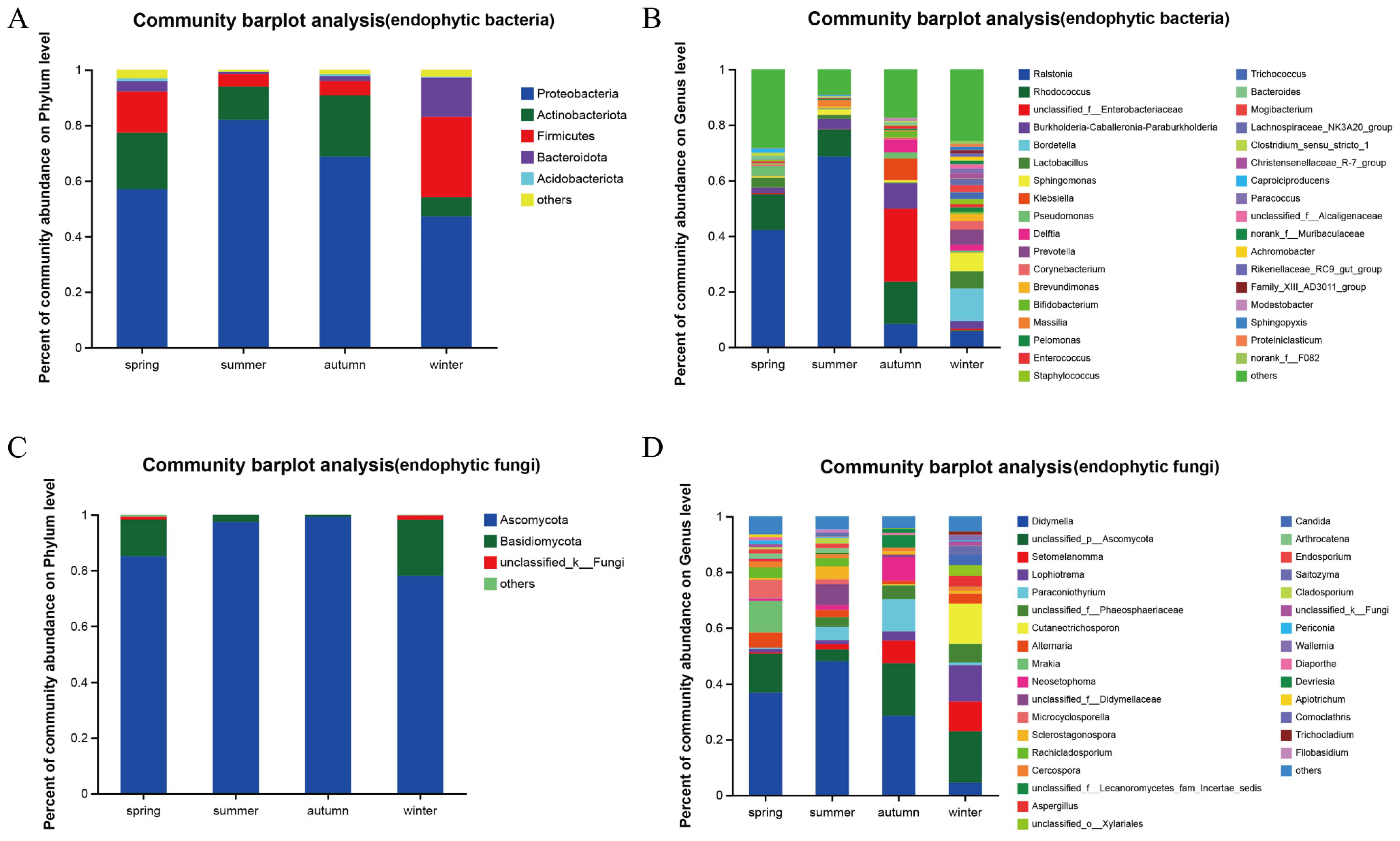
For this analysis, 2708 endophytic bacterial OTUs were compared with 36 phyla and 897 genera. At the phylum classification level (Figure 2A), Proteobacteria were predominant in the spring, summer, autumn and winter samples (56.98%, 81.95%, 68.75%, 47.22%); Actinobacteriota were subdominant in the spring, summer, and autumn samples (20.33%, 11.93%, 21.97%), the Firmicutes had a secondary dominance in winter and spring (28.75%, 14.78%). The proportion of the communities composed by Bacteroidota in winter is also relatively high (14.21%), and the proportion of Acidobacteriota in the four seasons is no more than 1.07%.
At the generic level, the spring samples contained 481 bacterial genera (Figure 2B) and Ralstonia (42.11%), Rhodococcus (12.86%), and Lactobacillus (3.60%) had a relative abundance greater than 3% and the relative abundance for the other genera was greater than 28.34%. The summer sample had 315 genera, including Ralstonia (68.60%), Rhodococcus (9.72%), Burkholderia-Caballeronia-Paraburkholderia groups (3.60%), and there were three genera with relative abundance greater than 3% and 9.14% for the other genera. The autumn samples contained 450 genera, with Unclassified_f_Enterobacteriaceae group (26.25%), Rhodococcus (15.20%) and Burkholderia-Caballeronia-Paraburkholderia group (9.18%), there were six genera with relative abundance greater than 3% and 17.14% for the other genera. The relative abundance of the 562 genera in the winter sample was greater than 3% for Bordetella (11.85%), Sphingomonas (6.65%), and Lactobacillus (6.15%) genera and 25.89% for the other genera. The total number of genera in the spring, summer, autumn and winter samples was 157, accounting for 17.50% of all species of endophytic bacteria and 90.21% of the total abundance of the community.
A total of 588 endophytic fungal OTUs were compared to eight phyla and 251 genera. At the phylum level (Figure 2C), Ascomycota was a major dominant phylum in stem tissues during spring, summer, autumn, and winter (85.14%, 97.46%, 99.26%, and 77.99%) and its relative abundance was higher in summer and autumn samples than in spring and winter samples. Basidiomycota was more abundant in winter and spring (20.20% and 13.09%) than in summer and autumn (2.50% and 0.73%). Unclassified_k_Fungi and unknown fungi were found in small amounts in spring and winter samples (1.07% and 0.70%, 1.58% and 0.22%, respectively), whereas no unknown fungi were found in summer samples.
At the genus level (Figure 2D), the spring, summer, autumn and winter samples had 84, 133, 83, and 140 genera, respectively. In the spring samples, six genera, including Didymella (36.79%), Unclassified_p_Ascomycota (14.04%), and Mrakia (11.43%), had a relative abundance greater than 3%, whereas that of the others was 6.44%. In the summer samples, seven genera, including Didymella (48.07%), Unclassified_f_Didymellaceae (14.04%), and Paraconiothyrium (4.86%), had a relative abundance greater than 3%, whereas that of the others was 4.71%. In the autumn samples, seven genera, including Didymella (28.50%), Unclassified_p_Ascomycota (18.86%), and Paraconiothyrium (11.58%), had a relative abundance greater than 3%, whereas that of the others was 4.19%. In the winter samples, ten genera, including Unclassified_p_Ascomycota (18.44%), Cutaneotrichosporon (14.45%), and Lophiotrema (13.10%), had a relative abundance greater than 3%, whereas that of the others was 5.43%. The spring, summer, autumn, and winter samples had 32 bacterial genera in common, accounting for 12.75% of all species of endophytic bacteria and 86.54% of the total abundance of the flora.
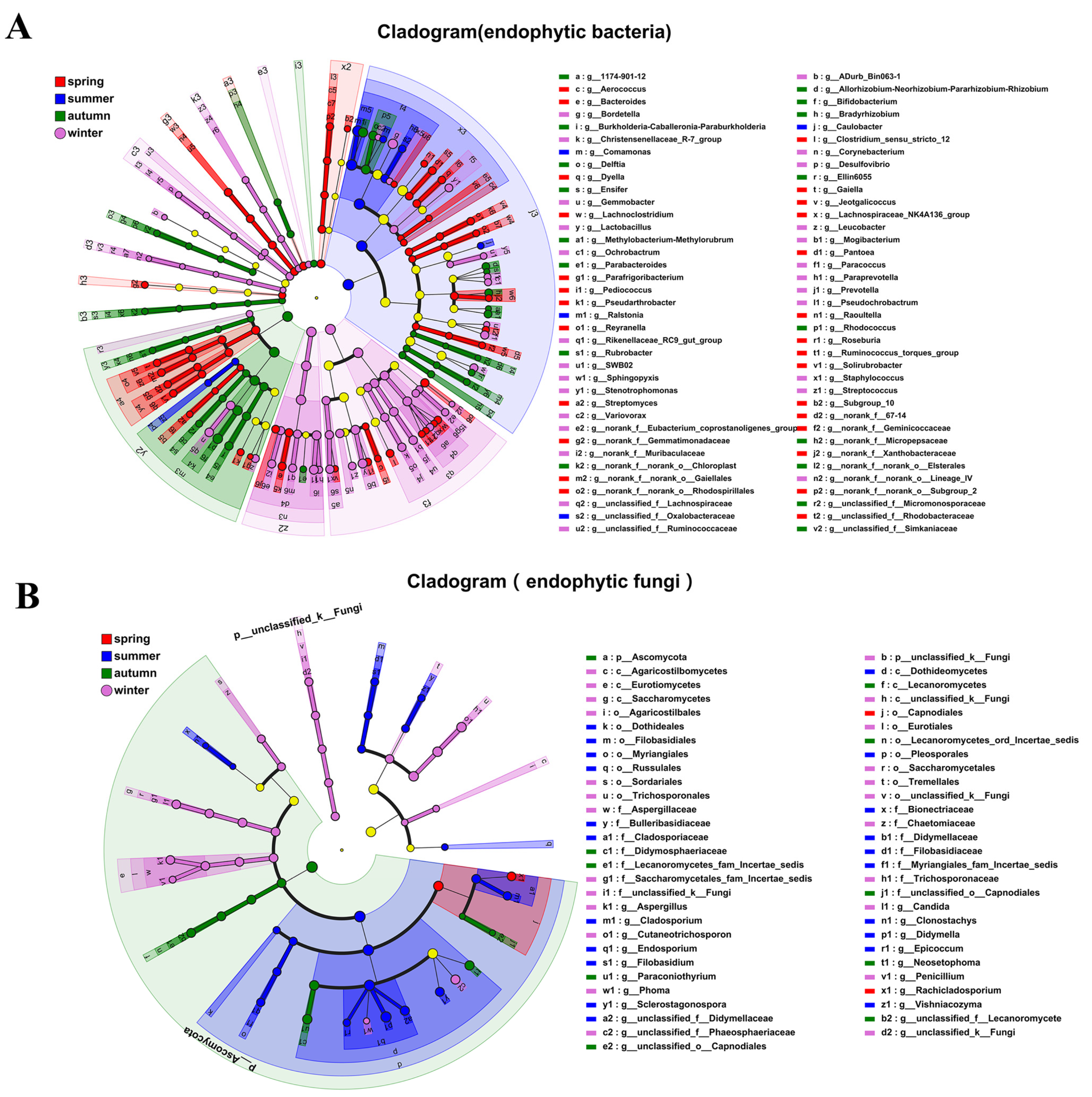
LEfSe multilevel-species hierarchical analysis showed that at the phylum level of endophytic bacteria Chloroflexi, Acidobacteriota, and Fusobacteriota were relatively dominant in spring; Proteobacteria were relatively dominant in summer; Actinobacteriota and Nitrospirota were relatively dominant in autumn; and Firmicutes, Bacteroidota, and Spirochaetota were relatively dominant in winter (Figure 3A). At the phylum level of endophytic fungi, there were no dominant phyla in spring or summer, Ascomycota was relatively dominant in autumn, and Basidiomycota and Unclassified_k_Fungi were relatively dominant in winter. At the genus level, 13, 3, 7, and 12 genera were relatively dominant in spring, summer, autumn and winter, respectively. Among the endophytic fungi, 1, 9, 3, and 5 genera were relatively dominant in spring, summer, autumn, and winter, respectively (Figure 3B).
LDA showed that 69 genera of endophytic bacteria, including Bacteroides, Ralstonia, Rhodococcus, and Prevotella, significantly affected the differences between the seasons. There were 26, 5, 16, and 22 differentially abundant genera in the spring, summer, autumn, and winter samples, respectively. These bacterial genera perform various functions in plants. For example, Frigoribacterium [30] and Pseudochrobactrum [31] have cold-tolerant functions, Variovorax [32] has growth-promoting functions, Streptomyces [33] has disease-resistance functions, and Desulfovibrio [34] and Stenotrophomonas [35] are able to degrade heavy metals and pesticide pollution (Figure 4A,B). Among the endophytic fungi, 19 genera such as Rachicladosporium, Didymella, Paraconiothyrium, and Candida had significant influences on the differences between different seasons. These fungi have different functions in plants. For example, Cladosporium, [36] Clonostachys, and Paraconiothyrium have antibacterial activity [37] (Figure 4C,D).

3.3. Effects of Environmental Factors on the Community Structure of Endophytic Bacteria in Actinidia arguta

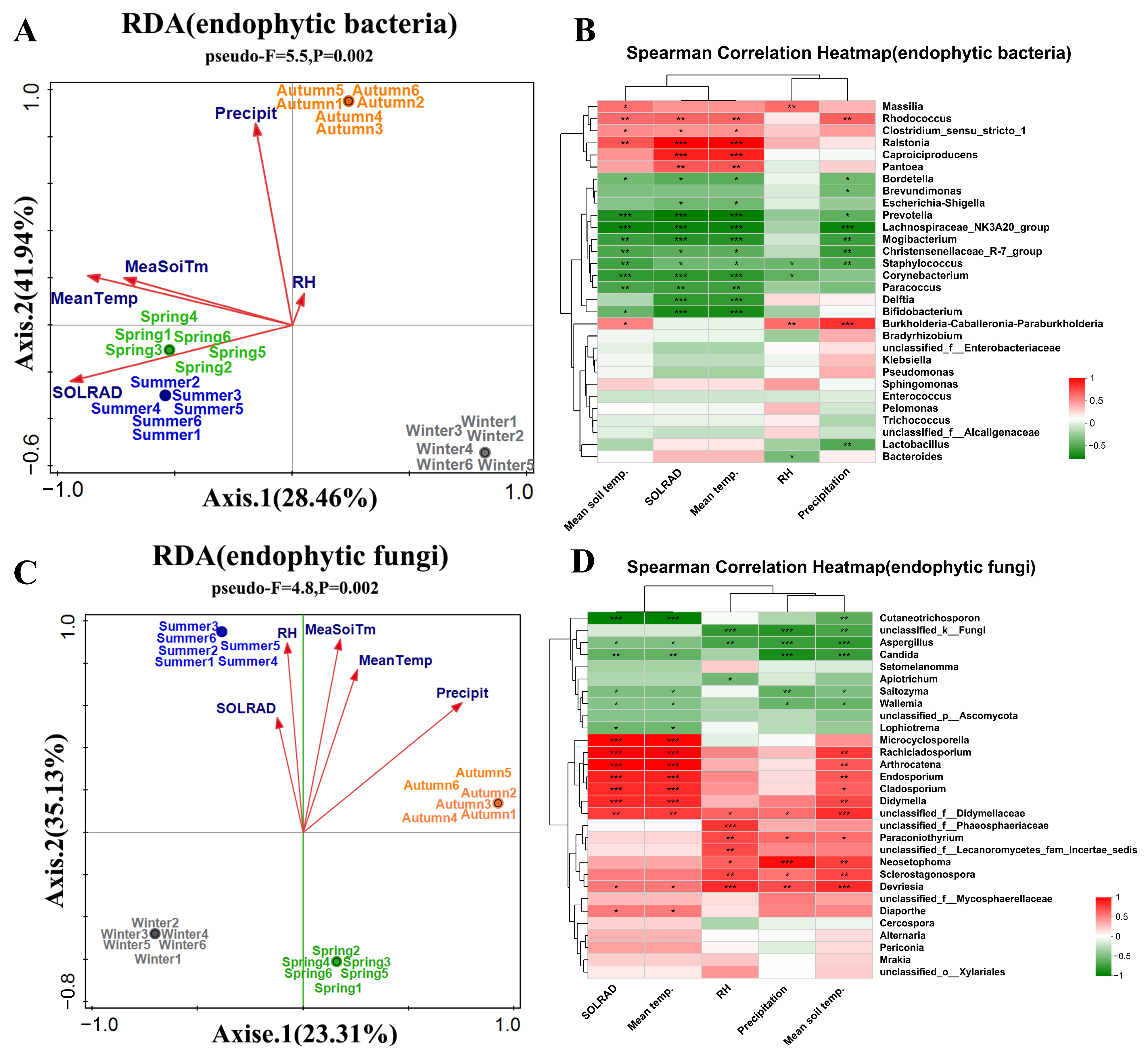
For endophytic bacteria, environmental factors exhibited a significant positive correlation with the community structure during spring, summer, and autumn, whereas a negative correlation was observed in winter (Figure 5A). The principal influencing factors included SOLRAD, Mean Temp, and Precipit. Specifically, MeaSoiTm and Mean Temp significantly and positively correlated with the abundance of eight bacterial genera, including Ralstonia, Rhodococcus, and Massilia, while precipitation markedly influenced the abundance of Rhodococcus and Burkholderia-Caballeronia-Paraburkholderia (Figure 5B).
Regarding endophytic fungi, environmental factors showed a positive correlation with the fungal community structure in summer and autumn, but this correlation was reversed in spring and winter (Figure 5C). The key influencing factors comprised RH, MeaSoiTm, Mean Temp, and Precipit. Genera such as Microcyclosporella, Rachicladosporium, and Arthrocatena were significantly positively correlated with mean temperature. Some genera, including Neosetophoma, Sclerostagonospora, and Devriesia were significantly influenced by RH and Precipit. Endosporium, Cladosporium, Didymella, among others, were significantly affected by MeaSoiTm (Figure 5D). These findings suggest that environmental factors such as MeaSoiTm, Mean Temp, Precipit, and SOLRAD significantly impact the community structure of endophytic bacteria and fungi across different seasons, with certain genera displaying clear positive correlations with specific environmental conditions.

3.4. Analysis of Functional Composition and Inter-Group Differences of Endophytic Bacteria in Actinidia arguta

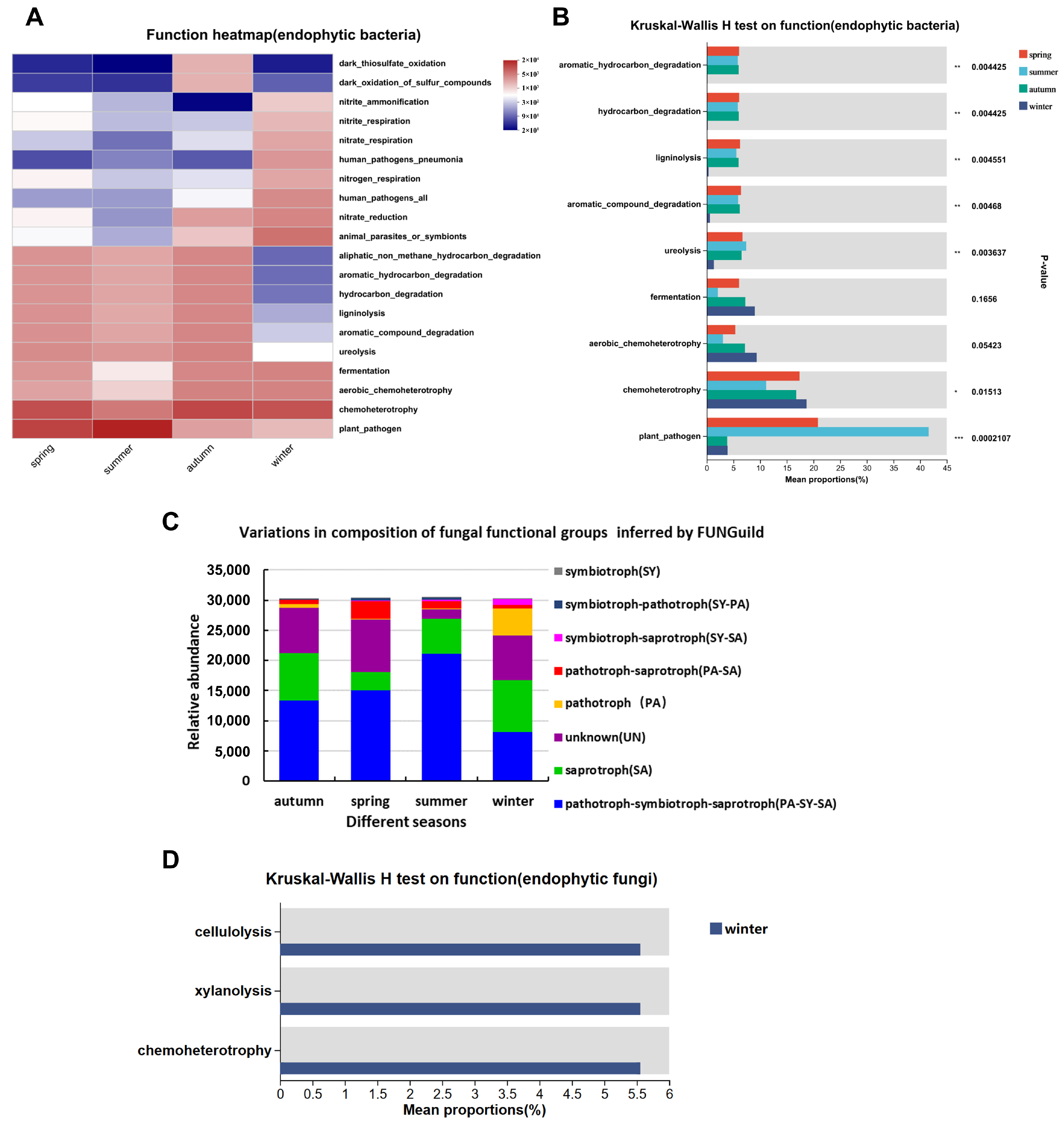
Utilizing FAPROTAX and FUNGuild, this study predicts the functional capabilities of endophytes in Actinidia arguta, exploring the potential interactions between microbial communities and the host plant. Functional analysis using the FAPROTAX database for the top 20 abundant endophytic bacterial genera revealed chemoheterotrophy, aerobic chemoheterotrophy, fermentation, and ureolysis as the most abundant functions. The functional similarity between spring and summer contrasted with that between autumn and winter, particularly for functions like aromatic-hydrocarbon degradation and ligninolysis, which were significantly lower in winter (p < 0.01) (Figure 6A). Conversely, fermentation and aerobic chemoheterotrophy were higher in winter but with no significant difference (p = 0.1656, p = 0.0542). Chemoheterotrophy was significantly higher in winter (p < 0.05), while plant pathogens were more abundant in summer than in spring, significantly so compared to autumn and winter (p < 0.01) (Figure 6B).
FUNGuild analysis showed significant differences in the nutritional patterns of endophytic fungi in different seasons, with PA-SY-SA (44.26%) as the main factor in spring (Figure 6C), followed by SA (25.96%) and UN (24.49%). In summer, PA-SY-SA (49.60%) was dominant, followed by UN (28.55%) and SA (10.19%). In autumn, PA-SY-SA (69.63%) was dominant, followed by SA (18.76%) and UN (5.40%). In winter, SA (28.44%) was dominant, followed by PA-SY-SA (27.07%) and UN (24.37%). The results of the survey of the common functions in the group are shown in Figure 6D, and there was no difference in the endogenous fungal function of the spring, summer, and autumn samples, whereas the functions of cellulolysis, xylanolysis, and chemoheterotrophy in winter were significantly different from the other seasons.
Analysis of the top 15 most abundant endophytic bacterial genera in Actinidia arguta (Table 5) revealed 10 genera belonging to Proteobacteria and 2 to Actinobacteria, with 1 each from Firmicutes, Bacteroidetes, and Acidobacteria. Ralstonia, dominant in spring and summer, saw a gradual increase in abundance, peaking in summer and declining towards winter, with a winter abundance of only 5.71%, indicating its importance in the microecosystem of Actinidia arguta. Rhodococcus was the second most dominant genus during spring and summer, displaying sensitivity to winter’s low temperatures and contributing to the ecological balance in warmer seasons. The Unclassified_f_Enterobacteriaceae genus dominated in autumn, but was suppressed in other seasons and sensitive to cold. Burkholderia-Caballeronia-Paraburkholderia detection was distributed across seasons, peaking in autumn. Bordetella detection was exclusive to winter, suggesting a specific role during the cold. Sphingomonas and Lactobacillus also showed higher winter abundance. Klebsiella, present only in spring and autumn, is considered cold-sensitive and suppressed by dominant genera in other seasons, aiding plant growth when nutrients are scarcer in autumn. Delftia, found only in autumn and winter, may help in stress resistance, while Prevotella, only present in winter, is cold-tolerant and possibly active during plant dormancy. Corynebacterium, absent in summer but abundant in winter, may play a role in osmotic stress resistance. Massilia, present in summer and autumn, is likely cold-sensitive.
Analysis of the ten most abundant endophytic fungal genera in Actinidia arguta (Table 6) shows that eight genera are from the phylum Ascomycota and two are from Basidiomycota. Didymella emerges as the predominant genus in spring and summer, maintaining the ecological balance of Actinidia arguta with growth-promoting functions. The unclassified_p_Ascomycota genus, dominant in winter and the second most prevalent in spring and autumn, is suppressed by Didymella in summer. Lophiotrema and Setomelanomma, increasing from spring to winter, may enhance plant stress resistance. Paraconiothyrium, detected from summer to winter with its highest content in autumn, is suggested to be cold-sensitive. Cutaneotrichosporon, found only in winter, is a cold-tolerant genus secreting lipids. Alternaria shows a rise and fall from winter to autumn, with its highest abundance in spring, indicating involvement in carbohydrate metabolism in winter and nitrogen metabolism in spring. Mrakia, present only in spring as the secondary dominant strain, likely aids the plant in resisting low temperatures through the production of cold-active enzymes and secondary metabolites.

4. Discussion

The Illumina MiSeq technology allows not only comprehensive and objective ascertainment of the relative composition of microbial communities in target environments but also aids in the study of the diversity of symbiotic microbial ecological functions [26]. This technique has been widely applied to the study of gut microbiome diversity [96], soil microbial diversity [97], and space microbiology [98], among other study areas. To date, research utilizing this technology on the endophytes of the Actinidia genus has primarily focused on analyzing the internal microbial community compositions of various tissues like fruits, pollen, and the sap of different kiwifruit varieties [99]. However, the study of endophytic bacteria within Actinidia arguta using this technology remains unexplored.
This study employed Illumina MiSeq sequencing technology to analyze, for the first time, the community structure and species abundance of endophytic bacteria and fungi in Actinidia arguta. The sequencing coverage exceeded 99.8%, showcasing the richness and diversity of the endophytic microbial community within Actinidia arguta. The endophytic bacteria were identified across 36 phyla and 897 genera, with the dominant phyla being Proteobacteria, Actinobacteriota, Firmicutes, and Bacteroidota. Endophytic fungi were identified in eight phyla and 251 genera, predominantly within Ascomycota and Basidiomycota. Cho et al. found similar dominant bacterial phyla in the overflowing sap of different kiwifruit varieties, aligning with our findings [100]. Kim et al. identified the dominant bacterial phyla in the sap of the CV kiwifruit variety [99]. Deliwoong reported the dominant bacterial phyla as Proteobacteria, Actinobacteria and Firmicutes, and the dominant fungal phyla as Ascomycota, Mortierellomycota, and Basidiomycota, with one extra phylum compared to our study, possibly due to environmental microbial differences [101]. To date, Actinidia arguta has represented the species with the richest microbial community within the Actinidia genus. The study of the overflowing sap in three kiwifruit varieties, including Hayward, identified endophytic bacteria within only 12 phyla and 150 genera. This conclusion lays a foundation for the further exploration of endophytic microbial resources in Actinidia arguta [100].
In natural ecosystems, plants coexist in a state of mutual dependence with microorganisms [102]. The dynamics of plant endophytic microbial communities are influenced by intrinsic factors such as host species [103], genotype [104], organ [105], developmental stage [106], and nutritional status [105], as well as by external environmental factors, including soil type [107], fertilization practices [101], and seasonal changes [108]. This study focused on the impacts of seasonal variations on the structure of endophytic communities. Analysis of alpha (p < 0.01) and beta (p < 0.01) diversity revealed significant differences in the structure and diversity of endophytic communities in Actinidia arguta across different seasons, underscoring seasonal change as a key determinant [109].
In the realm of studies on endophytic microbial diversity, most researchers posit that the diversity of endophytes during winter tends to decrease due to factors such as low temperatures and reduced solar radiation [110]. However, the diversity of both endophytic bacteria and endophytic fungi in Actinidia arguta is paradoxically highest during winter. Genera enriched specifically during the winter season include Bordetella, Prevotella, and Cutaneotrichosporon. Studies have shown that dophytes from Eucommia ulmoides bark [17] and Camellia yuhsienensis Hu roots [111] also displayed higher diversities in winter compared to other seasons. We hypothesize that because Actinidia arguta is highly cold-resistant, the endophytic microbes have gradually adapted to the cold winter conditions during their co-evolution with this plant. Furthermore, these endophytes likely play a significant role in assisting Actinidia arguta in coping with abiotic stress factors.
In terms of community structure, the differences between seasons are more pronounced [112]. A series of studies indicates that changes in climate factors such as solar radiation, temperature, rainfall, and humidity due to seasonal variations are crucial influencers of microbial communities [113]. Our research further substantiates this point; we have observed distinct differences in the community structure of endophytic microbes within Actinidia arguta across different seasons. These differences are influenced to varying degrees by solar radiation, temperature, soil temperature, rainfall, and air humidity. Moreover, during winter, the endophytic microbial community shows a negative correlation with these environmental factors. Analysis of the top thirty most abundant endophytic bacteria and fungi indicated that 66.67% of bacterial and 32% of fungal dominant genera are significantly influenced by environmental factors, such as temperature affecting genera like Ralstonia, Rhodococcus, and Massilia, while precipitation significantly impacts genera like Neosetophoma, Sclerostagonospora, and Devriesia. This further corroborates our previous findings that endophytic bacterial richness and diversity are greater during winter when solar radiation is weaker and temperatures are lower, demonstrating the strong environmental stress tolerance of endophytes in Actinidia arguta.
Endophytes maintain their growth by absorbing nutrients from plants, yet they also perform various functions that contribute to plant growth [4,5]. Utilizing FAPROTAX and FUNGuild to predict the functions of endophytes, it was found that the endophytic bacteria of Actinidia arguta primarily engage in functions such as chemoheterotrophy, oxidation, nitrate reduction, respiration, denitrification, and degradation, with significant seasonal variations. The nutritional modes of endophytic fungi in spring, summer, and autumn are mainly PA-SY-SA, while in winter, SA prevails. There are significant functional differences between the endophytic fungi of spring, summer, and autumn compared to winter, with winter exhibiting pronounced abilities in cellulolysis, xylanolysis, and chemoheterotrophy.
The primary functions of endophytes are directly related to the abundance of dominant genera across different seasons [5]. Ralstonia and Rhodococcus are the primary and secondary dominant genera in spring and summer, respectively, shaping the microbial structure of Actinidia arguta during these seasons [40] and potentially involved in biodegradation [41], bioremediation, and biotransformation [7]. The genus Unclassified_f_Enterobacteriaceae, dominant in autumn, is suppressed by Ralstonia and Rhodococcus and is sensitive to low temperatures. This genus is presumed to facilitate plant nutrient recirculation through chemoheterotrophy, ligninolysis, and fermentation, thus preparing the plant for winter [44]. Bordetella, found exclusively in winter and dominant during this season, is hypothesized to assist Actinidia arguta in coping with low temperatures by expressing antifreeze proteins [50]. Klebsiella, present in spring and autumn with a peak abundance of 7.72% in autumn, is considered cold-sensitive and is suppressed by dominant genera in other seasons, while aiding plant growth and enhancing disease resistance during the nutrient-scarce autumn [9]. Prevotella, exclusive to winter, is speculated to be cold-tolerant and active during plant dormancy, is severely suppressed by dominant genera in other seasons, and is thought to aid in plant adaptation to harsh winter climates through the degradation of proteins, sugars, and lignin [63,64]. Corynebacterium, absent in summer but abundant in winter, is believed to be suppressed by other genera in summer and helps the plant to withstand freezing stress in winter through osmotic stress tolerance [67].
Didymella is the dominant endophytic fungal genus in Actinidia arguta during spring, summer, and autumn, with its highest abundance in summer, suggesting that its primary role lies in promoting plant growth and aiding disease resistance [81]. The genus Unclassified_p_Ascomycota, prevalent in winter and the second most dominant genus in spring and autumn, exhibits low summer abundance, potentially due to suppression by Didymella, and is hypothesized to enhance plant stress resistance through the secretion of active secondary metabolites [83]. Cutaneotrichosporon, present only in winter as the second most dominant genus, is analyzed to be cold-tolerant, possibly secreting lipids to alter cellular fluid composition and enhance plant frost resistance. Mrakia, exclusive to spring and the third most dominant strain, is presumed to play a significant role in assisting the plant to withstand late cold spells during the pre-sprouting phase in spring [12,114].
Analyzing the endophytic microbial community structure and diversity of Actinidia arguta from a seasonal perspective not only enhances our understanding of its microecosystem and potential pathogenic microbes but also provides references for mining functional microbial strains within the plant [115]. This allows for the determination of optimal sampling seasons and the advance verification of culture conditions for targeted microbial genera, thereby increasing the likelihood of successful functional strain isolation [116].

5. Conclusions

Illumina MiSeq sequencing revealed a rich diversity of endophytic bacteria and fungi within Actinidia arguta, with OTUs belonging to 36 phyla and 897 genera and 8 phyla and 251 genera, respectively. Notable seasonal shifts were observed: Proteobacteria and Actinobacteriota dominated from spring to autumn, whereas Proteobacteria, Firmicutes, and Bacteroidota were prevalent in winter. In addition, season-specific genera such as Mrakia were identified in spring and Bordetella and Prevotella in winter. Temperature and rainfall significantly influenced microbial community structure. The functional roles of endophytes also varied with the seasons, particularly in winter, where endomicrobes demonstrated unique functions in cellulolysis and ureolysis, contributing to the growth and environmental stress resilience of Actinidia arguta.

Author Contributions

Conceptualization, J.A.; Methodology, W.L.; Software, B.Z. (Baoxiang Zhang); Validation, H.Q. and J.A.; Formal analysis, Y.W.; Investigation, S.F.; Data curation, Y.Y.; Writing—Original Draft Preparation, Y.L. (Yingxue Liu); Writing—Review & Editing, Y.L. (Yang Li), B.Z. (Boyu Zhai) and J.Z.; Visualization, P.X.; Supervision, C.L.; Project Administration, C.L.; Funding Acquisition, J.A. All authors have read and agreed to the published version of the manuscript.

Funding

This work was funded by the Science and Technology Development Program of Jilin Province: 20210203018SF.

Institutional Review Board Statement

Not applicable.

Informed Consent Statement

Not applicable.

Data Availability Statement

The data presented in this study are available on request from the corresponding author.

Acknowledgments

We thank the National Forest germplasm Resource Bank of Actinidia arguta and Schisandra Chinensis for providing the samples.

Conflicts of Interest

The authors declare no conflicts of interest.

References

  1. Petrini, O. Fungal endophytes of tree leaves. In Microbial Ecology of Leaves; Springer: New York, NY, USA, 1991; pp. 179–197. [Google Scholar]
  2. Gange, A.C.; Eschen, R.; Wearn, J.A.; Thawer, A.; Sutton, B.C. Differential effects of foliar endophytic fungi on insect herbivores attacking a herbaceous plant. Oecologia 2012, 168, 1023–1031. [Google Scholar] [CrossRef] [PubMed]
  3. Carvalho, T.L.; Balsemão-Pires, E.; Saraiva, R.M.; Ferreira, P.C.; Hemerly, A.S. Nitrogen signalling in plant interactions with associative and endophytic diazotrophic bacteria. J. Exp. Bot. 2014, 65, 5631–5642. [Google Scholar] [CrossRef] [PubMed]
  4. Santoyo, G.; Moreno-Hagelsieb, G.; Orozco-Mosqueda Mdel, C.; Glick, B.R. Plant growth-promoting bacterial endophytes. Microbiol. Res. 2016, 183, 92–99. [Google Scholar] [CrossRef] [PubMed]
  5. Singh, M.; Kumar, A.; Singh, R.; Pandey, K.D. Endophytic bacteria: A new source of bioactive compounds. 3 Biotech 2017, 7, 315. [Google Scholar] [CrossRef] [PubMed]
  6. Li, A.; Hu, T.; Luo, H.; Alam, N.U.; Xin, J.; Li, H.; Lin, Y.; Huang, J.; Huang, K.; Meng, Y.; et al. A carotenoid- and poly-β-hydroxybutyrate-free mutant strain of sphingomonas elodea atcc 31461 for the commercial production of gellan. mSphere 2019, 4, 10–1128. [Google Scholar] [CrossRef] [PubMed]
  7. Barik, M.; Das, C.P.; Verma, A.K.; Sahoo, S.; Sahoo, N.K. Metabolic profiling of phenol biodegradation by an indigenous rhodococcus pyridinivorans strain pdb9t n-1 isolated from paper pulp wastewater. Int. Biodeterior. Biodegrad. 2021, 158, 105168. [Google Scholar] [CrossRef]
  8. Oukala, N.; Aissat, K.; Pastor, V. Bacterial endophytes: The hidden actor in plant immune responses against biotic stress. Plants 2021, 10, 1012. [Google Scholar] [CrossRef]
  9. Wang, D.; Wang, H.; Li, J.; Zhang, W.; Pan, Y.; Liu, X. Investigating the Role of Endophytic Fungi in Gentiana scabra bge. by Cross-Growth Period Inoculation. Indian J. Microbiol. 2018, 58, 319–325. [Google Scholar] [CrossRef]
  10. Adhikari, P.; Pandey, A. Bioprospecting plant growth promoting endophytic bacteria isolated from Himalayan yew (Taxus wallichiana Zucc.). Microbiol. Res. 2020, 239, 126536. [Google Scholar] [CrossRef]
  11. Li, D.; Bodjrenou, D.M.; Zhang, S.; Wang, B.; Pan, H.; Yeh, K.W.; Lai, Z.; Cheng, C. The Endophytic Fungus Piriformospora indica Reprograms Banana to Cold Resistance. Int. J. Mol. Sci. 2021, 22, 4973. [Google Scholar] [CrossRef]
  12. Jaiswal, S.; Ojha, A.; Mishra, S.K. Assessment of Plant Growth-Promoting Parameters of Endophytes Isolated from Calotropis procera and Their Performance Under Irrigated and Non-irrigated Conditions. Curr. Microbiol. 2023, 81, 49. [Google Scholar] [CrossRef]
  13. Zhang, D.; Sun, W.; Xu, W.; Ji, C.; Zhou, Y.; Sun, J.; Tian, Y.; Li, Y.; Zhao, F.; Tian, Y. Antimicrobial and cytotoxic activity of endophytic fungi from lagopsis supina. J. Microbiol. Biotechnol. 2023, 33, 543–551. [Google Scholar] [CrossRef] [PubMed]
  14. Yao, X.; Chen, Z.; Wei, X.; Chen, S.; Nan, Z. A toxic grass achnatherum inebrians serves as a diversity refuge for the soil fungal community in rangelands of northern china. Plant Soil 2020, 448, 425–438. [Google Scholar] [CrossRef]
  15. Xu, G.; Liu, Y.; Cao, P.; Liu, X. Diversity of endophytic bacteria of Oxytropis glacialis in different ecological environments on the Tibetan Plateau. Plateau Sci. Res. 2020, 4, 20–29. (In Chinese) [Google Scholar] [CrossRef]
  16. Zhu, J.; Sun, X.; Tang, Q.Y.; Zhang, Z.D. Seasonal Dynamics and Persistency of Endophyte Communities in Kalidium schrenkianum Shifts Under Radiation Stress. Front. Microbiol. 2021, 12, 778327. [Google Scholar] [CrossRef] [PubMed]
  17. Liang, X.; Wan, D.; Tan, L.; Liu, H. Dynamic changes of endophytic bacteria in the bark and leaves of medicinal plant Eucommia ulmoides in different seasons. Microbiol. Res. 2023, 280, 127567. [Google Scholar] [CrossRef] [PubMed]
  18. Shen, Z.; Liu, X.; Yang, J.; Wang, Y.; Yao, K.; Huo, Q.; Fu, Y.; Wei, Y.; Guo, B. The temporal and spatial endophytic fungal community of Huperzia serrata: Diversity and relevance to huperzine A production by the host. BMC Microbiol. 2022, 22, 281. [Google Scholar] [CrossRef]
  19. Ou, T.; Xu, W.F.; Wang, F.; Strobel, G.; Zhou, Z.Y.; Xiang, Z.H.; Liu, J.; Xie, J. A Microbiome Study Reveals Seasonal Variation in Endophytic Bacteria Among different Mulberry Cultivars. Comput. Struct. Biotechnol. J. 2019, 17, 1091–1100. [Google Scholar] [CrossRef]
  20. Niu, Q.; Shen, J.; Liu, Y.; Nie, C.; Scripcenco, N.V.; Liu, D. Research progress on main active constituents and pharmacological activities of Actinidia arguta. Sci. Technol. Food Ind. 2019, 40, 333–338+344. (In Chinese) [Google Scholar] [CrossRef]
  21. Gong, D.S.; Sharma, K.; Kang, K.W.; Kim, D.W.; Oak, M.H. Endothelium-dependent relaxation effects of actinidia arguta extracts in coronary artery: Involvement of enos/akt pathway. J. Nanosci. Nanotechnol. 2020, 20, 5381–5384. [Google Scholar] [CrossRef]
  22. Sun, S.; Qi, X.; Wang, R.; Lin, M.; Fang, J. Evaluation of freezing tolerance inactinidiagermplasm based on relative electrolyte leakage. Hortic. Environ. Biotechnol. 2020, 61, 755–765. [Google Scholar] [CrossRef]
  23. Wen, X.; Qin, H.; Ai, J.; Wang, Y.; Han, X.; Li, C. Establishment and evaluation of resistance identification method for Pseudomonass yringae pv. actinidiae disease in Actinidia arguta germplasm resources. Plant Prot. 2021, 47, 193–199. (In Chinese) [Google Scholar] [CrossRef]
  24. Wang, R.; Zhang, Q.; Ju, M.; Yan, S.; Zhang, Q.; Gu, P. The endophytic fungi diversity, community structure, and ecological function prediction of sophora alopecuroides in ningxia, china. Microorganisms 2022, 10, 2099. [Google Scholar] [CrossRef] [PubMed]
  25. Wijayawardene, N.N.; Bahram, M.; Sánchez-Castro, I.; Dai, D.Q.; Ariyawansa, K.; Jayalal, U.; Suwannarach, N. Current insight into culture-dependent and culture-independent methods in discovering ascomycetous taxa. J. Fungi 2021, 7, 703. [Google Scholar] [CrossRef] [PubMed]
  26. Ren, F.; Dong, W.; Yan, D.H. Organs, cultivars, soil, and fruit properties affect structure of endophytic mycobiota of pinggu peach trees. Microorganisms 2019, 7, 322. [Google Scholar] [CrossRef] [PubMed]
  27. Xu, Y. Diversity analysis of cultured endophytic bacteria in Codonopsis pilosula and effects on promoting host growth. LanZhou Univ. Technol. 2019. (In Chinese) [Google Scholar] [CrossRef]
  28. Liu, C.; Zhao, D.; Ma, W.; Guo, Y.; Wang, A.; Wang, Q.; Lee, D.J. Denitrifying sulfide removal process on high-salinity wastewaters in the presence of Halomonas sp. Appl. Microbiol. Biotechnol. 2016, 100, 1421–1426. [Google Scholar] [CrossRef]
  29. Segata, N.; Izard, J.; Waldron, L.; Gevers, D.; Miropolsky, L.; Garrett, W.S.; Huttenhower, C. Metagenomic biomarker discovery and explanation. Genome Biol. 2011, 12, R60. [Google Scholar] [CrossRef]
  30. Wang, H.F.; Zhang, Y.G.; Chen, J.Y.; Guo, J.W.; Li, L.; Hozzein, W.N.; Zhang, Y.M.; Wadaan, M.A.M.; Li, W.J. Frigoribacterium endophyticum sp. Nov., an endophytic actinobacterium isolated from the root of anabasis elatior (c. A. Mey.) schischk. Int. J. Syst. Evol. Microbiol. 2015, 65, 1207–1212. [Google Scholar] [CrossRef]
  31. Liu, H.; Qin, Y.; Fu, Y. A Low Temperature Tolerant Bacterium Pseudochrobactrum A4 and Its Application in Crop Disease Control. Patent CN106544289B, 27 August 2019. (In Chinese). [Google Scholar]
  32. Finkel, O.M.; Salas-González, I. A single bacterial genus maintains root growth in a complex microbiome. Nature 2020, 587, 103–108. [Google Scholar] [CrossRef]
  33. Viaene, T.; Langendries, S.; Beirinckx, S.; Maes, M.; Goormachtig, S. Streptomyces as a plant’s best friend? FEMS Microbiol. Ecol. 2016, 92, fiw119. [Google Scholar] [CrossRef]
  34. Cabrera, G.; Pérez, R.; Gómez, J.M.; Abalos, A.; Cantero, D. Toxic effects of dissolved heavy metals on Desulfovibrio vulgaris and Desulfovibrio sp. Strains. J. Hazard. Mater. 2006, 135, 40–46. [Google Scholar] [CrossRef] [PubMed]
  35. Deng, S.; Chen, Y.; Wang, D.; Shi, T.; Wu, X.; Ma, X.; Li, X.; Hua, R.; Tang, X.; Li, Q.X. Rapid biodegradation of organophosphorus pesticides by Stenotrophomonas sp. G1. J. Hazard. Mater. 2015, 297, 17–24. [Google Scholar] [CrossRef] [PubMed]
  36. Salvatore, M.M.; Andolfi, A.; Nicoletti, R. The genus Cladosporium: A rich source of diverse and bioactive natural compounds. Molecules 2021, 26, 3959. [Google Scholar] [CrossRef] [PubMed]
  37. Wang, J.; Shao, S.; Liu, C.; Song, Z.; Liu, S.; Wu, S. The genus Paraconiothyrium: Species concepts, biological functions, and secondary metabolites. Crit. Rev. Microbiol. 2021, 47, 781–810. [Google Scholar] [CrossRef] [PubMed]
  38. Vailleau, F.; Genin, S. Ralstonia solanacearum: An arsenal of virulence strategies and prospects for resistance. Annu. Rev. Phytopathol. 2023, 61, 25–47. [Google Scholar] [CrossRef] [PubMed]
  39. Truchon, A.N.; Dalsing, B.L.; Khokhani, D.; MacIntyre, A.; McDonald, B.R.; Ailloud, F.; Klassen, J. Plant-pathogenic Ralstonia phylotypes evolved divergent respiratory strategies and behaviors to thrive in xylem. Mbio 2023, 14, e0318822. [Google Scholar] [CrossRef]
  40. De Oliveira, M.; Atalla, A.A.; Farias Frihling, B.E.; Cavalheri, P.S.; Migliolo, L.; Magalhaes Filho, F.J.C. Ibuprofen and caffeine removal in vertical flow and free-floating macrophyte constructed wetlands with heliconia rostrata and Eichornia crassipes. Chem. Eng. J. 2019, 373, 458–467. [Google Scholar] [CrossRef]
  41. Qiu, Z.; Wang, R.; Zhang, Y.; Wu, Q.; Xie, B.; Yang, J.; Chen, J.; Sun, Z. Research progress of Rhodococcus and its biodegradation. Food Sci. 2016, 37, 254–258. (In Chinese) [Google Scholar]
  42. Bell, K.S.; Philp, J.C.; Aw, D.W.; Christofi, N. The genus Rhodococcus. J. Appl. Microbiol. 1998, 85, 195–210. [Google Scholar] [CrossRef]
  43. Nazari, M.T.; Simon, V.; Machado, B.S.; Crestani, L.; Marchezi, G.; Concolato, G.; Ferrari, V.; Colla, L.M.; Piccin, J.S. Rhodococcus: A promising genus of actinomycetes for the bioremediation of organic and inorganic contaminants. J. Environ. Manag. 2022, 323, 116220. [Google Scholar] [CrossRef] [PubMed]
  44. Tapia, D.; Sanchez-Villamil, J.I.; Torres, A.G. Emerging role of biologics for the treatment of melioidosis and glanders. Expert Opin. Biol. Ther. 2019, 19, 1319–1332. [Google Scholar] [CrossRef] [PubMed]
  45. Li, Y.; Guo, P.; Sun, J. Isolation, identification, phylogeny and growth promoting characteristics of endophytic diazotrophs from tuber and root crops. Sci. Agric. Sin. 2017, 50, 104–122. (In Chinese) [Google Scholar]
  46. Kim, B.; Westerhuis, J.A.; Smilde, A.K.; Floková, K.; Suleiman, A.K.A.; Kuramae, E.E. Effect of strigolactones on recruitment of the rice root-associated microbiome. FEMS Microbiol. Ecol. 2022, 98, fiac010. [Google Scholar] [CrossRef] [PubMed]
  47. Luo, L.; Wang, L.; Deng, L.; Mei, X.; Liu, Y.; Huang, H.; Du, F.; Zhu, S.; Yang, M. Enrichment of Burkholderia in the rhizosphere by autotoxic ginsenosides to alleviate negative plant-soil feedback. Microbiol. Spectr. 2021, 9, e0140021. [Google Scholar] [CrossRef] [PubMed]
  48. Fu, L.; Ou, Y.; Shen, Z.; Wang, B.; Li, R.; Shen, Q. Stable microbial community and specific beneficial taxa associated. J. Microbiol. Biotechnol. 2019, 29, 1624–1628. [Google Scholar] [CrossRef]
  49. Gu, Y.; Banerjee, S.; Dini-Andreote, F.; Xu, Y.; Shen, Q.; Jousset, A.; Wei, Z. Small changes in rhizosphere microbiome composition predict disease outcomes earlier than pathogen density variations. ISME J. 2022, 16, 2448–2456. [Google Scholar] [CrossRef]
  50. Hingurao, K.; Nerurkar, A. A novel methylotrophic bacterial consortium for treatment of industrial effluents. Appl. Biochem. Biotechnol. 2018, 185, 691–704. [Google Scholar] [CrossRef]
  51. Tyler, C.A.; Kopit, L.; Doyle, C.; Yu, A.O.; Hugenholtz, J.; Marco, M.L. Polyol production during heterofermentative growth of the plant isolate Lactobacillus florum 2f. J. Appl. Microbiol. 2016, 120, 1336–1345. [Google Scholar] [CrossRef]
  52. Silva, E.; Marques, A.R.; Fialho, A.M.; Granja, A.T.; Sá-Correia, I. Proteins encoded by Sphingomonas elodea atcc 31461 rmla and ugpG genes, involved in gellan gum biosynthesis, exhibit both dtdp- and udp-glucose pyrophosphorylase activities. Appl Environ. Microbiol. 2005, 71, 4703–4712. [Google Scholar] [CrossRef]
  53. Zhang, W.; Chen, Z.; Wu, M.; Shi, Z.; Zhu, F.; Li, G.; Ma, T. Improved production of carotenoid-free welan gum in a genetic-engineered Alcaligenes sp. Atcc31555. Biotechnol. Lett. 2016, 38, 991–997. [Google Scholar] [CrossRef] [PubMed]
  54. Asaf, S.; Numan, M.; Khan, A.L.; Al-Harrasi, A. Sphingomonas: From diversity and genomics to functional role in environmental remediation and plant growth. Crit. Rev. Biotechnol. 2020, 40, 138–152. [Google Scholar] [CrossRef] [PubMed]
  55. Gupta, P.; Kumar, V.; Usmani, Z.; Rani, R.; Chandra, A.; Gupta, V.K. Implications of plant growth promoting Klebsiella sp. Cpsb4 and Enterobacter sp. Cpsb49 in luxuriant growth of tomato plants under chromium stress. Chemosphere 2020, 240, 124944. [Google Scholar] [CrossRef] [PubMed]
  56. Dutta, K.; Shityakov, S.; Das, P.P.; Ghosh, C. Enhanced biodegradation of mixed pahs by mutated naphthalene 1,2-dioxygenase encoded by Pseudomonas putida strain kd6 isolated from petroleum refinery waste. 3 Biotech 2017, 7, 365. [Google Scholar] [CrossRef] [PubMed]
  57. Yarte, M.E.; Gismondi, M.I.; Llorente, B.E.; Larraburu, E.E. Isolation of endophytic bacteria from the medicinal, forestal and ornamental tree Handroanthus impetiginosus. Environ. Technol. 2022, 43, 1129–1139. [Google Scholar] [CrossRef] [PubMed]
  58. Mello, I.S.; Targanski, S.; Pietro-Souza, W.; Frutuoso Stachack, F.F.; Terezo, A.J.; Soares, M.A. Endophytic bacteria stimulate mercury phytoremediation by modulating its bioaccumulation and volatilization. Ecotoxicol. Environ. Saf. 2020, 202, 110818. [Google Scholar] [CrossRef] [PubMed]
  59. Chen, T.; Chen, Z.; Ma, G.H.; Du, B.H.; Shen, B.; Ding, Y.Q.; Xu, K. Diversity and potential application of endophytic bacteria in ginger. Genet. Mol. Res. 2014, 13, 4918–4931. [Google Scholar] [CrossRef] [PubMed]
  60. Liu, Y.; Yao, J.; Ding, S.; Wu, D.; Zhang, P.; Tie, B.; Lou, S. Effects of adding Delftia sp. B9 on the morphological distribution of soil Cd and the uptake and accumulation of Cd in rice. J. Agric. Resour. Environ. 2023, 40, 1339–1348. (In Chinese) [Google Scholar] [CrossRef]
  61. Agafonova, N.V.; Doronina, N.V.; Kaparullina, E.N.; Fedorov, D.N.; Gafarov, A.B.; Sazonova, O.I.; Sokolov, S.L.; Trotsenko, Y.A. A novel delftia plant symbiont capable of autotrophic methylotrophy. Mikrobiologiia 2017, 86, 88–98. (In Russian) [Google Scholar] [CrossRef]
  62. Avgustin, G.; Wallace, R.J.; Flint, H.J. Phenotypic diversity among ruminal isolates of Prevotella ruminicola: Proposal of Prevotella brevis sp. Nov., Prevotella bryantii sp. Nov., and Prevotella albensis sp. Nov. And redefinition of Prevotella ruminicola. Int. J. Syst. Bacteriol. 1997, 47, 284–288. [Google Scholar] [CrossRef]
  63. Zorec, M.; Vodovnik, M.; Marinšek-Logar, R. Potential of selected rumen bacteria for cellulose and hemicellulose degradation. Food Technol. Biotechnol. 2014, 52, 210–221. [Google Scholar]
  64. Arafat, Y.; Ud Din, I.; Tayyab, M.; Jiang, Y.; Chen, T.; Cai, Z.; Zhao, H.; Lin, X.; Lin, W.; Lin, S. Soil sickness in aged tea plantation is associated with a shift in microbial communities as a result of plant polyphenol accumulation in the tea gardens. Front. Plant Sci. 2020, 11, 601. [Google Scholar] [CrossRef] [PubMed]
  65. Ueki, A.; Akasaka, H.; Satoh, A.; Suzuki, D.; Ueki, K. Prevotella paludivivens sp. Nov., a novel strictly anaerobic, gram-negative, hemicellulose-decomposing bacterium isolated from plant residue and rice roots in irrigated rice-field soil. Int. J. Syst. Evol. Microbiol. 2007, 57, 1803–1809. [Google Scholar] [CrossRef] [PubMed]
  66. Bodhankar, S.; Grover, M.; Hemanth, S.; Reddy, G.; Rasul, S.; Yadav, S.K.; Desai, S.; Mallappa, M.; Mandapaka, M.; Srinivasarao, C. Maize seed endophytic bacteria: Dominance of antagonistic, lytic enzyme-producing Bacillus spp. 3 Biotech 2017, 7, 232. [Google Scholar] [CrossRef] [PubMed]
  67. Michalko, J.; Medo, J.; Ferus, P.; Konôpková, J.; Košútová, D.; Hoťka, P.; Barta, M. Changes of endophytic bacterial community in mature leaves of Prunus laurocerasus L. During the seasonal transition from winter dormancy to vegetative growth. Plants 2022, 11, 417. [Google Scholar] [CrossRef] [PubMed]
  68. Merzaeva, O.V.; Shirokikh, I.G. Production of auxins by the endophytic bacteria of winter rye. Appl. Biochem. Microbiol. 2010, 46, 51–57. (In Russian) [Google Scholar] [CrossRef]
  69. Liaqat, F.; Eltem, R. Identification and characterization of endophytic bacteria isolated from in vitro cultures of peach and pear rootstocks. 3 Biotech 2016, 6, 120. [Google Scholar] [CrossRef] [PubMed]
  70. Cheng, D.; Tian, Z.; Feng, L.; Xu, L.; Wang, H. Diversity analysis of the rhizospheric and endophytic bacterial communities of Senecio vulgaris l. (asteraceae) in an invasive range. PeerJ 2019, 6, e6162. [Google Scholar] [CrossRef]
  71. Feng, G.D.; Yang, S.Z.; Li, H.P.; Zhu, H.H. Massilia putida sp. Nov., a dimethyl disulfide-producing bacterium isolated from wolfram mine tailing. Int. J. Syst. Evol. Microbiol. 2016, 66, 50–55. [Google Scholar] [CrossRef]
  72. Kumari, S.; Regar, R.K.; Manickam, N. Improved polycyclic aromatic hydrocarbon degradation in a crude oil by individual and a consortium of bacteria. Bioresour. Technol. 2018, 254, 174–179. [Google Scholar] [CrossRef]
  73. Du, Y.; Yu, X.; Wang, G. Massilia tieshanensis sp. Nov., isolated from mining soil. Int. J. Syst. Evol. Microbiol. 2012, 62, 2356–2362. [Google Scholar] [CrossRef]
  74. Zheng, B.X.; Bi, Q.F.; Hao, X.L.; Zhou, G.W.; Yang, X.R. Massilia phosphatilytica sp. Nov., a phosphate solubilizing bacteria isolated from a long-term fertilized soil. Int. J. Syst. Evol. Microbiol. 2017, 67, 2514–2519. [Google Scholar] [CrossRef] [PubMed]
  75. Faramarzi, M.A.; Fazeli, M.; Yazdi, M.T.; Adrangi, S.; Mohseni, F.A. Optimization of cultural conditions for production of chitinase by a soil isolate of Massilia timonae. Biotechnology 2009, 8, 93–99. [Google Scholar] [CrossRef]
  76. Liu, L.; Wang, S.; Peng, W.; Zhai, C.; Yi, L. Identification, genome sequence and production of polysaccharide hydrolases of Massilii eurypsychrophila 709. In Abstract Books of Eleventh China Symposium on Enzyme Engineering; Enzyme Engineering Committee of China Society of Microbiology: Wuhan, China, 2017. (In Chinese) [Google Scholar]
  77. Dodou, H.V.; de Morais Batista, A.H.; Sales, G.W.P.; de Medeiros, S.C.; Rodrigues, M.L.; Nogueira, P.C.N.; Silveira, E.R.; Nogueira, N.A.P. Violacein antimicrobial activity on Staphylococcus epidermidis and synergistic effect on commercially available antibiotics. J. Appl. Microbiol. 2017, 123, 853–860. [Google Scholar] [CrossRef]
  78. Han, X.; Satoh, Y.; Kuriki, Y.; Seino, T.; Fujita, S.; Suda, T.; Kobayashi, T.; Tajima, K. Polyhydroxyalkanoate production by a novel bacterium Massilia sp. Umi-21 isolated from seaweed, and molecular cloning of its polyhydroxyalkanoate synthase gene. J. Biosci. Bioeng. 2014, 118, 514–519. [Google Scholar] [CrossRef] [PubMed]
  79. Raj, G.; Shadab, M.; Deka, S.; Das, M.; Baruah, J.; Bharali, R.; Talukdar, N.C. Seed interior microbiome of rice genotypes indigenous to three agroecosystems of indo-burma biodiversity hotspot. BMC Genom. 2019, 20, 924. [Google Scholar] [CrossRef] [PubMed]
  80. Keinath, A.P. Survival of Didymella bryoniae in buried watermelon vines in south carolina. Plant Dis. 2002, 86, 32–38. [Google Scholar] [CrossRef] [PubMed]
  81. Kuo, J.; Chang, C.F.; Chi, W.C. Isolation of endophytic fungi with antimicrobial activity from medicinal plant Zanthoxylum simulans hance. Folia Microbiol. 2021, 66, 385–397. [Google Scholar] [CrossRef]
  82. Turbat, A.; Rakk, D.; Vigneshwari, A.; Kocsubé, S.; Thu, H.; Szepesi, Á.; Bakacsy, L.D.; Škrbić, B.; Jigjiddorj, E.A.; Vágvölgyi, C.; et al. Characterization of the plant growth-promoting activities of endophytic fungi isolated from Sophora flavescens. Microorganisms 2020, 8, 683. [Google Scholar] [CrossRef]
  83. Ma, N.; Yin, D.; Liu, Y.; Gao, Z.; Cao, Y.; Chen, T.; Huang, Z.; Jia, Q.; Wang, D. Succession of endophytic fungi and rhizosphere soil fungi and their correlation with secondary metabolites in Fagopyrum dibotrys. Front. Microbiol. 2023, 14, 1220431. [Google Scholar] [CrossRef]
  84. Kosawang, C.; Amby, D.B.; Bussaban, B.; McKinney, L.V.; Xu, J.; Kjær, E.D.; Collinge, D.B.; Nielsen, L.R. Fungal communities associated with species of Fraxinus tolerant to ash dieback, and their potential for biological control. Fungal Biol. 2018, 122, 110–120. [Google Scholar] [CrossRef]
  85. Gakuubi, M.M.; Ching, K.C.; Munusamy, M.; Wibowo, M.; Liang, Z.X.; Kanagasundaram, Y.; Ng, S.B. Enhancing the discovery of bioactive secondary metabolites from fungal endophytes using chemical elicitation and variation of fermentation media. Front. Microbiol. 2022, 13, 898976. [Google Scholar] [CrossRef]
  86. Azevedo, R.P.; Alves, N.M.; Costa, I.A.; Domingues, M.I.S.; Bandória, N.A.; de Figueiredo, U.J.; de Medeiros, F.H.V.; Silva, B.M.; Cardoso, P.G. Endophytic fungi assures tropical forage grass growth by water stress tolerances. Curr. Microbiol. 2021, 78, 4060–4071. [Google Scholar] [CrossRef] [PubMed]
  87. Nakashima, K.I.; Tomida, J.; Hirai, T.; Kawamura, Y.; Inoue, M. Paraconiothins a-j: Sesquiterpenoids from the endophytic fungus Paraconiothyrium brasiliense ecn258. J. Nat. Prod. 2019, 82, 3347–3356. [Google Scholar] [CrossRef] [PubMed]
  88. Zeng, Q.; Cui, H.; Yao, H.; Yuan, T. Five sesquiterpenes from Paraconiothyrium sp. And their anti-inflammatory activity. Chem. Biodivers. 2023, 20, e202300477. [Google Scholar] [CrossRef]
  89. Sathiyaseelan, A.; Saravanakumar, K.; Naveen, K.V.; Han, K.S.; Zhang, X.; Jeong, M.S.; Wang, M.H. Combination of Paraconiothyrium brasiliense fabricated titanium dioxide nanoparticle and antibiotics enhanced antibacterial and antibiofilm properties: A toxicity evaluation. Environ. Res. 2022, 212, 113237. [Google Scholar] [CrossRef] [PubMed]
  90. Shaigani, P.; Fuchs, T.; Graban, P.; Prem, S.; Haack, M.; Masri, M.; Mehlmer, N.; Brueck, T. Mastering targeted genome engineering of gc-rich oleaginous yeast for tailored plant oil alternatives for the food and chemical sector. Microb. Cell Factories 2023, 22, 25. [Google Scholar] [CrossRef] [PubMed]
  91. Tian, L.L.; Ren, H.; Xi, J.M.; Fang, J.; Zhang, J.Z.; Wu, Q.X. Diverse anti-inflammation and anti-cancer polyketides isolated from the endophytic fungi Alternaria sp. Mg1. Fitoterapia 2021, 153, 105000. [Google Scholar] [CrossRef] [PubMed]
  92. Ju, M.; Zhang, Q.; Wang, R.; Yan, S.; Li, Z.; Li, P.; Gu, P. Correlation in endophytic fungi community diversity and bioactive compounds of Sophora alopecuroides. Front. Microbiol. 2022, 13, 955647. [Google Scholar] [CrossRef]
  93. Hatamzadeh, S.; Rahnama, K.; White, J.F.; Oghaz, N.A.; Nasrollahnejad, S.; Hemmati, K. Investigation of some endophytic fungi from five medicinal plants with growth promoting ability on maize (Zea mays l.). J. Appl. Microbiol. 2023, 134, lxac015. [Google Scholar] [CrossRef]
  94. Tsuji, M. Cold-stress responses in the antarctic basidiomycetous yeast Mrakia blollopis. R. Soc. Open Sci. 2016, 3, 160106. [Google Scholar] [CrossRef] [PubMed]
  95. Yuivar, Y.; Alcaino, J.; Cifuentes, V.; Baeza, M. Characterization of gelatinase produced by antarctic Mrakia sp. J. Basic Microbiol. 2019, 59, 846–852. [Google Scholar] [CrossRef]
  96. Cha, T.; Kim, H.H.; Keum, J.; Kwak, M.J.; Park, J.Y.; Hoh, J.K.; Kim, C.R.; Jeon, B.H.; Park, H.K. Gut microbiome profiling of neonates using nanopore minion and illumina miseq sequencing. Front. Microbiol. 2023, 14, 1148466. [Google Scholar] [CrossRef] [PubMed]
  97. Zhang, P.; Guan, P.; Hao, C.; Yang, J.; Xie, Z.; Wu, D. Changes in assembly processes of soil microbial communities in forest-to-cropland conversion in changbai mountains, northeastern china. Sci. Total Environ. 2022, 818, 151738. [Google Scholar] [CrossRef] [PubMed]
  98. Chen, Y.; Wu, B. Current progression: Application of high-throughput sequencing technique in space microbiology. Biomed. Res. Int. 2020, 2020, 4094191. [Google Scholar] [CrossRef] [PubMed]
  99. Kim, M.J.; Do, H.; Cho, G.; Jeong, R.D.; Kwak, Y.S. Comparison of microbial community of rhizosphere and endosphere in kiwifruit. Plant Pathol. J. 2019, 35, 705–711. [Google Scholar] [CrossRef] [PubMed]
  100. Cho, G.; Kim, M.J.; Kwon, Y.; Kwak, Y.S. Comparison of endophytic microbial community in kiwifruit plant cultivars. Plant Pathol. J. 2018, 34, 341–346. [Google Scholar] [CrossRef] [PubMed]
  101. Rodríguez-Blanco, A.; Sicardi, M.; Frioni, L. Plant genotype and nitrogen fertilization effects on abundance and diversity of diazotrophic bacteria associated with maize (Zea mays L.). Biol. Fertil. Soils 2015, 51, 391–402. [Google Scholar] [CrossRef]
  102. Yan, L.; Zhu, J.; Zhao, X.; Shi, J.; Jiang, C.; Shao, D. Beneficial effects of endophytic fungi colonization on plants. Appl. Microbiol. Biotechnol. 2019, 103, 3327–3340. [Google Scholar] [CrossRef]
  103. Ding, T.; Melcher, U. Influences of plant species, season and location on leaf endophytic bacterial communities of non-cultivated plants. PLoS ONE 2016, 11, e0150895. [Google Scholar] [CrossRef]
  104. Ali, M.; Ali, Q.; Sohail, M.A.; Ashraf, M.F.; Saleem, M.H.; Hussain, S.; Zhou, L. Diversity and Taxonomic Distribution of Endophytic Bacterial Community in the Rice Plant and Its Prospective. Int. J. Mol. Sci. 2021, 22, 10165. [Google Scholar] [CrossRef] [PubMed]
  105. Chen, D.; Wang, Y.; Shi, W.; Zhao, J.; Hou, Q.; Zhang, H.; Jia, L.; Sun, K. Analysis of endophyte diversity of Rheum palmatum among different tissues and ages. Arch. Microbiol. 2022, 205, 14. [Google Scholar] [CrossRef] [PubMed]
  106. Mahmood, A.; Takagi, K.; Ito, K.; Kataoka, R. Changes in endophytic bacterial communities during different growth stages of cucumber (Cucumis sativus L.). World J. Microbiol. Biotechnol. 2019, 35, 104. [Google Scholar] [CrossRef] [PubMed]
  107. Edwards, J.; Johnson, C.; Santos-Medellín, C.; Lurie, E.; Podishetty, N.K.; Bhatnagar, S.; Eisen, J.A.; Sundaresan, V. Structure, variation, and assembly of the root-associated microbiomes of rice. Proc. Natl. Acad. Sci. USA 2015, 112, E911–E920. [Google Scholar] [CrossRef] [PubMed]
  108. Maela, M.P.; Serepa-Dlamini, M.H. Data on metagenomic profiles of bacterial endophyte communities associated with Dicoma anomala. Data Brief 2022, 42, 108112. [Google Scholar] [CrossRef] [PubMed]
  109. Lumibao, C.Y.; Borer, E.T.; Condon, B.; Kinkel, L.; May, G.; Seabloom, E.W. Site-specific responses of foliar fungal microbiomes to nutrient addition and herbivory at different spatial scales. Ecol. Evol. 2019, 9, 12231–12244. [Google Scholar] [CrossRef] [PubMed]
  110. Al Ashhab, A.; Meshner, S.; Alexander-Shani, R.; Dimerets, H.; Brandwein, M.; Bar-Lavan, Y.; Winters, G. Temporal and spatial changes in phyllosphere microbiome of acacia trees growing in arid environments. Front. Microbiol. 2021, 12, 656269. [Google Scholar] [CrossRef]
  111. Li, J.; Luo, Z.; Zhang, C.; Qu, X.; Chen, M.; Song, T.; Yuan, J. Seasonal Variation in the Rhizosphere and Non-Rhizosphere Microbial Community Structures and Functions of Camellia yuhsienensis Hu. Microorganisms 2020, 8, 1385. [Google Scholar] [CrossRef]
  112. Kowal, J.; Arrigoni, E.; Serra, J.; Bidartondo, M. Prevalence and phenology of fine root endophyte colonization across populations of Lycopodiella inundata. Mycorrhiza 2020, 30, 577–587. [Google Scholar] [CrossRef]
  113. de Souza, L.C.; Procópio, L. The adaptations of the microbial communities of the savanna soil over a period of wildfire, after the first rains, and during the rainy season. Environ. Sci. Pollut. Res. Int. 2022, 29, 14070–14082. [Google Scholar] [CrossRef]
  114. Turchetti, B.; De Francesco, G.; Mugnai, G.; Sileoni, V.; Alfeo, V.; Buzzini, P.; Yurkov, A.; Marconi, O. Species and temperature-dependent fermentative aptitudes of Mrakia genus for innovative brewing. Food Res. Int. 2023, 170, 113004. [Google Scholar] [CrossRef]
  115. HongYang, W.; ChuanZhi, K.; Sheng, W.; DaiQuan, J.; Zheng, P.; Yang, X.; YongXi, D.; Yan, Z.; DaHui, L.; LanPing, G. Research strategies for endophytes in medicinal plants based on high-throughput sequencing and traditional culture and isolation methods. Zhongguo Zhong Yao Za Zhi 2021, 46, 1910–1919. (In Chinese) [Google Scholar] [CrossRef]
  116. Kastanis, G.J.; Santana-Quintero, L.V.; Sanchez-Leon, M.; Lomonaco, S.; Brown, E.W.; Allard, M.W. In-depth comparative analysis of illumina(®) miseq run metrics: Development of a wet-lab quality assessment tool. Mol. Ecol. Resour. 2019, 19, 377–387. [Google Scholar] [CrossRef]
Figure 1. Beta−diversity analysis of endophytes in Actinidia argute. (A) The NMDS analysis based on the ANOSIM inter-group difference test in the beta-diversity analysis of the endophytic bacterial flora; (B) The NMDS analysis based on the ANOSIM inter-group difference test in the beta-diversity analysis of the endophytic fungal flora.
Life 14 00149 g001
Figure 2. Microbial community-structure analysis of endophytes based on genus and phylum levels. (A) Percent of community abundance at phylum level of endophytic bacteria. (B) Percent of community abundance at genus level of endophytic bacteria. (C) Percent of community abundance on phylum level of endophytic fungi. (D) Percent of community abundance at genus level of endophytic fungi.
Life 14 00149 g002
Figure 3. LEfSe multilevel phylogenetic tree diagram of endophytic fungi in Actinidia argute. (A,B) LEfSe multilevel-species hierarchical analysis of endophytic bacteria and fungi. From the inner to the outer circle of the LEfSe multilevel-species hierarchy tree represents the classification level of the flora from phylum to genus. The diameter of node circles is proportional to the relative abundance. The yellow nodes represent the flora with no significant differences between samples, while the red, blue, green, and pink nodes represent the flora with significant differences between samples. Red nodes indicate the flora with a comparative advantage in spring stem tissues, blue nodes indicate the flora with a comparative advantage in summer stem tissues, green nodes indicate the flora with a comparative advantage in autumn stem tissues, and pink nodes indicate the flora with a comparative advantage in winter stem tissues.
Life 14 00149 g003
Figure 4. LDA discriminant analysis of endophytic fungi in Actinidia argute. (A,B) Linear discriminant analysis of endophytic bacteria. (C,D) Linear discriminant analysis of endophytic fungi.
Life 14 00149 g004
Figure 5. Effect of environmental factors on the microbial community structure of endophytes in Actinidia arguta. (A,B) Redundancy analysis (RDA) was employed to examine the interrelationships among environmental factors, samples, and microbial communities. (C,D) Correlation Heatmap was utilized to analyze the correlations between environmental factors and selected species. The color bar in the legend represents data values through varying shades, with red indicating positive correlations and green indicating negative correlations. Note: In the figure, MeaSoiTm stands for soil temperature; SOLRAD stands for solar radiation; Mean Temp stands for temperature; RH stands for relative air humidity; and Precipit stands for quarterly precipitation; * means significant difference between samples (p < 0.05); ** means significant difference between samples (p ≤ 0.01); *** means significant difference between samples (p ≤ 0.001).
Life 14 00149 g005
Figure 6. Analysis of functional composition and inter-group differences of endophytic bacteria in Actinidia argute. (A) Inter-group analysis of FAPROTAX-function composition of endophytic bacteria; (B) Kruskal–Wallis H test on function of endophytic bacteria; (C) variations in composition of fungal functional groups inferred by FUNGuild; and (D) Kruskal–Wallis H test on function of endophytic fungi. Note: In the figure, PA represents pathotroph; SY represents symbiotroph; SA represents saprotroph; and UN represents unknown; * means significant difference between samples (p < 0.05); ** means significant difference between samples (p ≤ 0.01); *** means significant difference between samples (p ≤ 0.001).
Life 14 00149 g006
Table 1. Information ofabout sampling time and environmental factors.
Sampling SeasonSampling Date (Y.M.D)RH
(%)
Precipit
(mm)
SOLRAD
(W/m2)
Mean Temp
(°C)
MeaSoiTm
(°C)
Winter15 January 202173.6613.6080.05−16.09−4.54
Spring15 April 202156.3221.30183.606.504.45
Summer15 July 202178.4324.20245.0123.3824.50
Autumn15 October 202166.6922.70127.786.329.92
Note: In the table, MeaSoiTm stands for soil temperature; SOLRAD stands for solar radiation; Mean Temp stands for temperature; RH stands for relative air humidity; and Precipit stands for quarterly precipitation.
Table 2. Environmental information for the sample-collection points.
Sampling SeasonRH
(%)
Precipit
(mm)
SOLRAD
(W/m2)
Mean Temp
(°C)
MeaSoiTm
(°C)
Spring63.7559.20177.247.015.77
Summer80.77179.00213.8121.8222.61
Autumn75.99299.10109.386.3910.53
Winter72.2419.9088.99−13.27−2.92
Note: In the table, MeaSoiTm stands for soil temperature; SOLRAD stands for solar radiation; Mean Temp stands for temperature; RH stands for relative air humidity; and Precipit stands for quarterly precipitation.
Table 3. Alpha-diversity-index analysis of endophytic bacteria in Actinidia arguta based on the Student’s t-test.
Endophyte TypeIndex TypeSpring Stem (n = 6)Summer Stem (n = 6)Autumn Stem (n = 6)Winter Stem (n = 6)p Value
Endophytic BacteriaCoverage0.9990 ± 0.00020.9990 ± 0.00020.9966 ± 0.00210.9984 ± 0.00060.0009 ***
Chao347.73 ± 155.68232.73 ± 28.25454.46 ± 197.44467.86 ± 180.960.005 **
Shannon3.01 ± 0.711.7211 ± 0.302.81 ± 0.503.86 ± 1.230.008 **
Endophytic FungiCoverage0.9999 ± 0.00000.9999 ± 0.00001.0000 ± 0.00000.9999 ± 0.00000.001 ***
Chao31.75 ± 6.3168.74 ± 13.0732.58 ± 4.6856.94 ± 24.150.0009 ***
Shannon1.65 ± 0.401.95 ± 0.461.61 ± 0.52121.99 ± 0.400.436
Note: The values in the table represent the mean ± standard deviation; ** means significant difference between samples (p ≤ 0.01) and *** means significant difference between samples (p ≤ 0.001).
Table 4. Statistical differences between the endophytic microbial communities of Actinidia arguta across four seasons.
Endophyte TypeIndex Typep Value
(Autumn–Spring)
p Value
(Autumn–Summer)
p Value
(Winter–Autumn)
p Value
(Spring–Summer)
p Value
(Spring–Winter)
p Value
(Summer–Winter)
Endophytic BacteriaCoverage<0.01<0.01<0.1≥0.1≥0.1≥0.1
Chao<0.05<0.01≥0.1≥0.1≥0.1<0.1
Shannon≥0.1≥0.1≥0.1<0.1≥0.1<0.01
Endophytic FungiCoverage≥0.1<0.01≥0.1<0.01≥0.1≥0.1
Chao≥0.1<0.001<0.1<0.001≥0.1<0.1
Shannon≥0.1≥0.1≥0.1≥0.1≥0.1≥0.1
Table 5. Community structure and functional analysis of the top 15 most abundant endophytic bacterial genera in Actinidia arguta.
Endophytic Bacterial GenusPhylumSpring (%)Summer (%)Autumn (%)Winter (%)Functions
RalstoniaProteobacteria42.1166.688.355.71Some species within this genus are potential pathogens [38]; some possess unique protein synthesis pathways to adapt to specific lifestyles [39]; in some plants, an anomalous increase in Ralstonia may alter the overall ecosystem of the microbial community [40].
RhodococcusActinobacteria12.769.6115.180.34Biodegradation [41]; biosurfactants and bioflocculants production [42]; indicators in hydrocarbon deposits for crude oil exploration; enhancing the aroma of noni juice [40]; adaptation to harsh environments and tolerance to toxic substances [43]; capabilities in biormediation and biotransformation [7].
Unclassified_f_EnterobacteriaceaeProteobacteria0.450.2326.160.62Genera related to plant defense [44].
Burkholderia-Caballeronia-ParaburkholderiaProteobacteria1.993.589.272.64Certain species within the genus are potential plant pathogens [45]; some produce siderophores [46]; solubilize phosphates [47]; part of the ginsenoside-enriching microbial community [48].
BordetellaProteobacteria 11.89Suppressing pathogens and enhancing plant disease resistance [49]; a genus characterized by high protein expression [50]; potential for degrading fertilizers and pesticides [51].
SphingomonasProteobacteria0.481.950.826.72Involved in carbon, nitrogen, and sugar metabolism [52]; capable of synthesizing secondary metabolites such as welan gum and carotenoids to enhance plant stress resistance [53]; producing beneficial phytohormones [54].
KlebsiellaProteobacteria0.21 7.72 Promoting plant growth; biocontrol; degrading tebuconazole-class pesticides [9]; tolerance to the heavy metal Cd [55]; resilience to salinity and high temperatures [11].
PseudomonasProteobacteria3.590.342.150.66Degradation of polycyclic aromatic hydrocarbons like phenanthrene and chloroacetamide herbicides [56]; phosphate solubilization and production of IAA for growth promotion [57]; remediation of mercury contamination [58]; disease resistance [59].
DelftiaProteobacteria 4.611.98Remediation of Cd contamination [60]; promotion of plant growth [61].
PrevotellaBacteroidetes 5.51Possessing the capability to degrade proteins and peptides [62]; xylanolytic activity [63]; promoting growth [64]; hemicellulose decomposition [65].
CorynebacteriumAcidobacteria0.55 0.523.02Antagonizing pathogenic fungi [66]; tolerance to salinity and osmotic stress [67]; growth promotion [68].
BrevundimonasProteobacteria 0.540.312.67Production of indoleacetic acid (IAA), nitrogen fixation, phosphate solubilization, and siderophore production for growth promotion [69]; heavy metal tolerance [70].
MassiliaProteobacteria 2.380.41 Soil fumigants [71]; degradation of polycyclic aromatic hydrocarbons like phenanthrene and chloroacetamide herbicides [72]; heavy metal resistance [73]; phosphate solubilization [74]; chitinase production [75]; production of cold-active enzymes such as mannanase, amylase, cellulase, and other polysaccharide hydrolases [76]; production of secondary metabolites like violacein [77] and polyhydroxyalkanoates [78].
BifidobacteriumActinobacteria 2.080.66Probiotics commonly found in animal guts are also present on plants, including Actinidia arguta, with their functions yet to be determined [79].
Total proportion 65.2284.5576.5637.36
Table 6. Community structure and functional analysis of the top 10 most abundant endophytic fungal genera in Actinidia arguta.
Endophytic Fungi Genera PhylumSpring (%)Summer (%)Autumn (%)Winter (%)Function
DidymellaAscomycota36.7647.9628.554.41Can survive in the soil for more than six months without a host [80]; possesses antibacterial activity [81] and promotes growth [82].
Unclassified_p_AscomycotaAscomycota14.064.3118.8518.39Affects the production of plant secondary metabolites such as saponins and flavonoids [83].
SetomelanommaAscomycota0.261.818.0410.80Possesses biocontrol properties [84].
LophiotremaAscomycota1.351.413.3213.07Exhibits strong antibacterial effects [85].
ParaconiothyriumAscomycota 4.8811.480.93Has a wide range of hosts [37]; enhances plant drought resistance [86]; produces sesquiterpenoid compounds [87]; possesses biocontrol properties [88]; synthesizes titanium dioxide nanoparticles to reduce antibiotic use and lower the risk of antimicrobial resistance [89].
Unclassified_f__PhaeosphaeriaceaeAscomycota 3.434.966.66Unknown.
CutaneotrichosporonBasidiomycota 14.42Produces oleaginous yeasts [90].
AlternariaAscomycota5.391.791.213.55Produces antimicrobial multicopper compounds [91]; generates nitrogen metabolites, steroids, terpenes, pyrones, quinones, and phenolic compounds [92]; promotes growth [93]; produces substances with antimicrobial activity [13].
MrakiaBasidiomycota11.43 Some species are cold-tolerant yeasts that ferment various sugars at low temperatures in response to the cold [94]; some produce gelatinase [95].
NeosetophomaAscomycota0.751.798.52 Unknown.
Total proportion 70.0067.3884.9372.23
Disclaimer/Publisher’s Note: The statements, opinions and data contained in all publications are solely those of the individual author(s) and contributor(s) and not of MDPI and/or the editor(s). MDPI and/or the editor(s) disclaim responsibility for any injury to people or property resulting from any ideas, methods, instructions or products referred to in the content.