Article

Impact of Blood-Count-Derived Inflammatory Markers in Psoriatic Disease Progression

by
Oana Mirela Tiucă
1,2,3,
Silviu Horia Morariu
2,3,*,
Claudia Raluca Mariean
1,4,
Robert Aurelian Tiucă
1,5,6,
Alin Codrut Nicolescu
7 and
Ovidiu Simion Cotoi
4,8
1
Doctoral School of Medicine and Pharmacy, George Emil Palade University of Medicine, Pharmacy, Science, and Technology of Targu Mures, 540142 Targu Mures, Romania
2
Dermatology Department, George Emil Palade University of Medicine, Pharmacy, Science, and Technology of Targu Mures, 540142 Targu Mures, Romania
3
Dermatology Clinic, Mures Clinical County Hospital, 540342 Targu Mures, Romania
4
Pathophysiology Department, George Emil Palade University of Medicine, Pharmacy, Science, and Technology of Targu Mures, 540142 Targu Mures, Romania
5
Endocrinology Department, George Emil Palade University of Medicine, Pharmacy, Science, and Technology of Targu Mures, 540142 Targu Mures, Romania
6
Endocrinology Department, Mures Clinical County Hospital, 540139 Targu Mures, Romania
7
Agrippa Ionescu Emergency Clinical Hospital, 011773 Bucharest, Romania
8
Pathology Department, Mures Clinical County Hospital, 540011 Targu Mures, Romania
*
Author to whom correspondence should be addressed.
Life 2024, 14(1), 114; https://doi.org/10.3390/life14010114
Submission received: 4 December 2023 / Revised: 6 January 2024 / Accepted: 10 January 2024 / Published: 12 January 2024
(This article belongs to the Special Issue Dermatology: Inflammatory Disorders and Future Perspectives)

Abstract

:
Psoriasis is a chronic immune-mediated disease, linked to local and systemic inflammation and predisposing patients to a higher risk of associated comorbidities. Cytokine levels are not widely available for disease progression monitoring due to high costs. Validated low-cost and reliable markers are needed for assessing disease progression and outcome. This study aims to assess the reliability of blood-count-derived inflammatory markers as disease predictors and to identify prognostic factors for disease severity. Patients fulfilling the inclusion criteria were enrolled in this study. Patients were divided into three study groups according to disease severity measured by the Body Surface Area (BSA) score: mild, moderate, and severe psoriasis. White blood cell count (WBC), neutrophil-to-lymphocyte ratio (NLR), platelet-to-lymphocyte ratio (PLR), derived neutrophil-to-lymphocyte ratio (d-NLR), systemic immune index (SII), systemic inflammation response index (SIRI), and aggregate index of systemic inflammation (AISI) positively were correlated with disease severity (p < 0.005). d-NLR, NLR, and SII are independent prognostic factors for mild and moderate psoriasis (p < 0.05). d-NLR is the only independent prognostic factor for all three study groups. Moderate psoriasis is defined by d-NLR values between 1.49 and 2.19. NLR, PLR, d-NLR, MLR, SII, SIRI, and AISI are useful indicators of systemic inflammation and disease severity in psoriasis.

1. Introduction

Psoriasis is a chronic immune-mediated disease characterized by abnormal keratinocyte proliferation and the activation of innate and adaptative immune responses, with consequent immune cells infiltrating the skin. Even though it was initially considered a cutaneous disease, the concept of “psoriatic disease” [1] has been established, indicating that it extends beyond the skin level, affecting the joints, blood vessels, and the heart. As such, psoriasis is linked to local and systemic inflammation, predisposing patients to a higher risk of developing various comorbidities, such as arthritis, metabolic syndrome, inflammatory bowel syndrome, or cardiovascular disease [2].
The increased inflammatory state in psoriasis is partly due to cytokines and adipokines produced by the visceral adipose tissue [3], explaining, therefore, the increased risk of obesity these patients have, and partly due to Th-17 and consequent cytokines overexpression in the skin and joints of psoriasis patients [4]. An enhanced oxidative stress is linked to psoriasis, with increased glutathione-S transferase activity and decreased superoxide dismutase and glutathione peroxidase activity [5] in the psoriatic epidermis. Moreover, by magnifying inflammatory processes, psoriasis alters endothelial cell function by raising endothelin-1 and 2 plasmatic levels [6] and increases the risk of atherosclerosis and major cardiovascular events through the psoriatic march [7]. An increased and prolonged systemic inflammatory status in psoriasis may therefore be an important determinant in disease severity, progression, and outcome.
Clinically defined by erythema, scaling, and induration, psoriasis diagnosis is mainly based on clinical examinations and secondarily on anamnesis and histopathological criteria. Various tools have been developed for clinically grading psoriasis severity, such as the Psoriasis Area and Severity Index (PASI), the Body Surface Area score (BSA), or combined scores such as Physician’s Global Assessment (PGA) × BSA, modified PASI (mPASI), or psoriasis log-based area (PLASI). Nevertheless, each of these scales provides an accurate and reproducible psoriasis severity assessment [8,9].
Moreover, despite the rapidly growing knowledge regarding cytokines and interleukins’ involvement in psoriasis, they prove to be expensive and scarcely available to routinely assess inflammatory state in psoriasis. A clear-cut and prompt analysis of interleukin levels might be offered in the future by validated novel point-of-care devices using label-free electrochemical immunosensors, such as one determining IL-6 levels [10]. As such, a proper identification and comprehensive analysis of simple, low-cost, and widely available markers of systemic inflammation may provide much-needed additional data for a proper diagnosis, assessment, and treatment of patients with psoriasis.
Various blood-count-derived inflammatory markers have gained interest in recent years due to their high availability. The most extensively studied, the neutrophil-to-lymphocyte ratio (NLR) and platelet-to-lymphocyte ratio (PLR), were described with skin diseases such as hidradenitis suppurativa [11,12], atopic dermatitis [13], or bullous pemphigoid [14], but also related to various cancers [15,16], chronic obstructive pulmonary disease [17], and diabetes [18]. The aggregate index of systemic inflammation (AISI), a potential biomarker of pulmonary fibrosis [19] and hypertension-induced cardiovascular mortality [20], has never been assessed until now in relationship with psoriasis severity. The derived neutrophil-to-lymphocyte ratio (d-NLR) has proved its usefulness merely by referring to cancer prognosis [21,22,23]. The platelet-to-monocyte ratio (PMR) is a novel prognostic factor in liver cirrhosis [24], dyslipidemia [25], and T-cell lymphoma [26].
This study aims to assess the usefulness of blood-count-derived inflammatory markers as indicators of disease severity in psoriasis. Moreover, as far as we know, no study has assessed until now the reliability of AISI, d-NLR, and PMR as predictors of psoriasis severity.

2. Materials and Methods

2.1. Study Population

We conducted an observational retrospective study including patients diagnosed with psoriasis vulgaris at Mures Clinical County Hospital’s Dermatology Department between January 2017 and December 2022. Patients over the age of 18, diagnosed with psoriasis vulgaris during the aforementioned period, and with available data regarding disease severity and laboratory investigations were included in this study. The following were considered exclusion criteria: patients of pediatric age and diagnosed with other types of psoriasis, with no information regarding disease severity and laboratory investigation available; and patients with diabetes, active infections, malignant tumors, cardiovascular or liver disease at time of enrollment, and those who, three months before enrollment, had undergone systemic treatment with one of the following drugs: steroids, antimetabolites (methotrexate and azathioprine), calcineurin inhibitors (cyclosporine), or innovative drugs (any type of biologics and phosphodiesterase-4 inhibitors).

2.2. Data Collection

Patients’ demographic, clinical, and laboratory data were gathered using the hospital’s electronic databases. The Body Surface Area (BSA) score was used to assess psoriasis severity and interpreted in the following manner: mild (BSA < 5%), moderate (5% ≤ BSA < 10%), and severe (BSA > 10%). The laboratory parameters assessed were the following: complete white blood cell count (WBC), leucocyte subsets (neutrophils, lymphocytes, and monocytes) count, platelet count, and erythrocyte sedimentation rate (ESR). For patients who presented more than once to our clinic during the study period, data were collected based on the first presentation.

2.3. Biomarkers

Whole blood venous samples were collected in the morning, after an overnight fast, and analyzed with a Mindray BC-6200 automatic hematology analyzer (Mindray Medical International Limited, Shenzhen, China). The following blood-count-derived inflammatory markers were evaluated: neutrophil-to-lymphocyte ratio (NLR), derived neutrophil-to-lymphocyte ratio (d-NLR), monocyte-to-lymphocyte ratio (MLR), platelet-to-monocyte ratio (PMR), systemic immune index (SII), systemic inflammation response index (SIRI), and aggregate index of systemic inflammation (AISI). Table 1 depicts the formulas for the parameters mentioned above.

2.4. Study Outcome

In this paper, we sought to primarily determine whether blood-count-derived inflammatory markers may be used as prognostic factors of disease severity in psoriasis. Based on severity, patients were divided into three study groups: mild (BSA < 5%), moderate (5% ≤ BSA < 10%), and severe psoriasis (BSA > 10%). Second, we aimed to identify independent prognostic factors for disease severity. As far as we are aware, this is the first paper to analyze the overall relationship between d-NLR, PMR, AISI, and psoriasis severity.

2.5. Statistical Analysis

The MedCalc Statistic software for Windows, version 22.014, was used for the statistical analysis. A Shapiro–Wilk test was used to evaluate data normality. Data are presented as absolute counts and proportions for categorical variables, and as median or mean with standard deviations (SD) for continuous variables. The difference between study groups was assessed by the Mann–Whitney test for continuous variables, and by the Chi-square test for categorical ones. Correlations were assessed by Spearman’s correlation coefficient. One-way ANOVA was used to compare data in the three study groups. Post hoc Bonferroni analysis was used to assess multiple comparisons between groups. The performance of inflammatory markers in predicting disease severity was assessed using receiver operating characteristics (ROC) curve analysis and the area under the ROC curves (AUCs). The optimal cut-off values for relevant systemic inflammatory markers were determined using the Youden Index. Different parameters were compared using the DeLong Z test. Multivariate logistic regression was conducted to identify independent prognostic factors associated with psoriasis severity. The goodness of fit for the regression model was assessed using the Hosmer–Lemeshow test. p < 0.05 was considered statistically significant throughout all the analyses.

3. Results

3.1. Patients’ Clinical Profile

A total of 366 patients were included in this study. The majority were males (n = 219) and had a mean age of 54.48 ± 16.48. In terms of disease severity, 180 had mild disease, 143 had moderate psoriasis, and 43 presented with a severe course of disease. No statistically significant differences were identified between the three study groups in terms of age, gender, platelet, lymphocyte, and neutrophil count, nor for PMR and ESR values. On the other hand, WBC, neutrophil count, NLR, d-NLR, SII, SIRI, and AISI (p ≤ 0.001), PLR (p = 0.002), and MLR (p = 0.04), respectively, were significantly different between the three study groups (as seen in Table 2).
Subsequently, post hoc Bonferroni analysis (Table 3) revealed that patients with moderate psoriasis had significantly higher values of WBC (p = 0.004), neutrophil count (p = 0.003), NLR (p = 0.01), d-NLR (p = 0.02), and SII (p = 0.009) compared to those with mild disease and significantly lower neutrophil count (p = 0.01) and d-NLR (p = 0.007) values than those with severe disease.

3.2. Serological Markers and Disease Severity

Further analysis was conducted on the association between serological markers and disease severity. As the disease advanced, platelet and neutrophil count, PLR, NLR, d-NLR, PMR, SII, SIRI, and AISI increased. A constant gradual decrease was noted in the lymphocyte count. As depicted in Table 4, WBC, neutrophil count, PLR, NLR, d-NLR, MLR, SII, SIRI, and AISI significantly and positively correlated with disease severity. NLR exhibited the strongest correlation (r = 0.30). On the other hand, no correlation was identified for PMR, platelet, lymphocyte, and monocyte count.

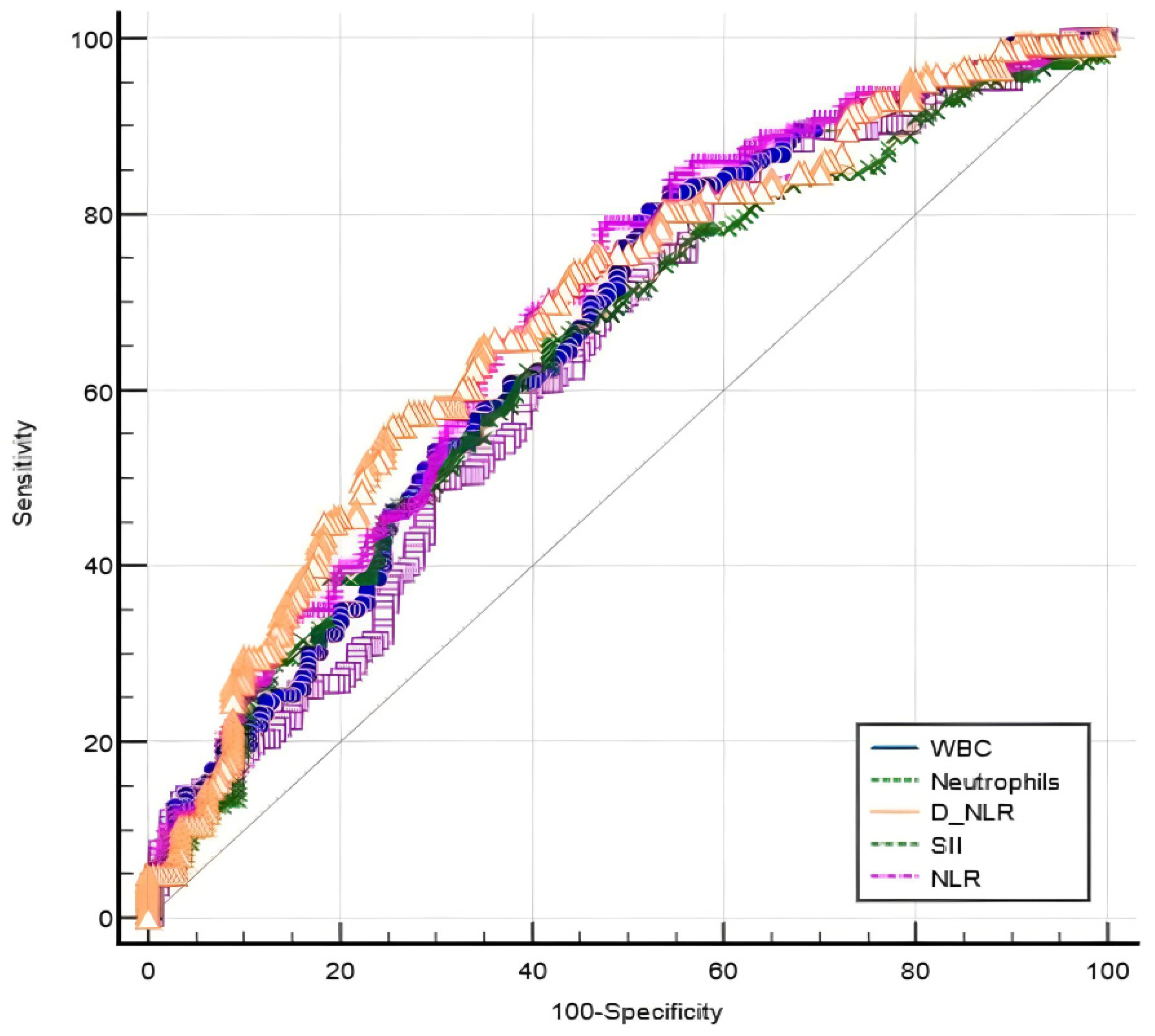
3.3. Performance of Blood-Count-Derived Inflammatory Markers in Evaluating Disease Severity

The diagnostic performance of different markers is shown in Table 5.
For discriminating between moderate and mild psoriasis, the AUC of WBC was 0.637 with a cut-off value of 6.25, while the neutrophil count AUC was 0.662 with a cut-off value of 3.64. On the same matter, the AUC of NLR was 0.687 with a cut-off value of 2.35, while d-NLR and SII predicted moderate psoriasis at, respectively, an AUC of 0.640 and a cut-off value of 65.73, and an AUC of 0.683 and a threshold value of 408.8. As for sensitivity, WBC was the highest, while specificity was the highest for NLR. Comparing AUCs of various markers for predicting a moderate course of disease, the AUC of NLR was the highest, but similar to those of WBC (p = 0.11), neutrophil count (p = 0.28), and SII (p = 0.81) and significantly different from that of d-NLR (p < 0.001) (Figure 1).
If referring to differentiating severe psoriasis from moderate disease, d-NLR significantly (p = 0.03) predicted severe psoriasis at an AUC of 0.598 and a cut-off value of 2.18. Nevertheless, regarding neutrophil count, due to the non-significant level (p = 0.55), the obtained AUC of 0.527 and the cut-off value of 5.66 cannot be considered reliable markers in discriminating between severe and moderate psoriasis.

3.4. The Dependability of Blood-Count-Derived Inflammatory Markers for Predicting Disease Severity

In a multivariate logistic regression model, patients with d-NLR (OR: 0.16, p < 0.001), NLR (OR: 4.13, p < 0.001), and SII (OR: 1, p = 0.046) above the cut-off values of 1.49, 2.35, and 408.8, respectively, have a higher risk of presenting with moderate psoriasis. Moreover, values for the aforementioned parameters below the cut-off value are significant predictors of mild psoriasis, as seen in Table 6. If referring to severe psoriasis, a higher level of d-NLR (OR: 0.69, p = 0.049) is an independent predictor of severe psoriasis.

4. Discussion

Early detection and an overall assessment of systemic inflammation are of great importance when referring to psoriasis. Psoriatic patients should benefit from an integrated, multidisciplinary-based therapeutical approach, taking into account the elevated risk of associated comorbidities this disease imposes.
Previous studies found that laboratory markers like NLR, PLR, MLR, and SII are associated with psoriasis or its severity, as well as with malignant tumors or other autoimmune diseases [27,28]. Currently, even though NLR and PLR are the most extensively studied, no consistent marker for assessing disease severity was reported, probably due to inconsistent study groups and racial variations, highlighting the need for more extensive studies on the subjects. NLR was reported to be higher in patients with psoriasis compared to controls [29] and a strong predictor of psoriatic arthritis [30]. Additionally, these markers can be used as indicators of all-cause mortality [31] and cardiovascular disease [32]. We have recently shown their reliability in assessing liver fibrosis [33] in psoriasis. NLR and PLR decreased in a similar form to the C-reactive protein (CRP) in Japanese patients treated with all kinds of biologics [34], and can, therefore, be useful in assessing the response to systemic treatment. Nevertheless, larger studies focusing on response to various kinds of biologics are needed in the future.
The present study identified that platelets and leucocytes subsets are potential contributors to psoriatic systemic inflammation. As depicted in Table 2 and Table 4, psoriatic severity is significantly positively correlated with WBC, neutrophil count, PLR, NLR, d-NLR, MLR, SII, SIRI, and AISI. Endothelial cells, epidermal keratinocytes, and dermal dendritic cells are involved in producing high levels of TNF-α, while Th17 cells, neutrophils, mast cells, and macrophages produce interleukin-17. Additionally, neutrophils promote increased production of reactive oxygen species [35] and are a major source of antimicrobial proteins [36] and lipocalin 2 [37], further sustaining systemic inflammation. Patients with psoriasis exhibit higher levels of advanced oxidation protein products [38,39] and catalase compared to controls [39], with additional differences between genders [40] supporting once more the need for a tailored therapeutical approach to each patient. Epidermal infiltration of neutrophils, a histopathological marker of psoriasis [41], with consequent activation, leads to the formation of neutrophil extracellular traps (NETs), which further interact directly with keratinocytes and heighten the IL-17A response [42]. In a study published by Rocha-Pereira et al. [43], psoriasis was characterized by an increased neutrophil count, with significantly higher levels in active psoriasis compared to inactive disease [43]. In our study, a constant gradual decrease was noted in the lymphocyte count as the severity increased. This paradoxical reaction is most likely the reverse effect of an increased influx of lymphocytes into the skin in patients with severe disease.
Moreover, as the disease progressed, platelet count increased in our study groups. Even though originally platelets were considered to interfere mainly with hemostasis, additional data highlight their involvement in inflammation progression and immunological processes [44]. Elevated platelet count may be due to bone marrow hyperproduction consecutive to their accumulation at inflammation sites, but also because TNF-α can directly activate platelets. Monocyte count was higher in severe forms compared to milder ones. They are responsible for producing interleukin-1 (IL-1) and interleukin-6 (IL-6), TNF-α [45], while immune cells derived from monocytes are key factors in interleukin-23 (IL-23)-driven psoriatic inflammation [46]. Nevertheless, it is worth mentioning that, when referring to our study and monocyte-derived markers, MLR varied significantly between the three study groups (p = 0.04), but PMR did not (p = 0.75). PMR values were the highest in patients presenting with mild disease, suggesting that this marker might not be reliable in assessing inflammatory status in psoriasis.
Thus, NLR, PLR, d-NLR, MLR SII, SIRI, and AISI seem to adequately reflect this involvement and, therefore, systemic inflammation in psoriasis. NLR exhibited the strongest correlation with disease severity (r = 0.30), followed by SII (r = 0.29), SIRI, and AISI (r = 0.28). PLR and MLR individually had a weak association with disease severity (r = 0.14 and r = 0.13, respectively). As such, composite markers, like SII, SIRI, and AISI, which incorporate various cell subsets (neutrophils, lymphocytes, monocytes, and/or platelets), may become more promising prognostic factors.
Patients with moderate psoriasis had significantly higher WBC, neutrophils count, NLR, d-NLR, and SII values compared to milder forms and lower neutrophils count and d-NLR values if compared to those with severe forms. In differentiating between moderate and milder forms of psoriasis, all the aforementioned parameters proved to be reliable and statistically significant taking into account the AUC (p < 0.001). Based on the AUC values, NLR is the most reliable factor in discriminating between moderate and mild psoriasis (AUC = 0.687) with a cut-off value of 2.35. Moderate psoriasis is defined by WBC above 6.25, neutrophil count above 3.64, NLR values above 2.35, d-NLR above 1.49, and SII above 408.8. Moreover, after running a multivariate regression analysis model, d-NLR (OR: 0.16), NLR (OR: 4.13), and SII proved to be significant (p < 0.001, p < 0.001, and p = 0.043, respectively) of a moderate course of disease. NLR is proven to be the strongest predictor of moderate psoriasis.
If talking about mild psoriasis, it should be defined by WBC below 6.25, neutrophil count below 3.64, NLR below 2.35, d-NLR below 1.49, and SII below 408.8. Regarding prognostic factors, our study identified that d-NLR (OR: 6.15), NLR (OR: 4.13), and SII (OR: 0.99) are significant indicators (p < 0.001, p < 0.001, and p = 0.046, respectively) of mild psoriasis.
On the other hand, when referring to severe psoriasis, our analysis identified that it can be differentiated from moderate psoriasis based on an AUC of 0.598 with a consequent calculated cut-off value of 2.18. With a p-value of 0.55, the neutrophil count cannot be considered a reliable marker in discriminating between severe and moderate psoriasis. Therefore, taking all of this into account, our study suggests that, referring to moderate psoriasis, it should be defined by d-NLR values between 1.49 and 2.18, with values below the lower limit of the interval indicating mild psoriasis, and higher values indicating severe psoriasis.
Out of all parameters overall identified as prognostic factors of disease severity, d-NLR is the only one linked to all three study groups. To the best of our knowledge, the reliability of d-NLR as an inflammatory marker in psoriasis has not been assessed until now. Unlike NLR, d-NLR also incorporates monocytes and other granulocytes, because it is calculated by dividing the neutrophil count by the difference between WBC and the neutrophil count (see Table 1). In an increased systemic inflammatory state, as in psoriasis, immature neutrophils can be released, leading to a rapid increase in neutrophil count. As such, compared to NLR, d-NLR reflects more accurately this negative inflammation, limits potential bias, and, in our study, proves to be the most reliable blood-count-derived inflammatory marker to predict disease severity.
Although our study showed a definite association between blood-count-derived inflammatory markers and disease severity in psoriasis, there are some limitations worth mentioning. First of all, it was a single-center retrospective study; this should be addressed in future studies by a prospective enrollment of the patients who should originate from multiple dermatologic centers, as such diminishing also the influence of geographical factors. Second, disease onset was not addressed; therefore, future work should additionally focus on this matter, as well as on patients’ behavioral patterns. Lastly, disease severity was assessed using only one scale, the BSA score. Our study was based on the BSA score and not the PASI score, because the latter one, even though frequently used, takes into account various parameters such as induration, erythema, and scaling, all dependent up to a certain point on the experience of the examiner, and these could not be controlled in this study, all the more so due to the retrospective manner of enrollment. Future ideas in this matter might refer to a comprehensive disease severity assessment using composite scores.

5. Conclusions

NLR, PLR, d-NLR, MLR SII, SIRI, and AISI are useful indicators of systemic inflammation and disease severity in psoriasis. Additionally, d-NLR accurately reflects the negative inflammation associated with this disease and proves to be the most reliable blood-count-derived inflammatory marker in predicting disease severity across all three study groups.
Referring to easily obtainable and low-cost markers, the results of our study comprehensively highlight the link between systemic inflammation and psoriasis progressions. Reporting for the first time the usefulness of d-NLR and AISI in appreciating and predicting disease severity, our results emphasize the usefulness of blood-count-derived inflammatory markers in a proper patient assessment monitoring disease course.

Author Contributions

Conceptualization O.M.T., S.H.M. and O.S.C.; Formal analysis O.M.T.; Methodology: O.M.T., C.R.M. and A.C.N.; Resources: C.R.M., R.A.T. and A.C.N.; Validation: O.S.C. and S.H.M.; Visualization: O.M.T. and R.A.T.; Writing–original draft: O.M.T., C.R.M. and R.A.T.; Writing–revision and editing: O.M.T. and A.C.N.; Supervision: O.S.C. and S.H.M. All authors have read and agreed to the published version of the manuscript.

Funding

This research received no external funding.

Institutional Review Board Statement

The study was conducted in accordance with the Declaration of Helsinki and approved by the Ethics Committee Mures Clinical County Hospital no. 3770/05.04.2023.

Informed Consent Statement

Informed consent was obtained from all subjects involved in the study.

Data Availability Statement

All data presented can be made available upon request.

Acknowledgments

This article is part of a Ph.D. thesis from the Doctoral School of Medicine and Pharmacy of the University of Medicine, Pharmacy, Science, and Technology George Emil Palade of Targu Mures, titled “The impact of systemic inflammation in modulating disease presentation in psoriasis”, which will be presented by Oana Mirela Tiucă by the fall of 2024.

Conflicts of Interest

The authors declare no conflict of interest.

References

  1. Scarpa, R.; Ayala, F.; Caporaso, N.; Olivieri, I. Psoriasis, psoriatic arthritis, or psoriatic disease? J. Rheumatol. 2006, 33, 210–212. [Google Scholar]
  2. Yamazaki, F. Psoriasis: Comorbidities. J. Dermatol. 2021, 48, 732–740. [Google Scholar] [CrossRef] [PubMed]
  3. Shibata, S.; Tada, Y.; Hau, C.; Tatsuta, A.; Yamamoto, M.; Kamata, M.; Karakawa, M.; Asano, Y.; Mitsui, H.; Sugaya, M.; et al. Adiponectin as an anti-inflammatory factor in the pathogenesis of psoriasis: Induction of elevated serum adiponectin levels following therapy: Correspondence. Br. J. Dermatol. 2011, 164, 667–670. [Google Scholar] [CrossRef] [PubMed]
  4. Dong, Q.; Li, D.; Xie, B.B.; Hu, L.H.; Huang, J.; Jia, X.X.; Tang, Y.L.; Liu, G.H.; Shen, N.N.; Yu, X.B. IL-17A and TNF-α inhibitors induce multiple molecular changes in psoriasis. Front. Immunol. 2022, 13, 1015182. [Google Scholar] [CrossRef] [PubMed]
  5. Campione, E.; Mazzilli, S.; Di Prete, M.; Dattola, A.; Cosio, T.; Lettieri Barbato, D.; Costanza, G.; Lanna, C.; Manfreda, V.; Gaeta Schumak, R.; et al. The Role of Glutathione-S Transferase in Psoriasis and Associated Comorbidities and the Effect of Dimethyl Fumarate in This Pathway. Front. Med. 2022, 9, 760852. [Google Scholar] [CrossRef]
  6. Trevisan, G.; Stinco, G.; Giansante, C.; Fiotti, N.; Vidimari, P.; Kokelj, F. Psoriasis and endothelins. Acta Derm. Venereol. Suppl. 1994, 186, 139–140. [Google Scholar]
  7. Boehncke, W.H.; Boehncke, S.; Tobin, A.M.; Kirby, B. The ‘psoriatic march’: A concept of how severe psoriasis may drive cardiovascular comorbidity: The ‘psoriatic march’. Exp. Dermatol. 2011, 20, 303–307. [Google Scholar] [CrossRef] [PubMed]
  8. Berth-Jones, J.; Grotzinger, K.; Rainville, C.; Pham, B.; Huang, J.; Daly, S.; Herdman, M.; Firth, P.; Hotchkiss, K. A study examining inter- and intrarater reliability of three scales for measuring severity of psoriasis: Psoriasis Area and Severity Index, Physician’s Global Assessment and Lattice System Physician’s Global Assessment: Validation of PASI, PGA and LS-PGA. Br. J. Dermatol. 2006, 155, 707–713. [Google Scholar] [CrossRef]
  9. Henseler, T.; Schmitt-Rau, K. A comparison between BSA, PASI, PLASI and SAPASI as measures of disease severity and improvement by therapy in patients with psoriasis. Int. J. Dermatol. 2008, 47, 1019–1023. [Google Scholar] [CrossRef]
  10. Cancelliere, R.; Cosio, T.; Campione, E.; Corvino, M.; D’Amico, M.P.; Micheli, L.; Signori, E.; Contini, G. Label-free electrochemical immunosensor as a reliable point-of-care device for the detection of Interleukin-6 in serum samples from patients with psoriasis. Front. Chem. 2023, 11, 1251360. [Google Scholar] [CrossRef]
  11. Gambichler, T.; Hessam, S.; Cramer, P.; Abu Rached, N.; Bechara, F.G. Complete blood collection-based systemic inflammation biomarkers for patients with hidradenitis suppurativa. Acad Dermatol. Venereol. 2022, 36, 1593–1596. [Google Scholar] [CrossRef] [PubMed]
  12. Öksüm Solak, E.; Baran Ketencioglu, B.; Cinar, S.L.; Kartal, D.; Borlu, M. The role of new inflammatory markers in determining disease activation and severity in patients with hidradenitis suppurativa. Int. J. Dermatol. 2023, 62, 1076–1081. [Google Scholar] [CrossRef]
  13. Bakic, M.; Klisic, A.; Karanikolic, V. Comparative Study of Hematological Parameters and Biomarkers of Immunity and Inflammation in Patients with Psoriasis and Atopic Dermatitis. Medicina 2023, 59, 1622. [Google Scholar] [CrossRef] [PubMed]
  14. Sun, C.; Li, X.; Qian, H.; Liang, G.; Xiang, R.; Zhao, C.; Li, Z.; Li, S.; Jing, K.; Wang, Y.; et al. Neutrophil-to-lymphocyte ratio and platelet-to-lymphocyte ratio are positively correlated with disease activity of bullous pemphigoid. Arch. Dermatol. Res. 2023, 315, 2383–2391. [Google Scholar] [CrossRef] [PubMed]
  15. Dolan, R.D.; Lim, J.; McSorley, S.T.; Horgan, P.G.; McMillan, D.C. The role of the systemic inflammatory response in predicting outcomes in patients with operable cancer: Systematic review and meta-analysis. Sci. Rep. 2017, 7, 16717. [Google Scholar] [CrossRef] [PubMed]
  16. Ying, H.-Q.; Deng, Q.-W.; He, B.-S.; Pan, Y.-Q.; Wang, F.; Sun, H.-L.; Chen, J.; Liu, X.; Wang, S.-K. The prognostic value of preoperative NLR, d-NLR, PLR and LMR for predicting clinical outcome in surgical colorectal cancer patients. Med. Oncol. 2014, 31, 305. [Google Scholar] [CrossRef] [PubMed]
  17. Paliogiannis, P.; Fois, A.G.; Sotgia, S.; Mangoni, A.A.; Zinellu, E.; Pirina, P.; Negri, S.; Carru, C.; Zinellu, A. Neutrophil to lymphocyte ratio and clinical outcomes in COPD: Recent evidence and future perspectives. Eur. Respir. Rev. 2018, 27, 170113. [Google Scholar] [CrossRef]
  18. Mertoglu, C.; Gunay, M. Neutrophil-Lymphocyte ratio and Platelet-Lymphocyte ratio as useful predictive markers of prediabetes and diabetes mellitus. Diabetes Metab. Syndr. Clin. Res. Rev. 2017, 11, S127–S131. [Google Scholar] [CrossRef]
  19. Zinellu, A.; Collu, C.; Nasser, M.; Paliogiannis, P.; Mellino, S.; Zinellu, E.; Traclet, J.; Ahmad, K.; Mangoni, A.A.; Carru, C.; et al. The Aggregate Index of Systemic Inflammation (AISI): A Novel Prognostic Biomarker in Idiopathic Pulmonary Fibrosis. J. Clin. Med. 2021, 10, 4134. [Google Scholar] [CrossRef]
  20. Xiu, J.; Lin, X.; Chen, Q.; Yu, P.; Lu, J.; Yang, Y.; Chen, W.; Bao, K.; Wang, J.; Zhu, J.; et al. The aggregate index of systemic inflammation (AISI): A novel predictor for hypertension. Front. Cardiovasc. Med. 2023, 10, 1163900. [Google Scholar] [CrossRef]
  21. Krenn-Pilko, S.; Langsenlehner, U.; Stojakovic, T.; Pichler, M.; Gerger, A.; Kapp, K.S.; Langsenlehner, T. The elevated preoperative derived neutrophil-to-lymphocyte ratio predicts poor clinical outcome in breast cancer patients. Tumor Biol. 2016, 37, 361–368. [Google Scholar] [CrossRef]
  22. Proctor, M.J.; McMillan, D.C.; Morrison, D.S.; Fletcher, C.D.; Horgan, P.G.; Clarke, S.J. A derived neutrophil to lymphocyte ratio predicts survival in patients with cancer. Br. J. Cancer 2012, 107, 695–699. [Google Scholar] [CrossRef] [PubMed]
  23. Chapman, J.W.; O’Callaghan, C.J.; Hu, N.; Ding, K.; A Yothers, G.; Catalano, P.J.; Shi, Q.; Gray, R.G.; O’Connell, M.J.; ACCENT Collaborative Group; et al. Innovative estimation of survival using log-normal survival modelling on ACCENT database. Br. J. Cancer 2013, 108, 784–790. [Google Scholar] [CrossRef]
  24. Zhou, J.; Li, X.; Wang, M.; Gu, C.; Liu, J. Platelet-to-Monocyte Ratio as a Novel Promising Agent for the Prognosis of Hepatitis B Virus-Associated Decompensated Cirrhosis. Can. J. Gastroenterol. Hepatol. 2023, 2023, 6646156. [Google Scholar] [CrossRef] [PubMed]
  25. Alfhili, M.A.; Alotaibi, G.A.; Alfaifi, M.; Almoghrabi, Y.; Alsughayyir, J. Association of Platelet-Monocyte Ratio with Dyslipidemia in Saudi Arabia: A Large, Population-Based Study. Life 2023, 13, 1685. [Google Scholar] [CrossRef] [PubMed]
  26. Zhang, Y.; Shi, Y.; Shen, H.; Shou, L.; Fang, Q.; Zheng, X.; Zhu, M.; Huang, X.; Huang, J.; Li, L.; et al. The value of a new prognostic model developed by lymphocyte-monocyte ratio and platelet-monocyte ratio in peripheral T-cell lymphoma. Cancer Cell Int. 2021, 21, 573. [Google Scholar] [CrossRef]
  27. Feng, F.; Tian, Y.; Liu, S.; Zheng, G.; Liu, Z.; Xu, G.; Guo, M.; Lian, X.; Fan, D.; Zhang, H. Combination of PLR, MLR, MWR, and Tumor Size Could Significantly Increase the Prognostic Value for Gastrointestinal Stromal Tumors. Medicine 2016, 95, e3248. [Google Scholar] [CrossRef] [PubMed]
  28. Imtiaz, F.; Shafique, K.; Mirza, S.; Ayoob, Z.; Vart, P.; Rao, S. Neutrophil lymphocyte ratio as a measure of systemic inflammation in prevalent chronic diseases in Asian population. Int. Arch. Med. 2012, 5, 2. [Google Scholar] [CrossRef] [PubMed]
  29. Paliogiannis, P.; Satta, R.; Deligia, G.; Farina, G.; Bassu, S.; Mangoni, A.A.; Carru, C.; Zinellu, A. Associations between the neutrophil-to-lymphocyte and the platelet-to-lymphocyte ratios and the presence and severity of psoriasis: A systematic review and meta-analysis. Clin. Exp. Med. 2019, 19, 37–45. [Google Scholar] [CrossRef]
  30. Kim, D.S.; Shin, D.; Lee, M.S.; Kim, H.J.; Kim, D.Y.; Kim, S.M.; Lee, M.G. Assessments of neutrophil to lymphocyte ratio and platelet to lymphocyte ratio in Korean patients with psoriasis vulgaris and psoriatic arthritis. J. Dermatol. 2016, 43, 305–310. [Google Scholar] [CrossRef]
  31. Adamstein, N.H.; MacFadyen, J.G.; Rose, L.M.; Glynn, R.J.; Dey, A.K.; Libby, P.; Tabas, I.A.; Mehta, N.N.; Ridker, P.M. The neutrophil–lymphocyte ratio and incident atherosclerotic events: Analyses from five contemporary randomized trials. Eur. Heart J. 2021, 42, 896–903. [Google Scholar] [CrossRef] [PubMed]
  32. Kvist-Hansen, A.; Kaiser, H.; Wang, X.; Krakauer, M.; Gørtz, P.M.; McCauley, B.D.; Zachariae, C.; Becker, C.; Hansen, P.R.; Skov, L. Neutrophil Pathways of Inflammation Characterize the Blood Transcriptomic Signature of Patients with Psoriasis and Cardiovascular Disease. Int. J. Mol. Sci. 2021, 22, 10818. [Google Scholar] [CrossRef] [PubMed]
  33. Tiucă, O.M.; Morariu, S.H.; Mariean, C.R.; Tiucă, R.A.; Nicolescu, A.C.; Cotoi, O.S. Predictive Performances of Blood-Count-Derived Inflammatory Markers for Liver Fibrosis Severity in Psoriasis Vulgaris. Int. J. Mol. Sci. 2023, 24, 16898. [Google Scholar] [CrossRef] [PubMed]
  34. Asahina, A.; Kubo, N.; Umezawa, Y.; Honda, H.; Yanaba, K.; Nakagawa, H. Neutrophil-lymphocyte ratio, platelet-lymphocyte ratio and mean platelet volume in Japanese patients with psoriasis and psoriatic arthritis: Response to therapy with biologics. J. Dermatol. 2017, 44, 1112–1121. [Google Scholar] [CrossRef]
  35. Chiang, C.C.; Cheng, W.J.; Korinek, M.; Lin, C.Y.; Hwang, T.L. Neutrophils in Psoriasis. Front. Immunol. 2019, 10, 2376. [Google Scholar] [CrossRef] [PubMed]
  36. Schonthaler, H.B.; Guinea-Viniegra, J.; Wculek, S.K.; Ruppen, I.; Ximénez-Embún, P.; Guío-Carrión, A.; Navarro, R.; Hogg, N.; Ashman, K.; Wagner, E.F. S100A8-S100A9 Protein Complex Mediates Psoriasis by Regulating the Expression of Complement Factor C3. Immunity 2013, 39, 1171–1181. [Google Scholar] [CrossRef] [PubMed]
  37. Shao, S.; Cao, T.; Jin, L.; Li, B.; Fang, H.; Zhang, J.; Zhang, Y.; Hu, J.; Wang, G. Increased Lipocalin-2 Contributes to the Pathogenesis of Psoriasis by Modulating Neutrophil Chemotaxis and Cytokine Secretion. J. Investig. Dermatol. 2016, 136, 1418–1428. [Google Scholar] [CrossRef] [PubMed]
  38. Yazici, C.; Köse, K.; Utaş, S.; Tanrikulu, E.; Taşlidere, N. A novel approach in psoriasis: First usage of known protein oxidation markers to prove oxidative stress. Arch. Dermatol. Res. 2016, 308, 207–212. [Google Scholar] [CrossRef]
  39. Bakic, M.; Klisic, A.; Kocic, G.; Kocic, H.; Karanikolic, V. Oxidative stress and metabolic biomarkers in patients with psoriasis. J. Med. Biochem. 2023, 42, 1–9. [Google Scholar]
  40. Klisic, A.; Bakic, M.; Karanikolic, V. Comparative Analysis of Redox Homeostasis Biomarkers in Patients with Psoriasis and Atopic Dermatitis. Antioxidants 2023, 12, 1875. [Google Scholar] [CrossRef]
  41. Yamanaka, K.; Yamamoto, O.; Honda, T. Pathophysiology of psoriasis: A review. J. Dermatol. 2021, 48, 722–731. [Google Scholar] [CrossRef] [PubMed]
  42. Shao, S.; Fang, H.; Dang, E.; Xue, K.; Zhang, J.; Li, B.; Qiao, H.; Cao, T.; Zhuang, Y.; Shen, S.; et al. Neutrophil Extracellular Traps Promote Inflammatory Responses in Psoriasis via Activating Epidermal TLR4/IL-36R Crosstalk. Front. Immunol. 2019, 10, 746. [Google Scholar] [CrossRef] [PubMed]
  43. Rocha-Pereira, P.; Santos-Silva, A.; Rebelo, I.; Figueiredo, A.; Quintanilha, A.; Teixeira, F. The inflammatory response in mild and in severe psoriasis. Br. J. Dermatol. 2004, 150, 917–928. [Google Scholar] [CrossRef] [PubMed]
  44. Gawaz, M. Platelets in inflammation and atherogenesis. J. Clin. Investig. 2005, 115, 3378–3384. [Google Scholar] [CrossRef] [PubMed]
  45. Costa, M.C.; Rocha, B.D.O.; Paixão, C.S.; Oliveira, M.D.F.S.P.D.; Mota, L.M.H.D.; Carvalho, L.P.D. Monocyte subpopulations study in patients with plaque psoriasis. Med. Hypotheses 2017, 104, 101–103. [Google Scholar] [CrossRef]
  46. Rendon, A.; Schäkel, K. Psoriasis Pathogenesis and Treatment. Int. J. Mol. Sci. 2019, 20, 1475. [Google Scholar] [CrossRef]
Figure 1. ROC comparison of WBC, neutrophils, NLR, d-NLR, and SII in predicting moderate psoriasis.
Life 14 00114 g001
Table 1. Formulas of analyzed biomarkers.
MarkerFormula
NLRNeutrophil count/lymphocyte count [×103/μL]
MLRMonocyte count/lymphocyte count [×103/μL]
d-NLRNeutrophil count/(WBC − neutrophil count) [×103/μL]
PLRPlatelet count/lymphocyte count [×103/μL]
PMRPlatelet count/monocyte count [×103/μL]
SII(Neutrophil count × platelet count)/lymphocyte count [×103/μL]
SIRI(Neutrophil count × monocyte count)/lymphocyte count [×103/μL]
AISI(Neutrophil count × monocyte count × platelet count)/lymphocyte count [×103/μL]
NLR, neutrophil-to-lymphocyte ratio; d-NLR, derived neutrophil-to-lymphocyte ratio; PLR, platelet-to-lymphocyte ratio; MLR, monocyte-to-lymphocyte ratio; SII, systemic immune inflammation index; SIRI, systemic inflammation response index; AISI, aggregate index of systemic inflammation.
Table 2. The characteristics of the study population.
Variables *All PatientsMild Disease (n = 180)Moderate
Disease (n = 143)
Severe Disease (n = 43)p-Value
Age54.48 ± 16.4853.86 ± 17.4657.51 ± 12.8954.35 ± 16.180.43
Gender
  Male219101 (56%)91 (64%)27 (63%)0.36
  Female14779 (44%)52 (36%)16 (37%)
WBC7.49 [7.15–7.83]6.77 [6.38–7.26]8.08 [7.60–8.73]7.98 [7.50–8.42]<0.001
Platelets238.25
[230.93–243.69]
231.70
[220-.95–240.22]
243.20
[221.87–265.52]
245
[236.51–259.56]
0.11
Neutrophils4.42 [4.22–4.69]3.80 [3.44–4.25]4.68 [4.39–5.26]4.96 [4.57–5.45]<0.001
Lymphocytes2.10 [1.97–2.23]2.22 [1.99–2.30]2.16 [1.91–2.34]1.97 [1.79–2.20]0.12
Monocytes0.51 [0.48–0.53]0.49 [0.45–0.52]0.52 [0.47–0.56]0.53 [0.48–0.57]0.59
PLR114.96
[110.45–121.10]
108.07
[100.89–115.10]
110.75
[96.66–126.26]
129.79
[117.02–135.15]
0.002
NLR2.05 [1.90–2.19]1.73 [1.59–1.88]2.23 [1.93–2.66]2.53 [2.20–2.79]<0.001
d-NLR1.55 [1.44–1.65]1.37 [1.29–1.49]1.45 [1.36–1.82]1.80 [1.62–1.95]0.001
MLR0.23 [0.22–0.25]0.22 [0.21–0.25]0.25 [0.23–0.28]0.24 [0.22–0.26]0.04
PMR475.02
[450.39–508.40]
479.08
[440.36–526.90]
467.59
[400–539.75]
476.42
[437.62–524.45]
0.75
SII478.52
[453.65–521.85]
404.61
[357.44–446.03]
541.21
[464.89–602.06]
560.46
[518.95–646.30]
<0.001
SIRI1.03 [0.93–1.09]0.86 [0.79–0.92]1.23 [1.01–1.44]1.27 [1.07–1.37]0.001
AISI255.83
[229–273.18]
196.96
[168.52–221.22]
287.76
[258.18–331.08]
293.10
[272.01–335.63]
0.001
ESR15
[12.8–17]
12.80
[11.80–15.80]
16.20
[12.20–20.98]
18
[14–20]
0.17
WBC, white blood cell count; PLR, platelet-to-lymphocyte ratio; NLR, neutrophil-to-lymphocyte ratio; d-NLR, derived neutrophil-to-lymphocyte ratio; MLR, monocyte-to-lymphocyte ratio; PMR, platelet-to-monocyte ratio; SII, systemic immune inflammation index; SIRI, systemic inflammation response index; AISI, aggregate index of systemic inflammation; ESR, erythrocyte sedimentation rate; * refer to Table 1 for measurement units.
Table 3. Pairwise comparison of blood-count-derived inflammatory markers.
Parameter *Mild PsoriasisModerate PsoriasisSevere Psoriasis
vs. Moderate Psoriasisvs. Severe Psoriasisvs. Mild Psoriasisvs. Severe Psoriasisvs. Mild Psoriasisvs. Moderate Psoriasis
WBC0.0040.320.0040.060.320.06
Neutrophils0.00310.0030.0110.01
PLR0.070.320.0710.321
NLR0.010.960.010.110.860.11
d-NLR0.0210.020.00710.007
MLR111111
SII0.0090.340.0090.120.340.12
SIRI0.1810.180.2610.26
AISI0.0610.060.3710.37
WBC, white blood cell count; PLR, platelet-to-lymphocyte ratio; NLR, neutrophil-to-lymphocyte ratio; d-NLR, derived neutrophil-to-lymphocyte ratio; MLR, monocyte-to-lymphocyte ratio; SII, systemic immune inflammation index; SIRI, systemic inflammation response index; AISI, aggregate index of systemic inflammation; * refer to Table 1 for measurement units.
Table 4. Correlation between disease severity and the analyzed markers.
Marker *rp-ValueMarkerrv-Value
WBC0.25<0.001MLR0.130.01
Neutrophil count0.26<0.001SII0.29<0.001
PLR0.140.005SIRI0.28<0.001
NLR0.30<0.001AISI0.28<0.001
d-NLR0.18<0.001ESR0.15<0.001
WBC, white blood cell count; PLR, platelet-to-lymphocyte ratio; NLR, neutrophil-to-lymphocyte ratio; d-NLR, derived neutrophil-to-lymphocyte ratio; MLR, monocyte-to-lymphocyte ratio; SII, systemic immune inflammation index; SIRI, systemic inflammation response index; AISI, aggregate index of systemic inflammation; ESR, erythrocyte sedimentation rate; * refer to Table 1 for measurement units.
Table 5. Predictive performance of blood-count-derived markers.
Moderate vs. Mild Disease
Parameter *AUC
(95% CI)
p-ValueCut-OffSe (%)Sp (%)Youden Index Jp-Value *
WBC0.637
[0.582–0.689]
<0.0016.2582.5242.220.250.11
Neutrophil count0.662
[0.607–0.713]
<0.0013.6480.4247.780.280.28
NLR0.687
[0.633–0.737]
<0.0012.3555.9474.440.30-
d-NLR0.640
[0.585–0.692]
<0.0011.4965.7358.330.24<0.001
SII0.683
[0.629–0.733]
<0.001408.879.0252.220.310.81
Severe vs. moderate disease
Parameter *AUC (95% CI)p-valueCut-OffSe (%)Sp (%)Youden Index Jp-value
Neutrophil count0.527
[0.453–0.601]
0.555.6676.7438.460.15-
d-NLR0.598
[0.524–0.669]
0.032.1890.7031.470.22-
Se: sensitivity; Sp: specificity; * Compared to NLR; WBC, white blood cell count; NLR, neutrophil-to-lymphocyte ratio; d-NLR, derived neutrophil-to-lymphocyte ratio; MLR, monocyte-to-lymphocyte ratio; SII, systemic immune inflammation index; * refer to Table 1 for measurement units.
Table 6. Predictors of disease severity in psoriasis.
Parameter *OR95% CIp-Value
Mild psoriasis
d-NLR6.152.53–14.91<0.001
NLR0.240.12–0.48<0.001
SII0.990.99–1.020.043
Moderate psoriasis
d-NLR0.160.07–0.39<0.001
NLR4.132.11–8.11<0.001
SII11–1.030.046
Severe psoriasis
d-NLR0.690.47–1.010.049
d-NLR, derived neutrophil-to-lymphocyte ratio; NLR, neutrophil-to-lymphocyte ratio; SII, systemic immune inflammation index; * refer to Table 1 for measurement units.
Disclaimer/Publisher’s Note: The statements, opinions and data contained in all publications are solely those of the individual author(s) and contributor(s) and not of MDPI and/or the editor(s). MDPI and/or the editor(s) disclaim responsibility for any injury to people or property resulting from any ideas, methods, instructions or products referred to in the content.

