Abstract
Many species belonging to the genus Ocimum are used for aromatic, medicinal, and cosmetic purposes. The essential oil (OFEO) obtained by hydrodistillation of the flowering aerial parts of Forsskal’s Basil “Ocimum forskolei Benth” growing in extreme environmental conditions in Mecca Region, Saudi Arabia was analyzed by GC-MS. The main constituents were phenylpropanoids (methyl eugenol 55.65% and eugenol 11.66%), monoterpene (linalool 9.75%), and sesquiterpenes (germacrene D 3.72% and β-caryophyllene 2.57%). The OFEO was tested against MCF7, HT29, and HCT116 cancer cells and compared with normal fibroblast cells (MRC5). The MTT assay showed that HCT116 was more sensitive to OFEO (IC50 5.34 μg/mL), which reduced the number of HCT116 colonies at 6 μg/mL, while causing complete colony death at 12 and 24 μg/mL. Western Blotting and qRT-PCR were used to evaluate the level change of different proteins with respect to GAPDH. OFEO upregulated the apoptotic protein (caspase 3), and downregulated the cell proliferation proteins (AKT and pAKT), cell cycle arrest (PCNA, Cyclin D1), and the anti-apoptotic Bcl2 proteins. OFEO was also tested against reference strains of Gram-negative and Gram-positive bacteria including Escherichia coli, Klebsiella pneumonia, Pseudomonas aeruginosa, and Staphylococcus aureus by using the well-diffusion and assessing their MICs, which ranged from 250 to 500 μg/mL.
1. Introduction
Plants belonging to the genus Ocimum are of great economic and medicinal value, as many species are used for their nutritional, pharmacological, and detoxicant properties [1,2,3]. Ocimum basilicum L., “Sweet basil”, the most nutritious species of this genus, and its cultivars are distributed worldwide and are largely used in the food and perfume industries [4,5]. Several studies were performed on Ocimum species essential oils, extracts, and pure constituents, revealing potent anti-proliferative and antimicrobial activities [6,7,8]. Besides the essential oil, some non-volatile secondary metabolites isolated from Ocimum species have shown anti-cancer activity [9,10]. Caffeic acid, isolated from O. gratissimum L., had an anti-proliferative effect against human cervical cancer cells (HeLa), and it dramatically increased the apoptosis of HeLa cells through the activation of various caspases [9]. Moreover, ursolic acid isolated from an O. basilicum extract, induced a significant decrease of cell percentage in the anaphase/telophase stages of the cell division cycle. Ursolic acid also showed effects on F-actin and microtubules [10]. Regarding the essential oil of Ocimum sp., it possesses a marked antimicrobial activity against a broad range of bacteria and fungi because of the presence of diverse classes of volatile constituents, such as monoterpenes, phenylpropanoids, and sesquiterpenes [11,12].
Cancer is a multifactorial disorder characterized by uncontrolled cell proliferation that outwits the body’s defenses and traditional anticancer treatments. It is globally one of the main causes of death: in 2020, 19.3 million new diagnosed cancer cases and 9.9 million cancer deaths were recorded [13]. Breast, lung, colorectal, prostate, and stomach cancers are the most frequent types [13]. New medicines and therapeutic systems have been developed for cancer treatment. The inhibition of cell death, the increase of DNA damage, the overexpression of P-glycoprotein, and the epithelial–mesenchymal transition are among the mechanisms by which tumor cells might gain innate or acquired multidrug resistance [14]. In the search for new anticancer drugs, plants are considered a rich source of possible metabolites to be used in anticancer therapy; it is no coincidence that many of the best anticancer substances are of natural origin [15].
Besides cancer, infectious diseases cause high mortality worldwide, mainly in developing countries, requiring the discovery of new sources of antimicrobial agents from medicinal plants [16,17,18,19,20,21,22,23]. Essential oils are a complex mixture of volatile secondary metabolites with diverse bioactivities, including both anticancer and antimicrobial ones [24].
In the present study, we selected the essential oil of Ocimum forskolei Benth, also known as Ocimum forsskaolii (Lamiaceae), to investigate its biological properties. This is an aromatic plant widely distributed in Saudi Arabia. It is a bushy-woody herb, with bright green leaves and lilac-pink flowers, which grows at altitudes from 300 to 1400 m. It is used by the native people in Saudi Arabia to aromatize butter and tea, as well as for its carminative and insect-repelling properties. Moreover, its flowers are a good source of propolis [25]. Previous studies of OFEO revealed the chemical diversity of volatile constituents depending on the geographical origin; the essential oil obtained from Omani sample of O. forskolei flower contains mainly estragole (59.4–65.2%), followed by linalool (25.0–28.1%) [26]. The Kenyan sample was rich in fenchone 49.86%, Limonene 14.08%, camphor 5.93% and α-Caryophyllene 4.46%, respectively [27], while the Ethiopian sample was rich in linalool 17.3%, methyl chavicol 19.3% and (E)-methyl cinnamate 33.0%, or myrcene 24.2% and eugenol 25.0%, respectively [28]. In previous study on O. forsskaolii, the silver nanoparticles of the aqueous extract of the leaves revealed an antimicrobial effect [7]. The most predominant classes of compounds in the genus Ocimum are phenylpropanoids, monoterpenes and sesquiterpenes [29,30,31,32,33]. The aim of the present study was to investigate the essential oil composition of the flowering aerial parts of O. forskolei growing wildly in Saudi Arabia and to assess its anticancer effect through cytotoxicity and molecular target validation using reverse transcription polymerase chain reaction (qRT-PCR) and Western Blot (WB). In addition, the essential oil of O. forskolei was tested against Gram-positive and Gram-negative microorganisms responsible for urinary tract infections, such as Staphylococcus aureus, Klebsiella pneumonia, Escherichia coli, and Pseudomonas aeruginosa.
2. Materials and Methods
2.1. Materials
All the chemicals were purchased from Sigma Aldrich, Riyadh, Saudi Arabia, unless otherwise mentioned.
2.2. Methods
2.2.1. Plant Material
The flowering aerial parts of O. forskolei were collected at Wadi Rahjan, near Makkah Al-Mukarramah in February 2021 (Google Earth GPS coordinates: 21°17′52″ N 40°05′23″ E); the plant was identified by one of the authors A. B., and a voucher specimen, number SA-IT-2021/1, was deposited at the Herbarium of Pharmacognosy Lab, Faculty of Pharmacy, Umm Al-Qura University.
2.2.2. Essential Oil Hydro-Distillation
Fresh plant material (200 g), previously cut into small pieces, was hydro-distilled with a Clevenger standard apparatus for 2 h (yield: 0.52%). Aliquots of the obtained essential oil were diluted to 10% in HPLC-grade n-hexane prior to the GC-MS injection, while the remaining parts were stored in a freezer at −18 °C until biological testing.
2.2.3. Gas Chromatography-Mass Spectrometry Analyses and Peak identification
Gas chromatography–electron impact mass spectrometry (GC-EIMS) analyses were performed with an Agilent 7890B gas chromatograph (Agilent Technologies Inc., Santa Clara, CA, USA) equipped with an Agilent HP-5MS (Agilent Technologies Inc., Santa Clara, CA, USA) capillary column (30 m × 0.25 mm; coating thickness 0.25 μm) and an Agilent 5977B single-quadrupole mass detector (Agilent Technologies Inc., Santa Clara, CA, USA). The working conditions of essential oil analysis were similar to our previous work [18]. The temperature of the oven was set to rise from 60 °C to 240 °C at 3 °C/min. Temperatures were set as follows: injector temperature, 220 °C; transfer-line temperature, 240 °C. The carrier gas was He with 1 mL/min flow. The acquisition was performed with the following parameters: full scan, with a scan range of 35–300 m/z; scan time: 1.0 s; threshold: 1 count. The identification of the constituents was based on the comparison of their retention times (tR) with those of pure reference samples and of their linear retention indices (LRIs) relative to the n-alkanes (C9–C25) series. The detected mass spectra were compared with those listed in the commercial libraries NIST 14 and ADAMS, as well as in a homemade mass-spectral library built up from pure substances and components of essential oils of known compositions and MS literature data [34,35,36,37,38].
2.2.4. Cell Culture
Three cancer cell lines, MCF7 (human breast adenocarcinoma), HT29 (human colorectal adenocarcinoma), and HCT116 (human colorectal carcinoma), were used, together with MRC5 (normal human fetal lung fibroblast). All cells were obtained from the ATCC. The three cancer cell lines were sub-cultured in RPMI-1640 media (10% FBS); while MRC5 cells were maintained in Eagle’s minimum essential medium (EMEM, 10% FBS), all at 37 °C, 5% CO2, and 100% relative humidity.
2.2.5. Cytotoxicity and Selectivity Studies
The cytotoxicity of OFEO was evaluated by MTT assay, as previously reported [39]. The cell lines were separately cultured in 96-well plates (3 × 10³/well), and incubated at 37 °C overnight. Final extract or doxorubicin concentrations were 0–100 μg/mL (DMSO 0.1%; v/v, n = 3). Plates were incubated for 72 h and then MTT was added to each well. The absorbance was read with a multi-plate reader (BIORAD, PR 4100, Hercules, CA, USA). Optical density of the purple formazan A550 was proportional to the number of viable cells. Extract concentration causing 50% inhibition (IC50) compared with control cell growth (100%) was determined using GraphPad Prism (San Diego, CA, USA). The selectivity index (SI) for the extract was calculated by dividing its IC50 against MRC5 cells/IC50 against the other tested cells.
2.2.6. Clonogenic Survival Assay
The clonogenic survival assay, which studies the ability of individual cancer cells to form colonies, was performed according to a previous report [40]. HCT116 cells were seeded in 2 mL of low-density medium (2 × 10²) in 6-well plates (n = 2). Plates were incubated at 37 °C overnight to allow attachment. Cells were treated with OFEO (0, 6, 12, and 24 μg/mL). After 72 h, the medium was discarded, and 2 mL of fresh medium was added. Plates were checked under the microscope every 2 days. After 14 days, clusters of at least 50 cells were considered a colony. The medium was aspirated and the cells were washed with cold phosphate-buffered saline (PBS), and then fixed with cold methanol for 5 min at room temperature. Cells were then stained with 0.5% v/v methylene blue in methanol: H2O (1:1) for 15 min. Colonies were washed with PBS and H2O. Next, plates were left to dry before the final counting of colonies.
2.2.7. Western Blotting
In this study, the level change of AKT, pAKT, PCNA, cyclin D1, Bcl2, and Caspase 3 proteins was investigated using Western Blotting. The effect of OFEO (0, 6, 12, and 24 μg/mL) on HCT116 cells was tested following a previous reported method [41]. Briefly, HCT116 cells (1 × 10⁶ cells/well in a 6-well plate) were treated for 24 h. The HCT116 cells were lysed with a mixture of RIPA lysis and extraction buffer in ice for 10 min. The lysates were centrifuged at 14,000× g for 10 min at 4 °C. BCA assay was used to quantify protein concentration. Samples with 20 μg of total proteins were separated on 10% SDS-PAGE gels and transferred to PVDF membranes. Membranes were blocked for 1 h in TBS-T containing 5% non-fat dry milk and incubated with antibodies at 4 °C overnight. After washing, blots were incubated with secondary antibodies (HRP-conjugated) for 1 h. Target proteins were detected using the LI-COR system for enhanced chemiluminescence. The following antibodies were used: AKT, pAKT, PCNA, cyclin D1, Bcl2, Caspase 3, and GAPDH in 1:1000 dilutions. Secondary anti-rabbit HPR conjugated antibodies (1:3000 dilution) were used.
2.2.8. Quantitative Real-Time PCR (qRT-PCR)
The HCT116 cells were treated with 12 μg/mL of OFEO for 24 h. Total RNA was extracted using a Purlink RNA isolation kit. Briefly, RNA (1 μg) was subjected to qRT-PCR using cDNA kits and a qPCR master mix kit according to the manufacturer’s instructions. Quantitative PCR (qPCR) was performed to evaluate the expression of eight genes, including total AKT, phosphorylated AKT (pAKT), PCNA, cyclin D1, Bcl2, and Caspase 3, while GAPDH was utilized as a housekeeping gene. The qPCR reactions were carried out in triplicate on an Applied Biosystems™ 7500 FAST Real-Time PCR Systems using SYBR green qPCR master mix with 100 ng cDNA template and 0.4 M of target-specific primers (Table 1). The following conditions were employed in the qPCR reaction: initial denaturation at 95 °C for 90 s, followed by denaturation at 95 °C for 20 s, annealing at 51 °C for 15 s, and extension at 60 °C for 30 s for 45 cycles. The expression of the genes was normalized against GAPDH, and the CT value was used to compute the relative gene expression level [42].

Table 1.
Sequence of primers used in qRT-PCR and their main role(s) in cancer.
2.2.9. Antibacterial Assay
The antibacterial activity of OFEO was evaluated against the following reference bacteria: Escherichia coli (ATCC 25922), Klebsiella pneumoniae (ATCC13883), Pseudomonas aeruginosa (ATCC 27853), and Staphylococcus aureus (ATCC 29213). All strains were stored in tubes containing glycerol (20%) at −20 °C, then sub-cultured on nutrient agar (Saudi Prepared Media Laboratory Company Ltd., SPML Ltd., Dammam, Saudi Arabia).
The well diffusion method was performed using Muller–Hinton agar (Saudi Prepared Media Laboratory Company Ltd., SPML Ltd. Dammam), which was previously described as a well-established method for investigating the antimicrobial activity of plant extracts [43]. Briefly, the bacterial inoculum (0.5 McFarland) for each strain was spread uniformly on Muller–Hinton agar (SPML Ltd. Dammam). The prepared solution of OFEO was added (100 μL) into wells (6 mm diameter) by cutting holes/wells in the agar gel. Each well was made 20 mm apart from the others. Antibiotic discs (Amikacin and Amoxicillin, Bioanalyse Tibbi Malz. San. ve Tic. Ltd., Ankara, Turkey) were used as positive controls. The plates were then incubated for 24 h at 37 °C. After incubation, bacterial growth inhibitions were determined by measuring the zone of inhibition (in millimeters) surrounding the treated wells and compared with that of the positive control. The experiment was repeated in triplicate.
The minimum inhibitory concentration (MIC) assay was performed by using a microdilution method on a 96-microliter plate [44]. Briefly, microdilutions were performed for each bacterial isolate used in this study, which was prepared in nutrient broth and standardized to be diluted to 0.5 MacFarland (equal to 1 × 10⁸). The first well contained 100 μL of OFEO (starting inoculum 1000 mg/mL) + 100 μL nutrient broth, which were prepared to start the dilution. The MIC value was obtained employing a microdilution method by adding the starting concentration (1000 μg/mL) from each prepared solution which was then diluted for the subsequent wells (500, 250 and 125 μg/mL) by mixing, and then 100 μL was transferred from each starting well into the subsequent well, which initially contained 100 μL nutrient broth. Dilution continued for each well (three subsequent wells). Then, 100 μL of bacterial suspension (1 × 10⁸) that was prepared for each bacterial isolate was added to each well (in triplicate) including the organism control (inoculum with nutrient broth only) to compare the growth turbidity; negative control wells were included in triplicate and contained the nutrient broth with 1000 μg/mL OFEO. Positive control wells were prepared using 1000 μg/mL of ciprofloxacin solution (≥98% HPLC, Sigma-Aldrich). The 96-well plates were then incubated aerobically at 37 °C for 24 h, and the antimicrobial effect of the solutions was clearly evidenced by a clear/turbidity effect.
2.2.10. Statistical Analysis
Graphpad Prism was used to perform multiple comparison tests ANOVA (one-way) with Tukey’s post-hoc for the assessment of statistical differences.
3. Results
3.1. OFEO Analysis
To the best of our knowledge, the chemical composition of the O. forskolei essential oil (OFEO) from samples growing wildly in Saudi Arabia was here reported for the first time, and analysis results are reported in Table 2. Overall, 33 compounds were detected, covering 100% of the whole composition. Phenylpropanoids, representing 68.7%, were undoubtedly the major detected chemical class, with methyl eugenol (55.6%) and eugenol (12.1%) as the most abundant constituents. Sesquiterpenes were also found in relevant percentages, even though their hydrocarbon form prevailed (14.2%). Among this class, the most representative components were germacrene D and β-caryophyllene, both detected in amounts below 5%. Among sesquiterpenes, also the sulfide derivative mint sulfide was identified, and it was detected in the genus Ocimum for the first time.
Table 2.
Complete chemical composition of the EO of O. forskolei obtained from the flowering aerial parts.
Finally, another important class was that of oxygenated monoterpenes, covering 10.0% of the whole composition. This class, however, was almost totally represented by linalool (9.8%), which was the third highest constituent found in the analyzed EO. The chromatogram of GC analysis is shown in Figure 1.
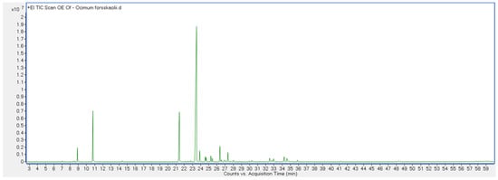
Figure 1.
Chromatogram of GC analysis of OFEO.
3.2. Cytotoxicity and Selectivity Studies
The cytotoxicity of OFEO was evaluated against MCF7, HT29, and HCT116 cell lines and exhibited variable activities (IC50: 5.34–17.09) (Table 3). The colon cancer cells (HT29 and HCT116) were three-fold more sensitive to OFEO compared with the breast cancer cells (MCF7). The IC50 of OFEO was comparable to that of doxorubicin against HCT116 cells. The extract showed selectivity against the three cancer cells compared with MRC5, while it was more selective against HCT116 cells (Table 4).
Table 3.
Cytotoxic activity of OFEO and doxorubicin against three cancer cell lines and normal one.
Table 4.
Selectivity of OFEO and doxorubicin against the cancer cells compared with normal MRC5 fibroblasts.
3.3. Clonogenicity Assay
The clonogenicity of OFEO extract was tested against the most sensitive cell line, i.e., HCT116 cells. The concentrations tested were IC50, 2 × IC50 and 4 × IC50 (i.e., 6, 12, and 24 μg/mL). The extract decreased the number of HCT116 colonies at 6 μg/mL, while it caused complete colony death at 12 and 24 μg/mL Figure 2.
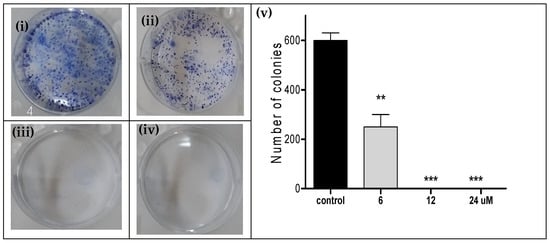
Figure 2.
Clonogenicity of HCT116 cells following 72 h treatment in a six-well plate with (i) vehicle control, (ii) OFEO (6 μg/mL), (iii) OFEO (12 μg/mL), and (iv) OFEO (24 μg/mL). (v): Bar chart showing the four treatments (x-axis) against the number of HCT116 colonies (y-axis). Experiments were repeated three times. Statistical differences compared with untreated control cells were assessed by one-way ANOVA with the Tukey’s post-hoc multiple comparison test (p < 0.010 (**), p < 0.001 (***) were taken as significant).
3.4. Western Blotting
Following the identification of HCT116 cell line as the most sensitive to the OFEO, the possible underlying cell proliferation/survival, cell cycle, and apoptosis molecular mechanisms of cell death were investigated. Immunoblotting can be used to identify the up- and down-regulated proteins based on different concentrations of tested essential oil. Figure 3 represents the relative gene expression of studied genes related to GAPDH as reference protein. Caspase 3 was up-regulated, while the AKT, pAKT, PCNA, Bcl2, and cyclinD1 were down-regulated.
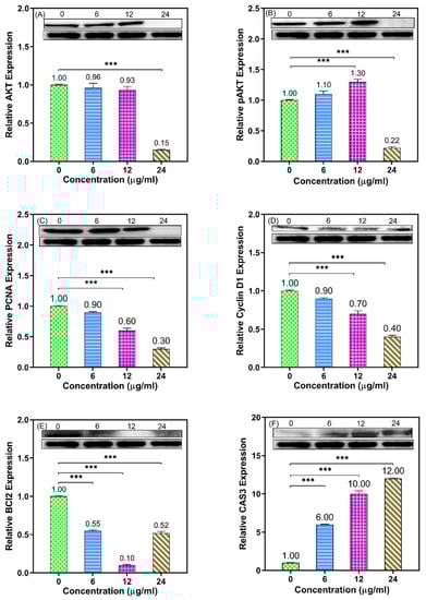
Figure 3.
Western blotting analysis showing the effect of OFEO (0, 6, 12, and 24 μg/mL; 24 h) in cultures of HCT116 regarding eight proteins: AKT (A), pAKT (B), PCNA (C), CyclinD1 (D), Bcl2 (E), and Caspase 3 (F) compared with GAPDH as the reference protein (bottom blots). Bar graphs represent the quantitative densitometric value of the expressed protein vs. GAPDH and refer to mean values calculated from three experiments (*** p < 0.001 vs. control).
3.5. Quantitative Real-Time PCR (qRT-PCR)
To confirm whether pre-treatment with OFEO influences the gene expression of cell cycle and apoptosis pathways in HCT116 cells, qRT-PCR was performed. As shown in Figure 4, the results revealed that OFEO at 12 ug/mL (24 h) upregulated the expression levels of Caspase 3, while it downregulated pAKT, PCNA, Bcl2, and Cyclin D1, all compared with GAPDH. These results indicated that treatment of HCT116 cells with OFEO inhibited the cell proliferation, caused cell cycle arrest, and induced apoptosis in HCT116 cells.
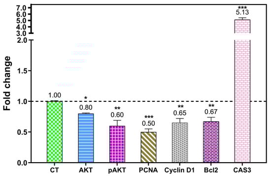
Figure 4.
qRT-PCR results showing fold change in the gene expression of (decrease): AKT, phosphorylated AKT, PCNA, cyclin D1, and Bcl2. Caspase 3 increased in HCT116 cells treated with OFEO (12 ug/mL, 24 h). Gene expression was related to GAPDH as a housekeeping gene, and was normalized to the untreated cells (control, CT = 1-fold change as shown by the dashed line). p < 0.100 (*), p < 0.010 (**), and p < 0.001 (***) were taken as significant.
3.6. Antibacterial Activity
3.6.1. Well-Diffusion Method
The zones of inhibitions were recorded and illustrated in Table 5. The diameter of inhibition of the growth occurred from the diffusion of the solution applied into the agar media. We compared the zone of inhibition of OFEO solution with the positive control diameter. The OFEO showed medium inhibition for E. coli, K. pneumonia, P. aeruginosa, and S. aureus (15, 15, 15, and 13 mm, respectively) and with Amikacin (18, 20, and 22 mm).
Table 5.
Means of inhibition zone in mm for each bacterial strain with the three solutions and positive control (discs of Amikacin for G −ve bacteria and Amoxicillin for G +ve bacteria).
3.6.2. Minimum Inhibitory Concentration (MIC) Assay
The MIC is the lowest concentration of the solutions prepared of OFEO that showed inhibition of the growth of the tested bacterial strains and appeared as a clear solution. Microdilution of the solution concentration of OFEO started from 1000 μg/mL and then was subsequently diluted to make 500, 250, and 125 μg/mL. OFEO inhibited the growth of all bacterial strains at 250 μg/mL except P. aeruginosa, which was inhibited at 500 μg/mL as shown in Table 6.
Table 6.
The mean of the MIC assay for the OFEO solution with the bacterial reference strains used in this study. Each concentration is shown in μg/mL.
4. Discussion
The investigated plant grows in the Makkah region in extreme environmental conditions, characterized by a long, sweltering, and arid summer, with maximum temperatures around 45 °C in June and minimum temperatures around 29 °C in the same month; the winter is short, comfortable, dry, and mostly clear. By means of GC-MS, three main classes of compounds (phenylpropanoids, monoterpenes, and sesquiterpenes) were identified in the OFEO. Methyl eugenol, eugenol and linalool were the major constituents, and also detected in other Ocimum species, including O. basilicum, O. gratissimum, O. campechianum, and O. tenuiflorum [29,30]. Sesquiterpenes were the second most abundant class. Some of them, such as β-caryophyllene and germacrene D, are also common in other Ocimum species, such as O. suave [31]. The sesquiterpene mint sulfide was the most unusual component characterized in this EO, being identified for the first time in the genus Ocimum. However, previous studies on this species revealed evident compositional variation, which could be attributed to the geographical and environmental diversity [26,27,28].
This genus has multiple traditional uses in addition to its reported pharmacological properties [1,2,3]. The inhibition of cancer cell proliferation and survival are some of the main strategies of anticancer drugs [14]. In the present study, the cytotoxic effect of OFEO against one breast and two colon cancer cells was studied using the MTT method. The resulting IC50 ranged between 5.34 and 17.09 μg/mL. The colon cancer cells (HT29 and HCT116) were more sensitive to OFEO compared with the breast cancer cells (MCF7). Most importantly, OFEO showed selectivity against the three cancer cells (especially against HCT116 cells) compared with the normal cells (MRC5). Next, the HCT116 cell line was subjected to clonogenic assay, which confirmed that OFEO has a dose-dependent clonogenic effect on this cell line. Further mechanistic studies using Western Blotting and qRT-PCR showed that OFEO inhibited cell proliferation, caused cell cycle arrest, and induced apoptosis in HCT116 cells. This was evident as OFEO significantly upregulated the expression level of Caspase 3, while downregulating AKT, pAKT, PCNA, Cyclin D1, and Bcl2, all compared with GAPDH. Previously, a non-volatile metabolite, caffeic acid, isolated from O. gratissimum L. showed both anti-proliferative and apoptotic effects against HeLa cells [9]. However, ursolic acid, isolated from an O. basilicum extract, showed cell cycle effects [10]. The main constituent methyl eugenol constitutes 55.65% of the whole oil, and the activity could be attributed mainly to this compound, and methyl eugenol or the essential oils/extracts containing it were proofed for their anticancer activity; the ethanol extract Rosa persica roots exhibited anticancer activity against human glioblastoma (U-87-MG) and human breast cancer (MDA-MB-231) cell lines [45], and another study revealed that the combination of methyl eugenol and myricetin enhanced the anticancer activity, cell cycle arrest, and apoptosis induction of cis-platin against HeLa cervical cancer cell lines [46]. Some studies on eugenol have also revealed a potential apoptotic and anti-angiogenic effect in gastric carcinogenesis [47]. Eugenol decreased Bcl-2 and Bcl-xL expression, and increased the expression of Bax, Bid, Bad, Apaf-1, cytochrome C, and caspase-9. Eugenol induced autophagy and apoptosis in triple-negative breast cancer cells (MDA-MB-231) via pi3k/akt/foxo3a pathway inhibition [48]. Some studies have also revealed the anticancer effect of eugenol on the HCT116 cell line [49]. In the present study, the markers that upregulate the apoptosis and downregulate the proliferation were determined by Western Blot.
MIC can be used as a semi-quantitative assay, indicating the minimum concentration capable to inhibit the growth of each bacterial strain. The well diffusion method and MIC assay performed in the present study showed that OFEO had a certain antimicrobial effect on Gram-negative strains and a good inhibition activity against the Gram-positive bacteria (S. aureus). In this study OFEO was shown to inhibit growth of all bacteria at 250 μg/mL, except P. aeruginosa, which required 500 μg/mL, indicating good antimicrobial activity of this oil. The essential oil of Ocimum sp. has previously been reported to possess marked antimicrobial activity against a broad range of bacteria and fungi due to the presence of diverse classes of volatile constituents, such as monoterpenes (linalool, 1,8-cineole, geranial and neral), phenylpropanoids (methyl cinnamate, methyl chavicol, eugenol), and sesquiterpenes (β-selinene) [12,32,33]. Thus, OFEO has cytotoxic antiproliferative, cell cycle arrest, and apoptotic effects on HCT116 colon cancer cells. The antimicrobial effect observed on Gram-negative bacteria (E. coli and K. pneumonia) and Gram-positive bacteria (S. aureus) warrant further phytochemical and pharmacological investigations on the essential oil [2].
5. Conclusions
OFEO is rich in secondary metabolites, which possess many biological and therapeutical properties that indicate its possible use for the treatment of various ailments. It is mainly composed of small molecules, which have the ability to interact with cellular enzymes, causing upregulation of apoptotic protein and downregulation of proliferative and cell cycle proteins. Since this plant is used as a food additive, it could be recommended for use as an adjuvant cancer therapy, as well as a supplement for the prevention of malignant tumors. In addition, OFEO could be used to compose certain anti-microbial formulas to be used locally and systematically for the treatment of infectious diseases. As potential future research, OFEO could be investigated in combination with classical anticancer drugs in order to reduce their side effects and enhance their therapeutic effects.
As a limitation of our work, we can highlight some technical issues related to the formulations and the lipophilic character of the essential oil, which should imply the use of advanced formulation technologies, such as micro and nano formulations.
Author Contributions
Conceptualization, A.B. and A.N.A.; methodology, A.N.A., N.A.O., M.E.E. and M.Z.E.-R.; software, Y.P., validation, H.M.N.; formal analysis, G.F. and Y.P.; investigation, A.N.A., L.Y.; M.E.E., G.F., Y.P. and M.Z.E.-R.; resources, A.B.; data curation, R.A.A. and H.M.N.; writing—original draft preparation, A.B.; writing A.N.A., N.A.O., G.F., Y.P. and M.Z.E.-R.; review and editing G.F.; visualization, R.A.A.; supervision, A.B.; project administration A.B.; funding acquisition, A.B. All authors have read and agreed to the published version of the manuscript.
Funding
This research was funded by Deanship of Scientific Research at Umm Al-Qura University for supporting this work by grant code 22UQU4320529DSR05.
Institutional Review Board Statement
Not applicable.
Informed Consent Statement
Not applicable.
Data Availability Statement
All relevant data are contained in the present manuscript.
Acknowledgments
The authors would like to thank the Deanship of Scientific Research at Umm Al-Qura University for supporting this work by grant code 22UQU4320529DSR05.
Conflicts of Interest
The authors declare no conflict of interest.
Abbreviations
The following abbreviations were used: OFEO, Ocimum forskolei essential oil; MCF-7 Michigan Cancer Foundation-7 breast cancer; HT-29, human colon cancer cell line; HCT116, human colorectal carcinoma cell line; MRC-5 (Medical Research Council cell strain 5) fibroblasts; MTT assay, 3-(4,5-dimethylthiazol-2-yl)-2,5-diphenyltetrazolium bromide; qRT-PCR, Real-Time Quantitative Reverse Transcription PCR.
References
- Martiz, R.M.; Patil, S.M.; Abdulaziz, M.; Babalghith, A.; Al-Areefi, M.; Al-Ghorbani, M.; Mallappa Kumar, J.; Prasad, A.; Mysore Nagalingaswamy, N.P.; Ramu, R. Defining the Role of Isoeugenol from Ocimum tenuiflorum against Diabetes Mellitus-Linked Alzheimer’s Disease through Network Pharmacology and Computational Methods. Molecules 2022, 27, 2398. [Google Scholar] [CrossRef] [PubMed]
- AlQathama, A.; Ezuruike, U.F.; Mazzari, A.L.; Yonbawi, A.; Chieli, E.; Prieto, J.M. Effects of selected Nigerian medicinal plants on the viability, mobility, and multidrug-resistant mechanisms in liver, colon, and skin cancer cell lines. Front. Pharmacol. 2020, 11, 546439. [Google Scholar] [CrossRef] [PubMed]
- Sakr, S.A.; Al-Amoudi, W.M. Effect of leave extract of Ocimum basilicum on deltamethrin induced nephrotoxicity and oxidative stress in albino rats. J. Appl. Pharm. Sci. 2012, 2, 22–27. [Google Scholar] [CrossRef]
- Leelapornpisid, P.; Wickett, R.R.; Chansakaow, S.; Wongwattananukul, N. Potential of native Thai aromatic plant extracts in antiwrinkle body creams. J. Cosmet. Sci. 2015, 66, 219–231. [Google Scholar]
- Nadeem, H.R.; Akhtar, S.; Ismail, T.; Qamar, M.; Sestili, P.; Saeed, W.; Azeem, M.; Esatbeyoglu, T. Antioxidant Effect of Ocimum basilicum Essential Oil and Its Effect on Cooking Qualities of Supplemented Chicken Nuggets. Antioxidants 2022, 11, 1882. [Google Scholar] [CrossRef] [PubMed]
- Abdelhady, M.I.; Motaal, A.A. A cytotoxic C-glycosylated derivative of apigenin from the leaves of Ocimum basilicum var. thyrsiflorum. Rev. Bras. Farmacogn. 2016, 26, 763–766. [Google Scholar] [CrossRef][Green Version]
- Sharmin, E.; Kafyah, M.T.; Alzaydi, A.A.; Fatani, A.A.; Hazazzi, F.A.; Babgi, S.K.; Alqarhi, N.M.; Sindi, A.A.H.; Akram, D.; Alam, M.; et al. Synthesis and characterization of polyvinyl alcohol/corn starch/linseed polyol-based hydrogel loaded with biosynthesized silver nanoparticles. Int. J. Biol. Macromol. 2020, 163, 2236–2247. [Google Scholar] [CrossRef]
- Manosroi, J.; Dhumtanom, P.; Manosroi, A. Anti-proliferative activity of essential oil extracted from Thai medicinal plants on KB and P388 cell lines. Cancer Lett. 2006, 235, 114–120. [Google Scholar] [CrossRef]
- Chang, W.C.; Hsieh, C.H.; Hsiao, M.W.; Lin, W.C.; Hung, Y.C.; Ye, J.C. Caffeic acid induces apoptosis in human cervical cancer cells through the mitochondrial pathway. Taiwan J. Obstet. Gynecol. 2010, 49, 419–424. [Google Scholar] [CrossRef]
- Kehkashan Arshad, Q.; Ahsana, D.; Bina, S.S.; Nurul, K.; Huma, A.; Shakil, A.; Shaista, E.; Shazia, H.; Sabira, B. Anticancer Activity of Ocimum basilicum and the Effect of Ursolic Acid on the Cytoskeleton of MCF-7 Human Breast Cancer Cells. Lett. Drug Des. Discov. 2010, 7, 726–736. [Google Scholar] [CrossRef]
- Rao, B.R.R.; Kothari, S.K.; Rajput, D.K.; Patel, R.P.; Darokar, M.P. Chemical and biological diversity in fourteen selections of four Ocimum species. Nat. Prod. Commun. 2011, 6, 1934578X1100601134. [Google Scholar] [CrossRef]
- Padalia, R.C.; Verma, R.S.; Chauhan, A.; Goswami, P.; Singh, V.R.; Verma, S.K.; Darokar, M.P.; Singh, N.; Saikia, D.; Chanotiya, C.S. Essential Oil Composition and Antimicrobial Activity of Methyl cinnamate-Linalool Chemovariant of Ocimum basilicum L. from India. Rec. Nat. Prod. 2017, 11, 193–204. [Google Scholar]
- Sung, H.; Ferlay, J.; Siegel, R.L.; Laversanne, M.; Soerjomataram, I.; Jemal, A.; Bray, F. Global Cancer Statistics 2020: GLOBOCAN Estimates of Incidence and Mortality Worldwide for 36 Cancers in 185 Countries. CA Cancer J. Clin. 2021, 71, 209–249. [Google Scholar] [CrossRef]
- El-Readi, M.Z.; Al-Abd, A.M.; Althubiti, M.A.; Almaimani, R.A.; Al-Amoodi, H.S.; Ashour, M.L.; Wink, M.; Eid, S.Y. Multiple Molecular Mechanisms to Overcome Multidrug Resistance in Cancer by Natural Secondary Metabolites. Front. Pharmacol. 2021, 12, 658513. [Google Scholar] [CrossRef] [PubMed]
- Banerjee, S.; Wang, Z.; Mohammad, M.; Sarkar, F.H.; Mohammad, R.M. Efficacy of selected natural products as therapeutic agents against cancer. J. Nat. Prod. 2008, 71, 492–496. [Google Scholar] [CrossRef] [PubMed]
- Bloom, D.E.; Cadarette, D. Infectious disease threats in the twenty-first century: Strengthening the global response. Front. Immunol. 2019, 10, 549. [Google Scholar] [CrossRef] [PubMed]
- Dal Piaz, F.; Bader, A.; Malafronte, N.; D’Ambola, M.; Petrone, A.M.; Porta, A.; Hadda, T.B.; De Tommasi, N.; Bisio, A.; Severino, L. Phytochemistry of compounds isolated from the leaf-surface extract of Psiadia punctulata (DC.) Vatke growing in Saudi Arabia. Phytochemistry 2018, 155, 191–202. [Google Scholar] [CrossRef]
- El-Readi, M.Z.; Eid, H.H.; Ashour, M.L.; Eid, S.Y.; Labib, R.M.; Sporer, F.; Wink, M. Variations of the chemical composition and bioactivity of essential oils from leaves and stems of Liquidambar styraciflua (Altingiaceae). J. Pharm. Pharmacol. 2013, 65, 1653–1663. [Google Scholar] [CrossRef]
- Di Stasi, M.; Donadio, G.; Bader, A.; De Leo, M.; Braca, A. Two new triterpenes from Commicarpus grandiflorus (A. Rich.) Standl. aerial parts exudate. Nat. Prod. Res. 2022. [Google Scholar] [CrossRef]
- Cowan, M.M. Plant products as antimicrobial agents. Clin. Microbiol. Rev. 1999, 12, 564–582. [Google Scholar] [CrossRef]
- Youssef, F.S.; Eid, S.Y.; Alshammari, E.; Ashour, M.L.; Wink, M.; El-Readi, M.Z. Chrysanthemum indicum and Chrysanthemum morifolium: Chemical Composition of Their Essential Oils and Their Potential Use as Natural Preservatives with Antimicrobial and Antioxidant Activities. Foods 2020, 9, 1460. [Google Scholar] [CrossRef] [PubMed]
- Amin, S.M.; Hassan, H.M.; El Gendy, A.E.N.G.; El-Beih, A.A.; Mohamed, T.A.; Elshamy, A.I.; Bader, A.; Shams, K.A.; Mohammed, R.; Hegazy, M.E.F. Comparative chemical study and antimicrobial activity of essential oils of three Artemisia species from Egypt and Saudi Arabia. Flavour Fragr. J. 2019, 34, 450–459. [Google Scholar] [CrossRef]
- El-Said, H.; Ashgar, S.S.; Bader, A.; AlQathama, A.; Halwani, M.; Ascrizzi, R.; Flamini, G. Essential Oil Analysis and Antimicrobial Evaluation of Three Aromatic Plant Species Growing in Saudi Arabia. Molecules 2021, 26, 959. [Google Scholar] [CrossRef] [PubMed]
- Mohammed, H.A.; Eldeeb, H.M.; Khan, R.A.; Al-Omar, M.S.; Mohammed, S.A.A.; Sajid, M.S.M.; Aly, M.S.A.; Ahmad, A.M.; Abdellatif, A.A.H.; Eid, S.Y.; et al. Sage, Salvia officinalis L., Constituents, Hepatoprotective Activity, and Cytotoxicity Evaluations of the Essential Oils Obtained from Fresh and Differently Timed Dried Herbs: A Comparative Analysis. Molecules 2021, 26, 5757. [Google Scholar] [CrossRef] [PubMed]
- Gushash, A.S. Plants in the Mountains of Sarat and Hejaz; Sarawat Designer and Printers: Madinah, Saudi Arabia, 2006; Volume 2, pp. 15–19. [Google Scholar]
- Fatope, M.O.; Marwah, R.G.; Al Hadhrami, N.M.; Onifade, A.K.; Williams, J.R. Identification of the Chemotypes of Ocimum forskolei and Ocimum basilicum by NMR Spectroscopy. Chem. Biodivers. 2008, 5, 2457–2463. [Google Scholar] [CrossRef] [PubMed]
- Demissew, S. A description of some essential oil bearing plants in Ethiopia and their indigenous uses. J. Essent. Oil Res. 1993, 5, 465–479. [Google Scholar] [CrossRef]
- Odalo, J.O.; Omolo, M.O.; Malebo, H.; Angira, J.; Njeru, P.M.; Ndiege, I.O.; Hassanali, A. Repellency of essential oils of some plants from the Kenyan coast against Anopheles gambiae. Acta Trop. 2005, 95, 210–218. [Google Scholar] [CrossRef] [PubMed]
- Jayaramaiah, R.H.; Anand, A.; Beedkar, S.D.; Dholakia, B.B.; Punekar, S.A.; Kalunke, R.M.; Gade, W.N.; Thulasiram, H.V.; Giri, A.P. Functional characterization and transient expression manipulation of a new sesquiterpene synthase involved in β-caryophyllene accumulation in Ocimum. Biochem. Biophys. Res. Commun. 2016, 473, 265–271. [Google Scholar] [CrossRef] [PubMed]
- Benitez, N.P.; Meléndez León, E.M.; Stashenko, E.E. Eugenol and methyl eugenol chemotypes of essential oil of species Ocimum gratissimum L. and Ocimum campechianum Mill. from Colombia. J. Chromatogr. Sci. 2009, 47, 800–803. [Google Scholar] [CrossRef]
- Runyoro, D.; Ngassapa, O.; Vagionas, K.; Aligiannis, N.; Graikou, K.; Chinou, I. Chemical composition and antimicrobial activity of the essential oils of four Ocimum species growing in Tanzania. Food Chem. 2010, 119, 311–316. [Google Scholar] [CrossRef]
- van Vuuren, S.; Viljoen, A. Plant-based antimicrobial studies-methods and approaches to study the interaction between natural products. Planta Med. 2011, 77, 1168–1182. [Google Scholar] [CrossRef] [PubMed]
- Stanojevic, L.P.; Marjanovic-Balaban, Z.R.; Kalaba, V.D.; Stanojevic, J.S.; Cvetkovic, D.J.; Cakic, M.D. Chemical Composition, Antioxidant and Antimicrobial Activity of Basil (Ocimum basilicum L.) Essential Oil. J. Essent. Oil Bear. Plants 2017, 20, 1557–1569. [Google Scholar] [CrossRef]
- NIST; Wiley Technology. NIST/EPA/NIH Mass Spectral Library, 1st ed.; Wiley: Hoboken, NJ, USA, 2014; p. 48. [Google Scholar]
- Adams, R. Identification of Essential Oil Components by Gas Chromatography–Mass Spectroscopy; Allured Publishing Corp.: Carol Stream, IL, USA, 1995. [Google Scholar]
- Bader, A.; AlQathama, A.; Cioni, P.L.; Ceccarini, L.; Abdelhady, M.I.; Al-Shareef, W.; Ascrizzi, R.; Flamini, G. Essential Oil Biodiversity of Achillea ligustica All. Obtained from Mainland and Island Populations. Plants 2022, 11, 1054. [Google Scholar] [CrossRef]
- Bader, A.; Flamini, G.; Cioni, P.L.; Morelli, I. The composition of the root oil of Salvadora persica L. J. Essent. Oil Res. 2002, 14, 128–129. [Google Scholar] [CrossRef]
- Bader, A.; Caponi, C.; Cioni, P.L.; Flamini, G.; Morelli, I. Acorenone in the essential oil of flowering aerial parts of Seseli tortuosum L. Flavour Frag. J. 2003, 18, 57–58. [Google Scholar] [CrossRef]
- Shaheen, U.; Ragab, E.A.; Abdalla, A.N.; Bader, A. Triterpenoidal saponins from the fruits of Gleditsia caspica with proapoptotic properties. Phytochemistry 2018, 145, 168–178. [Google Scholar] [CrossRef]
- Khalid, A.; Algarni, A.S.; Homeida, H.E.; Sultana, S.; Javed, S.A.; Abdalla, H.; Alhazmi, H.A.; Albratty, M.; Abdalla, A.N. Phytochemical, Cytotoxic, and Antimicrobial Evaluation of Tribulus terrestris L., Typha domingensis pers., and Ricinus communis L.: Scientific Evidences for Folkloric Uses. Evid.-Based Complement. Altern. Med. 2022, 2022, 6519712. [Google Scholar] [CrossRef]
- Almaimani, R.A.; Aslam, A.; Ahmad, J.; El-Readi, M.Z.; El-Boshy, M.E.; Abdelghany, A.H.; Idris, S.; Alhadrami, M.; Althubiti, M.; Almasmoum, H.A.; et al. In vivo and in vitro enhanced tumoricidal effects of metformin, active vitamin D3, and 5-fluorouracil triple therapy against colon cancer by modulating the PI3K/Akt/PTEN/mTOR network. Cancers 2022, 14, 1538. [Google Scholar] [CrossRef]
- Abdalla, A.N.; Di Stefano, M.; Poli, G.; Tuccinardi, T.; Bader, A.; Vassallo, A.; Abdallah, M.E.; El-Readi, M.Z.; Refaat, B.; Algarni, A.S.; et al. Co-Inhibition of P-gp and Hsp90 by an Isatin-Derived Compound Contributes to the Increase of the Chemosensitivity of MCF7/ADR-Resistant Cells to Doxorubicin. Molecules 2021, 27, 90. [Google Scholar] [CrossRef]
- Valgas, C.; Souza, S.M.D.; Smânia, E.F.; Smânia, A., Jr. Screening methods to determine antibacterial activity of natural products. Braz. J. Microbiol. 2007, 38, 369–380. [Google Scholar] [CrossRef]
- Balouiri, M.; Sadiki, M.; Ibnsouda, S.K. Methods for in vitro evaluating antimicrobial activity: A review. J. Pharm. Anal. 2016, 6, 71–79. [Google Scholar] [CrossRef] [PubMed]
- Koohestanian, A.; Tatari, M.; Samadi Kazemi, M.; Asgharzade, A.; Taghizadeh, S.F. Phytochemicals, Antioxidant Activity, and Biological Activities of Rosa persica Root. Erwerbs-Obstbau 2022. [Google Scholar] [CrossRef]
- Yi, J.L.; Shi, S.; Shen, Y.L.; Wang, L.; Chen, H.Y.; Zhu, J.; Ding, Y. Myricetin and methyl eugenol combination enhances the anticancer activity, cell cycle arrest and apoptosis induction of cis-platin against HeLa cervical cancer cell lines. Int. J. Clin. Exp. Pathol. 2015, 8, 1116. [Google Scholar] [PubMed]
- Manikandan, P.; Murugan, R.S.; Priyadarsini, R.V.; Vinothini, G.; Nagini, S. Eugenol induces apoptosis and inhibits invasion and angiogenesis in a rat model of gastric carcinogenesis induced by MNNG. Life Sci. 2010, 86, 936–941. [Google Scholar] [CrossRef] [PubMed]
- Abdullah, M.L.; Al-Shabanah, O.; Hassan, Z.K.; Hafez, M.M. Eugenol-induced autophagy and apoptosis in breast cancer cells via PI3K/AKT/FOXO3a pathway inhibition. Int. J. Mol. Sci. 2021, 22, 9243. [Google Scholar] [CrossRef]
- Fadilah, F.; Andrajati, R.; Yanuar, A.; Arsianti, A. In-vitro anticancer activity combination of eugenol and simple aromatic benzoate compounds against human colon HCT-116 cells and WiDr cells. J. Pharm. Sci. Res. 2017, 9, 637. [Google Scholar]


Disclaimer/Publisher’s Note: The statements, opinions and data contained in all publications are solely those of the individual author(s) and contributor(s) and not of MDPI and/or the editor(s). MDPI and/or the editor(s) disclaim responsibility for any injury to people or property resulting from any ideas, methods, instructions or products referred to in the content. |
© 2023 by the authors. Licensee MDPI, Basel, Switzerland. This article is an open access article distributed under the terms and conditions of the Creative Commons Attribution (CC BY) license (https://creativecommons.org/licenses/by/4.0/).