Article

Prostaglandin D2 Added during the Differentiation of 3T3-L1 Cells Suppresses Adipogenesis via Dysfunction of D-Prostanoid Receptor P1 and P2

by
Michael N. N. Nartey
1,2,
Mitsuo Jisaka
1,3,4,*,
Pinky Karim Syeda
3,
Kohji Nishimura
1,3,4,5,
Hidehisa Shimizu
1,3,4,5 and
Kazushige Yokota
1,3,4
1
The United Graduate School of Agricultural Sciences, Tottori University, 4-101 Koyama-Minami, Tottori 680-8553, Japan
2
Council for Scientific and Industrial Research-Animal Research Institute, Achimota, Accra P.O. Box AH20, Ghana
3
Department of Life Science and Biotechnology, Shimane University, 1060 Nishikawatsu-Cho, Matsue 690-8504, Japan
4
Institute of Agricultural and Life Sciences, Academic Assembly, Shimane University, 1060 Nishikawatsu-Cho, Matsue 690-8504, Japan
5
Interdisciplinary Center for Science Research, Shimane University, 1060 Nishikawatsu-Cho, Matsue 690-8504, Japan
*
Author to whom correspondence should be addressed.
Life 2023, 13(2), 370; https://doi.org/10.3390/life13020370
Submission received: 11 December 2022 / Revised: 20 January 2023 / Accepted: 27 January 2023 / Published: 29 January 2023
(This article belongs to the Special Issue New Updates in Adipocytes and Adipose Tissue)

Abstract

:
We previously reported that the addition of prostaglandin, (PG)D2, and its chemically stable analog, 11-deoxy-11-methylene-PGD2 (11d-11m-PGD2), during the maturation phase of 3T3-L1 cells promotes adipogenesis. In the present study, we aimed to elucidate the effects of the addition of PGD2 or 11d-11m-PGD2 to 3T3-L1 cells during the differentiation phase on adipogenesis. We found that both PGD2 and 11d-11m-PGD2 suppressed adipogenesis through the downregulation of peroxisome proliferator-activated receptor gamma (PPARγ) expression. However, the latter suppressed adipogenesis more potently than PGD2, most likely because of its higher resistance to spontaneous transformation into PGJ2 derivatives. In addition, this anti-adipogenic effect was attenuated by the coexistence of an IP receptor agonist, suggesting that the effect depends on the intensity of the signaling from the IP receptor. The D-prostanoid receptors 1 (DP1) and 2 (DP2, also known as a chemoattractant receptor-homologous molecule expressed on Th2 cells) are receptors for PGD2. The inhibitory effects of PGD2 and 11d-11m-PGD2 on adipogenesis were slightly attenuated by a DP2 agonist. Furthermore, the addition of PGD2 and 11d-11m-PGD2 during the differentiation phase reduced the DP1 and DP2 expression during the maturation phase. Overall, these results indicated that the addition of PGD2 or 11d-11m-PGD2 during the differentiation phase suppresses adipogenesis via the dysfunction of DP1 and DP2. Therefore, unidentified receptor(s) for both molecules may be involved in the suppression of adipogenesis.

1. Introduction

Obesity caused by overnutrition used to be considered a problem specific to high-income countries, but it is now an increasing problem even in low- and middle-income countries, where the prevalence of obesity previously tended to be low [1]. Obesity is a problem because it causes the development of cardiovascular disease, type 2 diabetes, cancer, osteoarthritis, occupational disorders, and sleep apnea [1]. For instance, as early as 2001, it was predicted that because cardiovascular disease survival rates had become higher in industrialized countries than in non-industrialized countries, the number of patients with cardiovascular disease associated with obesity would increase, particularly in industrialized countries [2]. In addition, the current number of patients with type 2 diabetes associated with obesity is also predicted to increase, particularly in low- and middle-income countries. As a result, increases in the prevalence of nephropathy, atherosclerosis, neuropathy, and retinopathy due to type 2 diabetes are expected in these countries [2]. Thus, obesity, which is a major cause of health problems and a reduced quality of life, has gone from being a relatively minor public health problem primarily affecting affluent societies to a major threat to public health worldwide. To prevent obesity with excess adipose tissue, which has become such a major threat to public health, it is important to understand the mechanisms of adipogenesis. Immortal preadipocyte cell lines, such as 3T3-L1 cells, have been established with the goal of elucidating the mechanisms of adipogenesis [3,4]. The stages of adipogenesis comprise growth, differentiation, and maturation, and incubating confluent 3T3-L1 preadipocytes in medium containing 3-isobutyl-1-methylxanthine (IBMX), dexamethasone, and insulin (MDI) during differentiation can initiate adipogenesis.
Peroxisome proliferator-activated receptors (PPAR)s are ligand-activated transcription factors that belong to the nuclear hormone receptor superfamily and function as heterodimers with the retinoid X receptor (RXR). Three types of PPARs were isolated, PPARα, PPARδ (also known as PPARβ, NUC1, and FAAR), and PPARγ, each with different reported physiological functions [5,6]. PPARγ has two protein isoforms, PPARγ1 and PPARγ2, which result from alternative promoter usage and differential splicing at the 5′ end of the gene [7,8]. Multiple lines of evidence have been reported regarding the importance of PPARγ, one of the master regulators of adipogenesis, during the process of differentiation from preadipocytes to adipocytes, as described below. The findings of the initial in vitro studies showed that the ligand-dependent activation of ectopically expressed PPARγ2 accelerates adipogenesis in fibroblast cell lines [9]. Furthermore, the findings of the studies in which PPARγ antagonists and dominant-negative forms of the receptor were used showed that the loss of the receptor function is related to a decreased ability to differentiate adipocytes [10,11,12]. Although it was difficult to establish the importance of adipogenesis in vivo because the genetic disruption of PPARγ confers an embryonic lethal phenotype [13,14], the data obtained from chimeric animals generated using an improved transgenic technique confirmed the importance of PPARγ during adipogenesis in mice [13,14].
PPARγ can upregulate the expression of a variety of adipocyte-specific genes related to adipogenesis, including adiponectin and lipoprotein lipase (LPL) [15,16]. Adiponectin is known as a fat-derived hormone, and genetic mutations in the adiponectin gene that result in low plasma adiponectin levels have been reported to be associated with metabolic syndrome, including insulin resistant, diabetes, and atherosclerotic disease [17]. LPL is known as an enzyme that hydrolyzes triacylglycerol (TAG) in lipoprotein particles into fatty acids and monoglycerol [18]. In addition, LPL has been reported to be involved in the uptake mechanism of TAG in medium into 3T3-L1 cells, resulting in the accumulation of TAG in the cells [19]. Adiponectin and LPL have a functional PPAR-responsive element (PPRE) in their promoters, and the PPARγ/RXR heterodimer was shown to bind directly to these PPREs and regulate their expression [15,16]. Therefore, these two genes are frequently used as adipogenic marker genes, associated with PPARγ expression levels.
The biosynthesis of prostaglandins (PG)s occurs via multiple enzymatically controlled reactions. This process is initiated by the release of arachidonic acid synthesized from linoleic acid, one of the ingested n-6 polyunsaturated fatty acids (n-6 PUFA), from membrane phospholipids via the catalytic action of phospholipases. Subsequently, arachidonic acid is then converted to PGG2, and then PGH2, through the action of PG endoperoxide synthase, cyclooxygenase (COX). COX has two distinct isozymes, COX-1 and -2, which are differentially regulated [20]. The unstable PGH2 intermediates generated via COX-1 or -2 are substrates for certain PG synthases and catalyze the formation of bioactive PGs [21]. PGs are broadly classified into two categories: anti-adipogenic PGs and pro-adipogenic PGs. Anti-adipogenic PGs include PGE2 [22,23,24] and PGF [25,26], which inhibit adipogenesis by activating EP4 and FP receptors, respectively [23,24,25,26]. Pro-adipogenic PGs include the PGJ2 derivatives, 15-deoxy-Δ12,14-PGJ2 and Δ12-PGJ2 [27,28,29,30], and PGI2 [31,32]. In addition to the IP receptor being a known receptor for PGI2 [32], the activation of the IP receptor is suggested to activate PPARγ in HEK293 cells [33].
PGD2 is biosynthesized via lipocalin-type PGD synthase (L-PGDS), which is preferentially expressed in adipocytes from PGH2 produced from arachidonic acid via COXs [21] and is also known as a precursor to PGJ2 derivatives [34]. In addition to the increased transcriptional activity of PPARγ induced by PGD2 [27,28], the addition of PGD2 during the maturation phase promotes MDI-induced adipogenesis by binding and activating D-prostanoid 1 (DP1) and/or 2 receptor (DP2, also known as a chemoattractant receptor-homologous molecule expressed on TH 2 cells) [35,36]. Based on these reports, PGD2 is considered to be pro-adipogenic. Nevertheless, L-PGDS is involved in PGD2 biosynthesis and is preferentially expressed in adipocytes. Antisense L-PGDS reduces the endogenous expression of L-PGDS and promotes adipogenesis when stably expressed in 3T3-L1 preadipocytes [37]. Conversely, stably overexpressed L-PGDS impairs adipogenesis in 3T3-L1 preadipocytes [38]. Furthermore, the altered L-PGDS expression in 3T3-L1 preadipocytes is reflected by the abundance of PGD2 and PGJ2 derivatives [37,38]. These findings regarding L-PGDS support the notion that incubating 3T3-L1 cells with PGD2 during the differentiation phase inhibits MDI-induced adipogenesis. Furthermore, PGD2 undergoes nonenzymatic dehydration and is readily converted to PGJ2 derivatives [34], whereas the chemically stable PGD2 analog, 11-deoxy-11-methylene-PGD2 (11d-11m-PGD2) with an exocyclic methylene, instead of being in an 11-keto group, is considered to resist spontaneous conversion to PGJ2 derivatives. In fact, we were able to generate an antibody against PGD2 using 11d-11m-PGD2 [34]. Based on the previous reports, we considered 11d-11m-PGD2 to facilitate the analysis of the mechanism through which PGD2 affects MDI-induced adipogenesis. Therefore, in the present study, we aimed to determine whether the incubation of 3T3-L1 cells with PGD2 or 11d-11m-PGD2 during the differentiation phase inhibits MDI-induced adipogenesis during the maturation phase. In addition, the crosstalk between PGD2 or 11d-11m-PGD2 and IP signaling that promotes the pro-adipogenic effect was also analyzed.

2. Materials and Methods

2.1. Materials

The following were obtained from the respective suppliers: Dulbecco’s modified Eagle medium containing 25 mM HEPES, penicillin G potassium salt, streptomycin sulfate, dexamethasone, fatty acid-free bovine serum albumin, and recombinant human insulin (Sigma-Aldrich Corp., St. Louis, MO, USA); L-ascorbic acid phosphate magnesium salt n-hydrate, 3-isobutyl-1-methylxanthine (IBMX), and Triglyceride E-Test Kits (Wako Pure Chemical Industries Ltd., Osaka, Japan); fetal bovine serum (FBS) (MP Biomedicals, Solon, OH, USA); PGD2, 11d-11m-PGD2, MRE-269, BW245C, and 15R-15-methyl-PGD2 (15R-15m-PGD2) (Cayman Chemical (Ann Arbor, MI, USA); M-MLV reverse transcriptase (Ribonuclease H minus, point mutant) and polymerase chain reaction (PCR) Master Mix (Promega Corp., Madison, WI, USA).

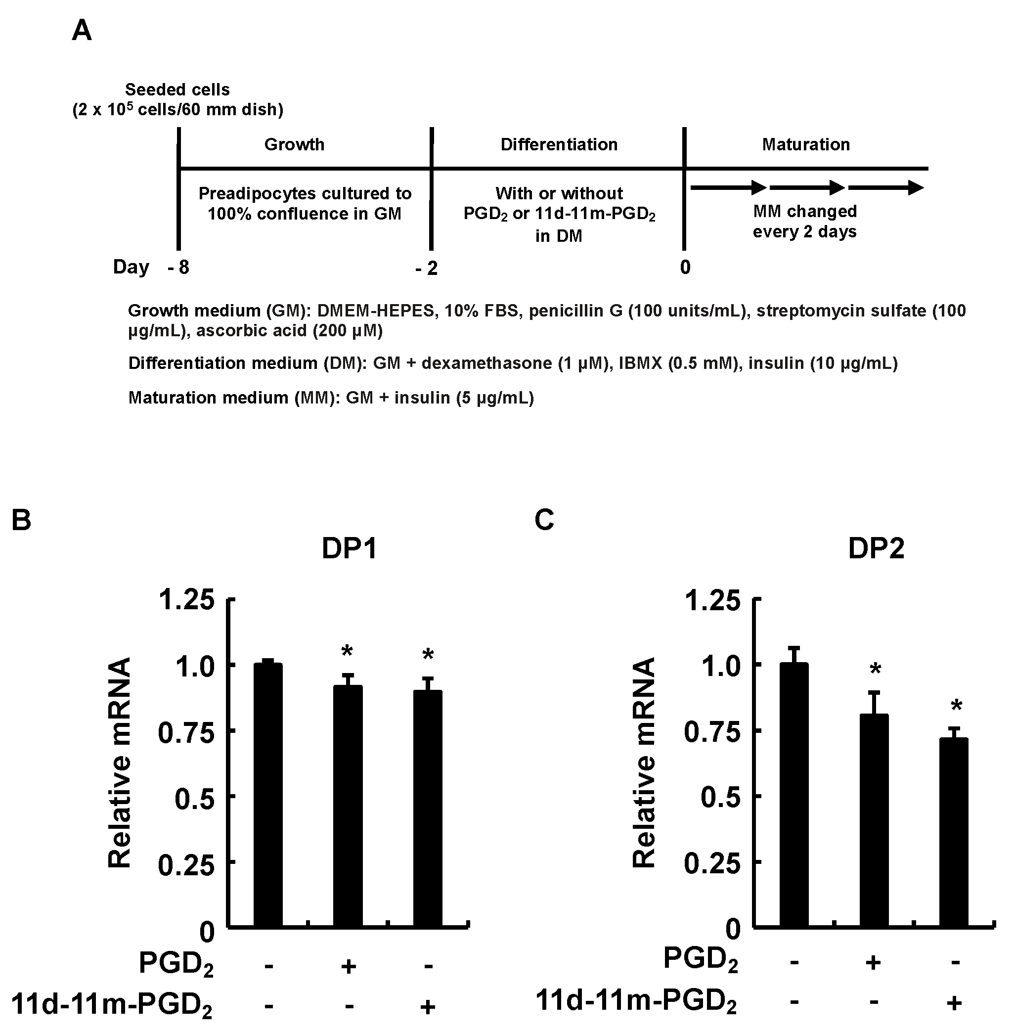
2.2. Cell Culture of 3T3-L1 Cells and Induction of Adipogenesis

Mouse 3T3-L1 pre-adipogenic cells (JCRB9014; JCRB Cell Bank, Osaka, Japan) at the growth phase were seeded at a density of 1 × 10⁵ or 2 × 10⁵ in 35 or 60 mm dishes containing 2 or 4 mL, respectively, in a growth medium (GM) comprising DMEM containing HEPES supplemented with 10% fetal bovine serum (FBS), penicillin G (100 units/mL), streptomycin sulfate (100 μg/L), and ascorbic acid (200 μM). Then, they were incubated at 37 °C under 7% CO2 until they reached confluence. Confluent monolayers were incubated with differentiation medium (DM) comprising GM supplemented with dexamethasone (1 μM), IBMX (0.5 mM), and insulin (10 μg/mL) for 48 h to induce differentiation into adipocytes. The cells were then cultured for 6 to 10 days in a maturation medium (MM; GM supplemented with 5 μg/mL of insulin). The medium was replaced with fresh MM, every 2 days for 10 days, to promote fat storage in the adipocytes during maturation. We examined the effects of various agents on adipogenesis during differentiation by incubating confluent cell monolayers in DM, supplemented with test compounds, for 48 h, followed by the standard maturation protocol. The test compounds were dissolved in ethanol and added to the DM to a final ethanol concentration of 0.2%.

2.3. Quantitation of Intracellular Triacylglycerols and Proteins

The cultured mature adipocytes were harvested, suspended in phosphate-buffered saline (PBS) without Ca2+ and Mg2+ (PBS [-]), supplemented with 0.05% trypsin and 0.53 mM EDTA, and incubated at 37 °C for 5 min. The cell suspensions were washed with PBS (-), divided into two portions, and homogenized in 25 mM Tris-HCl buffer (pH 7.4) containing 1 mM EDTA and 1 N NaOH. Intracellular triacylglycerol (TAG) accumulation was quantified in one portion using Triglyceride E-Test Kits (Wako Pure Chemical Industries Ltd., Osaka, Japan). The cellular proteins were precipitated in the other portion with chilled 6% trichloroacetic acid to remove interfering substances, and then, they were quantified using the Lowry method, with fatty-acid-free bovine serum albumin as the standard. The fat contents were normalized to the protein contents and are expressed as the relative amounts of accumulated lipids in the results.

2.4. Quantitative Analysis of Gene Expression

The total RNA (1 μg) extracted from the cells on day 6 of the maturation phase using acid guanidium thiocyanate/phenol/chloroform was reverse transcribed (RT) using M-MLV reverse transcriptase (Ribonuclease H Minus Point Mutant). The single-stranded cDNA was synthesized using oligo-(dT)15 and a random 9-mer (Promega Corp., Madison, WI, USA) as primers in the RT reaction. The amount of transcript was determined via qRT-PCR using TB GreenTM Premix Ex TaqTM II (Tli RNaseH Plus) kits (Takara Bio Co., Inc., Kusatsu, Japan) and a Thermal Cycler DiceTM Real Time System (Takara Bio Co., Inc., Kusatsu, Japan) according to the threshold cycle (CT) and ΔΔCT methods described by the manufacturer. Table 1 shows the oligonucleotides used herein. The cycling program comprised 95 °C for 30 s, 40 cycles at 95 °C for 5 s and 60 °C for 30 s, followed by 95 °C for 15 s and 60 °C for 30 s. Amounts of target gene transcripts were normalized to those of β-Actin. The accession numbers of the target genes are as follows: PPARγ, NM_011146; adiponectin, NM_009605.5; LPL, NM_008509.2; DP1, NM_008962.4; DP2, XM_006526696.5; β-Actin, NM_007393.

2.5. Statistical Analyses

All of the results are expressed as means ± standard deviation (SD). The data were statistically analyzed via Student t-tests using Excel for Mac (Microsoft Corp., Redmond, WA, USA). The values were considered statistically significant at p < 0.05.

3. Results

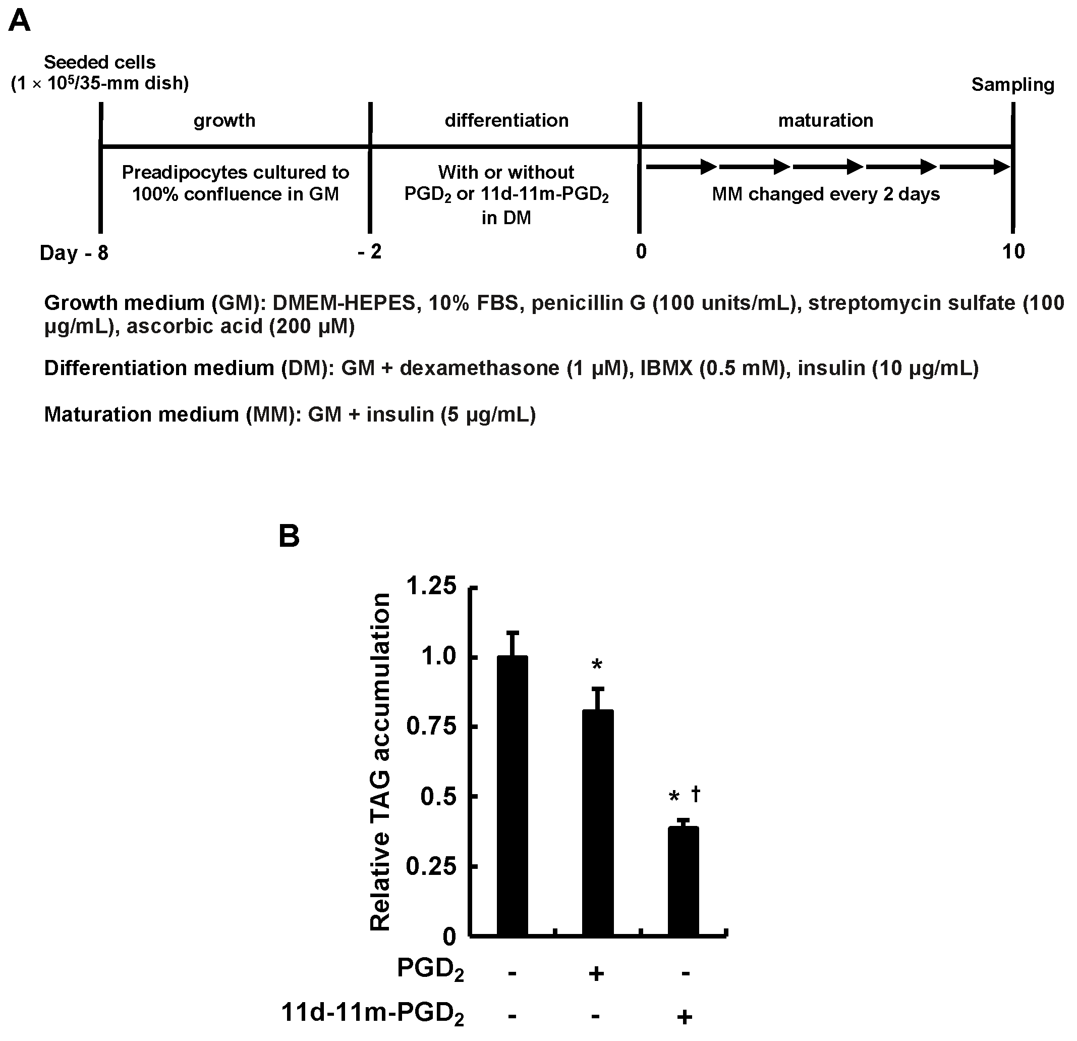
3.1. Incubating 3T3-L1 Cells with PGD2 or 11d-11m-PGD2 during the Differentiation Phase Suppressed MDI-Induced Adipogenesis

We initially examined how the 3T3-L1 cells responded to the addition of PGD2 or 11d-11m-PGD2 during the differentiation phase, which were pro-adipogenic when added during the maturation phase [36] (Figure 1A). We evaluated the adipogenic differentiation of the 3T3-L1 cells based on the MDI-induced intracellular TAG accumulation. In contrast to our previous findings regarding the pro-adipogenic effects of the addition of PGD2 or 11d-11m-PGD2 to cells during the maturation phase [36], both PGs attenuated the MDI-induced intracellular TAG accumulation (Figure 1B). Furthermore, 11d-11m-PGD2 was more anti-adipogenic than PGD2 (Figure 1B). These results suggest that PGD2 and 11d-11m-PGD2 inhibit the fate-deciding mechanism working in adipogenesis during the differentiation phase.

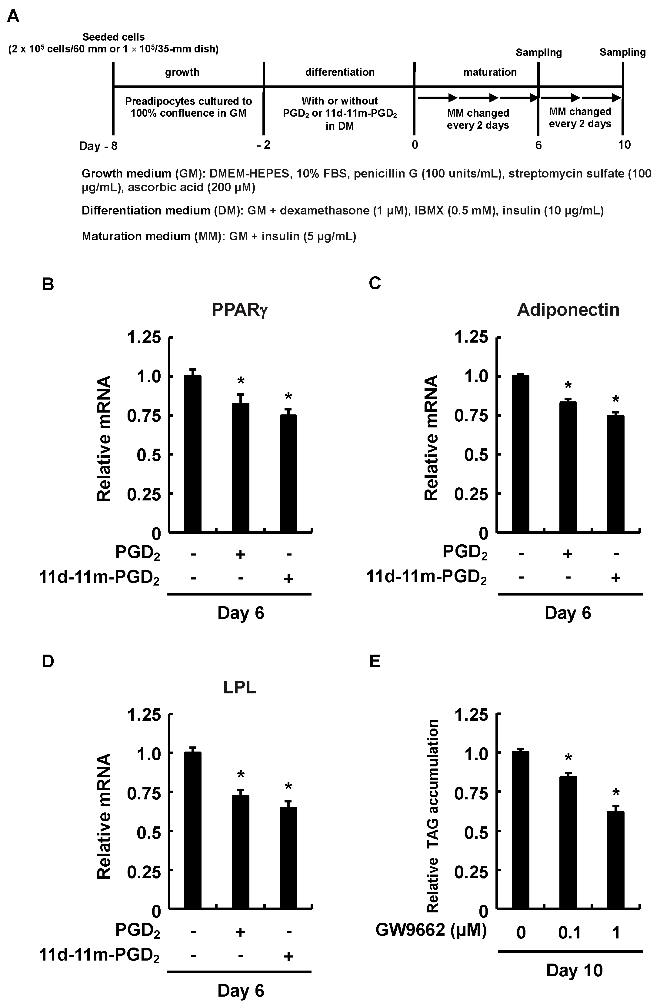
3.2. Downregulated PPARγ Expression Affected the Inhibitory Effects of Addition of PGD2 or 11d-11m-PGD2 during the Differentiation Phase on MDI-Induced Adipogenesis

We further confirmed whether the reduced level of intracellular TAG accumulation during the maturation phase was reflected in the expression levels of PPARγ, known as one of the master regulators of adipogenesis [39], and the adiponectin and LPL regulated downstream of PPARγ [15,16]. We found that the gene expression levels of PPARγ, adiponectin, and LPL in the 3T3-L1 cells peaked by day six in the maturation phase [40] (Figure 2A). In the maturation phase, the expression level of PPARγ in the 3T3-L1 cells was significantly reduced on day six after the addition of PGD2 or 11d-11m-PGD2 during the differentiation phase (Figure 2B). The decreased expression of PPARγ also affected the expression levels of its regulated genes, adiponectin and LPL (Figure 2C,D). In particular, LPL is involved in the mechanism by which 3T3-L1 cells take up TAG from the media [19], suggesting that the attenuation of the PPARγ-LPL pathway was reflected in the level of intracellular TAG accumulation. In fact, the addition of the PPARγ antagonist, GW9662, during the differentiation phase of 3T3-L1 inhibited MDI-induced intracellular TAG accumulation during the maturation phase (Figure 2E). Taken together, these results indicate that the addition of PGD2 or 11d-11m-PGD2 during the differentiation phase suppresses the MDI-induced intracellular TAG accumulation through the downregulation of the PPARγ expression during the maturation phase.

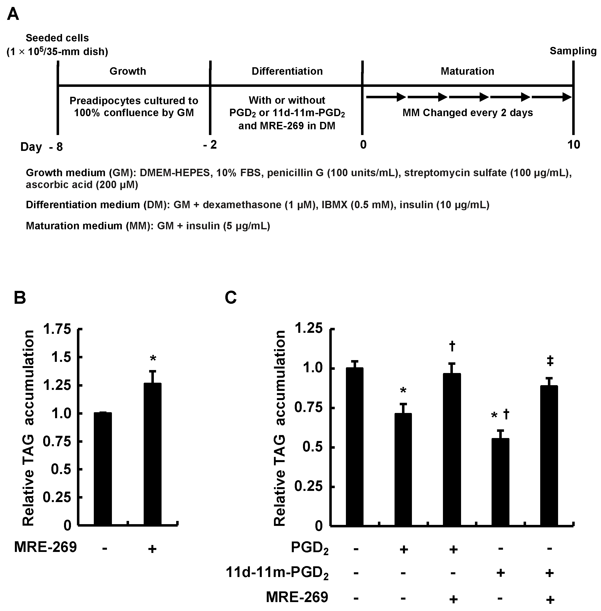
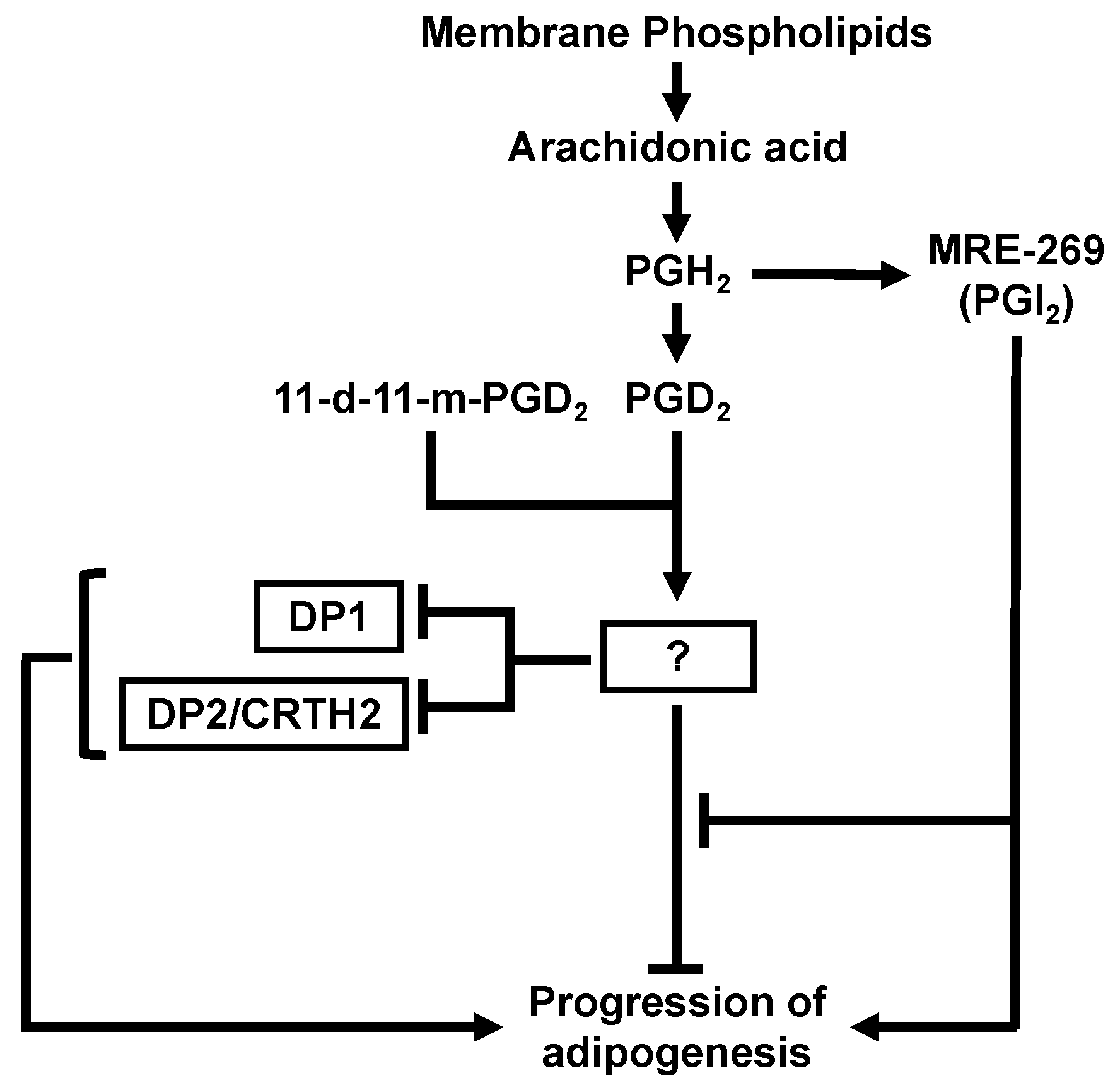
3.3. MRE-269 Attenuated the Inhibitory Effects of Addition of PGD2 or 11d-11m-PGD2 during the Differentiation Phase on MDI-Induced Adipogenesis

To investigate the relationship between the activation of the IP receptor, an origin of the pro-adipogenic signaling of PGI2, and the anti-adipogenic effects of PGD2 or 11d-11m-PGD2, we explored how the coexistence of MRE-269—a highly selective agonist for the IP receptor—with PGD2 or 11d-11m-PGD2 during the differentiation phase affects the adipogenesis that follows (Figure 3A). In addition to promoting adipogenesis by itself (Figure 3B), in this study, MRE-269 abrogated the anti-adipogenic effects of PGD2 or 11d-11m-PGD2 (Figure 3C). These findings indicate that the anti-adipogenic effects of PGD2 and 11d-11m-PGD2 are attenuated depending on the signal from the IP receptor. That is, the levels of PGI2 production and IP receptor expression may influence the anti-adipogenic effects of PGD2 and 11d-11m-PGD2.

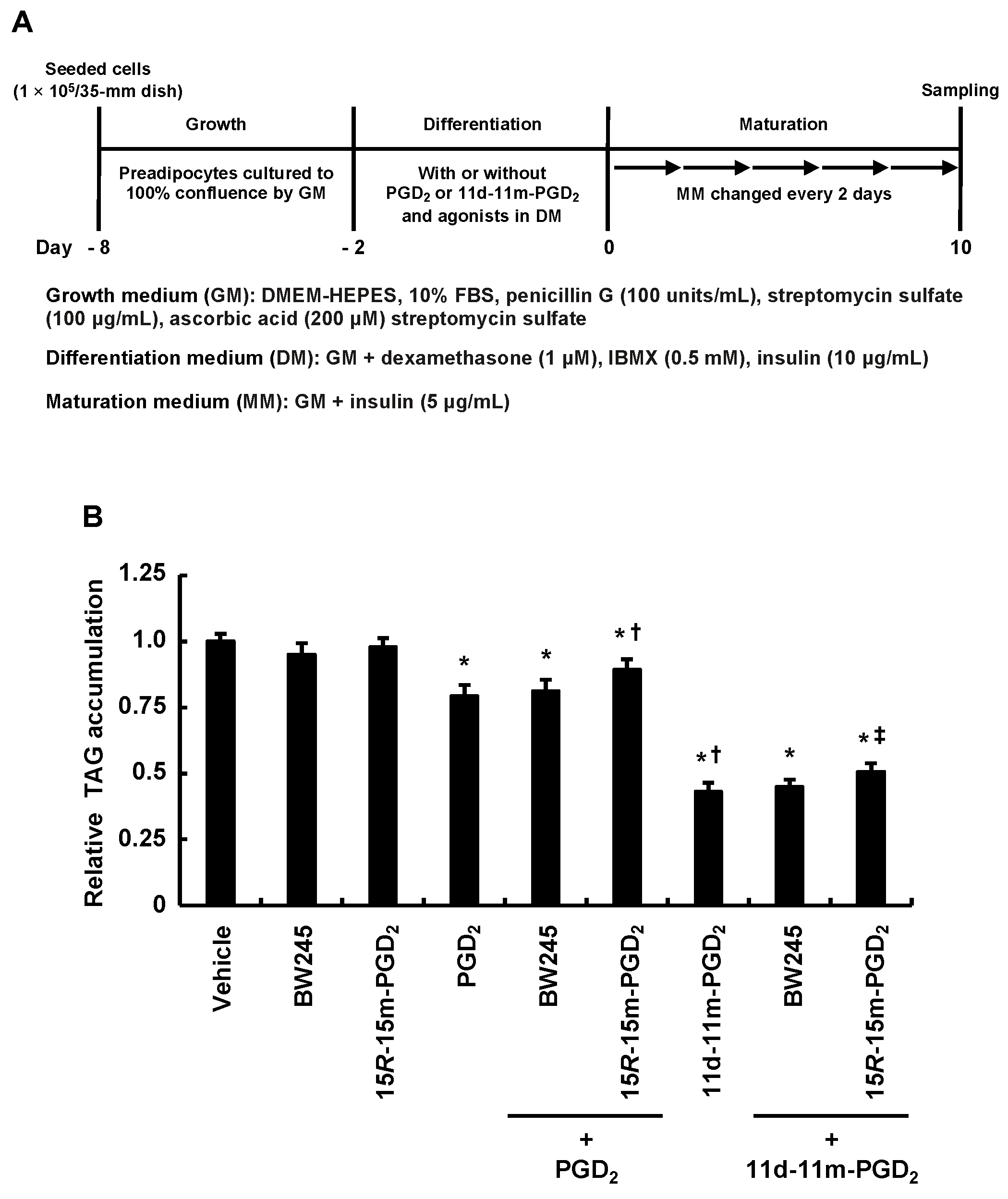
3.4. DP2 Agonist, but Not DP1 Agonist, Partially Alleviated the Inhibitory Effects of Addition of PGD2 or 11d-11m-PGD2 during the Differentiation Phase on MDI-Induced Adipogenesis

We analyzed the inhibitory effects of PGD2 and 11d-11m-PGD2 on MDI-induced adipogenesis using selective agonists for PGD2 receptors, DP1 and DP2, during the differentiation phase, because the activation of these receptors promotes adipogenesis during the maturation phase [35,36] (Figure 4A). The selective DP1 agonist, BW245C, and the selective DP2 agonist, 15R-15m-PGD2, were used at the previously reported concentrations [35,36]. The selective DP1 agonist, BW245C, did not affect the anti-adipogenic effects of PGD2 or 11d-11m-PGD2 (Figure 4B). In contrast, the selective DP2 agonist, 15R-15m-PGD2, slightly but significantly recovered the accumulation of the intracellular TAG that had been decreased by PGD2 and 11d-11m-PGD2 (Figure 4B). These results suggest that PGD2 and 11d-11m-PGD2 exert anti-adipogenic effects by inhibiting the DP1 and DP2 activation induced by BW245C and 15R-15m-PGD2, respectively, during the differentiation phase. That is, PGD2 and 11d-11m-PGD2 may reduce the sensitivity of DP1 to BW245C and DP2 to 15R-15m-PGD2.

3.5. Incubation of 3T3-L1 Cells with PGD2 or 11d-11m-PGD2 during the Differentiation Phase Suppressed Expression of DP1 and DP2 during the Maturation Phase

Finally, we evaluated the effects of the addition of PGD2 or 11d-11m-PGD2 during the differentiation phase on the expression of DP1 and DP2 during the maturation phase (Figure 5A). The incubation of the 3T3-L1 cells with PGD2 or 11d-11m-PGD2 for two days in the differentiation phase significantly reduced the DP1 and DP2 expression on day six in the maturation phase (Figure 5B,C). As DP1 and DP2 contribute to the promotion of adipogenesis during the maturation phase [36], these results suggest that the addition of PGD2 and 11d-11m-PGD2 during the differentiation phase exerts anti-adipogenic effects by decreasing the DP1 and DP2 expression during the maturation phase.

4. Discussion

Figure 6 summarizes the present findings. We found that the addition of PGD2 and 11d-11m-PGD2 during the differentiation phase suppressed adipogenesis during the following maturation phase. These anti-adipogenic effects were disabled by the signal from the IP receptor. In addition, the anti-adipogenic effects of PGD2 and 11d-11m-PGD2 may be related to the decreased expression of PPARγ during the maturation phase, which may be caused by the reduced sensitivity of the PGD2 receptors, DP1 and DP2, during the differentiation phase, as well as by the decreased expression of DP1 and DP2 during the maturation phase. Taken together, PGD2 exerts pro-adipogenic effects when its binding to DP1 and DP2 is prioritized during the maturation phase [36], but exerts anti-adipogenic effects, presumably by desensitizing DP1 and DP2, during the differentiation phase. The desensitizing effects may be mediated by the preferential binding of PGD2 to unidentified receptor(s), other than DP1 and DP2, which causes the dysfunction of DP1 and DP2 during the differentiation phase, leading to the suppression of adipogenesis in the maturation phase.
The more effective inhibition of adipogenesis via 11d-11m-PGD2 in 3T3-L1 preadipocytes may be because PGD2 is easily converted to PGJ2 derivatives through non-enzymatic dehydration [34], whereas 11d-11m-PGD2 is chemically stable with an exocyclic methylene group in place of an 11-keto group. PGJ2 derivatives from PGD2 may be more pro-adipogenically effective than PGD2 during the differentiation phase. In addition, the conversion of PGD2 to PGJ2 derivatives would reduce the concentration of PGD2 acting on the cells. Nevertheless, we found that adipogenesis was inhibited more potently by 11d-11m-PGD2 than PGD2 because it is more chemically stable and may bind to the target PGD2 receptors more strongly than PGD2.
We previously showed that DP1 and DP2 are expressed during the differentiation phase [36]. However, in addition to the possibility that PGD2 and 11d-11m-PGD2 do not act on DP1 or DP2 during the differentiation phase, based on the results of this study, we propose that unidentified receptors for PGD2 and 11d-11m-PGD2 are involved in the suppression of MDI-induced adipogenesis. Such receptors should have higher affinity for PGD2 and 11d-11m-PGD2 than DP1 and DP2, or should be more abundantly expressed than DP1 and DP2 during the differentiation phase. This would cause the inhibitory effect of PGD2 on adipogenesis to take precedence over its pro-adipogenic effects mediated by DP1 and DP2. In addition, in the present study, the inhibitory effect of PGD2 on adipogenesis was stronger, even though ~50% of the initial PGD2 would have been transformed to PGJ2 derivatives within 6 h at 37 °C in MM [34]. Therefore, PGD2 being bound to unidentified receptor(s) would lead to the inhibition of MDI-induced adipogenesis before its conversion into PGJ2 derivatives.
Neither PGD2 nor 11d-11m-PGD2 suppressed the MDI-induced adipogenesis promoted by MRE-269. A positive feedback loop between PPARγ and cytosine–cytosine–adenosine–adenosine–thymidine (CCAAT) box motif/enhancer binding protein alpha (C/EBPα) functions during adipocyte differentiation [41]. MRE-269 activates PPARγ via the IP receptor [33], suggesting that the PPARγ-C/EBPα loop is activated by MRE-269 and PGI2. The activation of this loop could alleviate the downregulation of PPARγ caused by PGD2 and 11d-11m-PGD2 and restore the MDI-induced adipogenesis. Thus, the balance between the signaling from PGD2 and PGI2 may influence adipogenesis.
According to the findings of the Signaling Pathway Program [42], C/EBPα should bind to the DP1 and DP2 promoters. In addition, C/EBPα forms a regulatory positive feedback loop with PPARγ during adipogenesis [41]. Based on these findings, we predict that PPARγ can regulate DP1 and DP2 expression via C/EBPα. This prediction can be supported by our previous finding that the expression of PPARγ, DP1, and DP2 in the 3T3-L1 cells changed, following a similar trend within ten days of the maturation phase [36]. Furthermore, this finding also suggests that DP1 and DP2 may contribute to adipogenesis during the maturation phase. If DP1 and DP2 also function in a pro-adipogenic manner during the differentiation phase, the addition of PGD2 and 11d-11m-PGD2 during that period would promote adipogenesis. The present results, however, reveal that the incubation of the 3T3-L1 cells with PGD2 or 11d-11m-PGD2 for two days in the differentiation phase has anti-adipogenic effects and, furthermore, significantly reduces the expression of PPARγ, DP1, and DP2 on day six in the maturation phase. Thus, if PPARγ activation leads to increased DP1 and DP2 expression during the differentiation phase, then the addition of PGD2 and 11d-11m-PGD2 during that period would be more pro- than anti-adipogenic. Our present finding that MRE-269 canceled the inhibitory effects of PGD2 and 11d-11m-PGD2 on adipogenesis during the differentiation phase may also have been due to the increase in DP1 and DP2 expression via the PPARγ activation induced by MRE-269. Taken together, the PPARγ activated by MRE-269 and the IP receptor will increase the expression of C/EBPα, which may promote the upregulation of DP1 and DP2 expression by binding to the DP1 and DP2 promoters. Based on this scheme, we can propose that unidentified PGD2 receptor(s) exist at least during the differentiation phase and suppress the expression or transcriptional activity of C/EBPα and/or PPARγ, leading to decreases in the DP1 and DP2 expression levels. However, this requires further investigation, in addition to how PGD2 and 11d-11m-PGD2 reduce sensitivity to DP1 and DP2 ligands.
Six PG receptors—DP1, DP2, PPARγ, the IP receptor, the FP receptor, and the EP4 receptor—have been reported to be expressed in the differentiation phase of 3T3-L1 cells [23]. Among these, the FP and EP4 receptors, specific to PGF and PGE2, respectively, exhibit anti-adipogenic effects [32]. Therefore, it could be possible that PGD2 or 11d-11m-PGD2 inhibit adipogenesis via signaling from the FP and/or EP4 receptors if the compounds can act on these receptors. The FP receptor reduces the transcriptional activity of PPARγ via its phosphorylation through the activation of mitogen-activated protein kinase (MAPK) [43]. The EP4 receptor produces PGF by increasing its COX-2 expression [44]. That is, the activation of the EP4 receptor may lead to a decrease in the transcriptional activity of PPARγ, as well as the FP receptor. As PPARγ has an auto-loop mechanism with C/EBPα [41], decreased PPARγ transcriptional activity simultaneously leads to the suppression of its own expression via decreased C/EBPα expression [41]. As a result, the activation of the FP and EP4 receptors would lead to a decrease in the expression of PPARγ, which is similar to the results of the present study. However, if PGD2 and 11d-11m-PGD2 can bind to EP4 and FP receptors, the addition of PGD2 and 11d-11m-PGD2 during the maturation phase should inhibit MDI-induced adipogenesis. Our previous results show that the addition of PGD2 and 11d-11m-PGD2 during the maturation phase promotes MDI-induced adipogenesis [36]. This means that PGD2 and 11d-11m-PGD2 may not bind to EP4 and/or FP receptors. As described above, it is likely that PGD2 and 11d-11m-PGD2 do not bind to the previously reported PG receptors expressed in 3T3-L1 cells and are more likely to be a ligand for other receptors.
We focused on insulin signaling as a mechanism by which PGD2 and 11d-11m-PGD2 inhibit MDI-induced adipogenesis via receptors other than the PG receptors previously reported. Insulin signaling is essential for adipogenesis [45], and insulin was also added during the differentiation phase in the present study. Insulin leads the insulin receptor and its substrate, insulin receptor substrate-1 (IRS-1), to phosphorylate tyrosine residues [46], through which it signals phosphatidylinositol-3 kinase (PI3K) to Akt, ultimately leading to differentiation from preadipocytes to adipocytes [47]. Thus, if PGD2 and 11d-11m-PGD2 can be involved in signaling to dephosphorylate phosphorylated tyrosine residues in the insulin receptor and IRS-1, the differentiation from preadipocytes to adipocytes should be inhibited. The leukocyte common antigen-related phosphatase receptor (LAR, also known as protein tyrosine phosphatase-RF (PTP-RF)), a transmembrane receptor-type PTP, is a receptor that dephosphorylates the tyrosine-residue-phosphorylated insulin receptor and IRS-1, and has been reported to inhibit the differentiation from preadipocytes to adipocytes as a PTP [48]. However, unlike transmembrane receptor-type protein tyrosine kinases, transmembrane receptor-type PTPs, including LAR, have reduced PTP activity upon the ligand-mediated dimerization of their receptors [49]. That is, if PGD2 and 11d-11m-PGD2 are ligands for LAR, PGD2 may enhance insulin signaling and thereby promote adipogenesis by suppressing PTP activity through LAR dimerization. Therefore, as PGD2 and 11d-11m-PGD2 were shown to suppress MDI-induced adipogenesis in the present study, it is unlikely that PGD2 and 11d-11m-PGD2 are ligands for transmembrane receptor-type PTPs, including LAR.
Continuing to focus on insulin signaling, in addition to the phosphorylation of tyrosine residues, IRS-1 has also been reported to be a phosphorylated serine site. For instance, when human IRS-1 is phosphorylated at serine 312 (serine 307 in mouse IRS-1), its interaction with the insulin receptor is disrupted [47]. In addition, phosphorylation at serine 616 in human IRS-1 (serine 612 in mouse IRS-1) results in the attenuation of its interaction with PI3K. Thus, the phosphorylation of these serine residues in IRS-1 causes insulin resistance. Consistent with these results, if PGD2 and 11d-11m-PGD2 can bind to the receptors that activate molecules that phosphorylate serine residues of IRS-1, as described above, then the differentiation from preadipocytes to adipocytes should be inhibited by this pathway. However, as we have not yet clarified which intracellular signaling pathways are activated by PGD2 and 11d-11m-PGD2 in 3T3-L1 cells during the differentiation phase, leading to the downregulation of PPARγ expression and anti-adipogenic effects during the maturation phase, we cannot predict which molecules will function as receptor(s) for PGD2 and 11d-11m-PGD2 in the present situation. Therefore, in the future, the clarification of the intracellular signaling pathway induced by PGD2 and 11d-11m-PGD2 and the identification of its receptor are expected, using the decreased expression of PPARγ as an indicator.

5. Conclusions

PGD2 can promote and inhibit adipogenesis, depending on the affinity or expression of DP1 and DP2 and unidentified receptor(s). Therefore, the unknown receptor(s) for PGD2 and the regulatory mechanisms of DP1 and DP2 expression should be identified to understand the effects of PGs on the adipogenic program that drives the differentiation of white adipocytes. In addition, in the investigation of the actions of PGD2, 11d-11m-PGD2 would be a more practical choice.

Author Contributions

Conceptualization, M.J. and K.Y.; formal analysis, M.N.N.N. and K.Y.; investigation, M.N.N.N., M.J., P.K.S., K.N., H.S. and K.Y.; writing—original draft preparation, H.S.; writing—review and editing, M.N.N.N., M.J., P.K.S., K.N. and H.S.; supervision, M.J. and K.Y.; project administration, M.N.N.N.; funding acquisition, K.Y. All authors have read and agreed to the published version of the manuscript.

Funding

This study was supported by Grants-in-Aid for Scientific Research (C) (Grant Number JP25450128 to K.Y.) from the Ministry of Education, Culture, Sports, Science and Technology (MEXT) of Japan.

Institutional Review Board Statement

Not applicable.

Informed Consent Statement

Not applicable.

Data Availability Statement

The data presented in this study are available on request from the corresponding author.

Acknowledgments

The authors are grateful to the Faculty of Life and Environmental Sciences at Shimane University for providing financial support for the publication of this report.

Conflicts of Interest

The authors declare no conflict of interest.

References

  1. Seidell, J.C.; Halberstadt, J. The global burden of obesity and the challenges of prevention. Ann. Nutr. Metab. 2015, 66 (Suppl. S2), 7–12. [Google Scholar] [CrossRef]
  2. Visscher, T.L.; Seidell, J.C. The public health impact of obesity. Annu. Rev. Public. Health 2001, 22, 355–375. [Google Scholar] [CrossRef] [Green Version]
  3. Green, H.; Kehinde, O. Sublines of mouse 3T3 cells that accumulate lipid. Cell 1974, 1, 113–116. [Google Scholar] [CrossRef]
  4. Green, H.; Kehinde, O. An established preadipose cell line and its differentiation in culture. II. Factors affecting the adipose conversion. Cell 1975, 5, 19–27. [Google Scholar] [CrossRef]
  5. Rangwala, S.M.; Lazar, M.A. Transcriptional control of adipogenesis. Annu. Rev. Nutr. 2000, 20, 535–559. [Google Scholar] [CrossRef] [PubMed]
  6. Rosen, E.D.; Spiegelman, B.M. Molecular regulation of adipogenesis. Annu. Rev. Cell Dev. Biol. 2000, 16, 145–171. [Google Scholar] [CrossRef]
  7. Zhu, Y.; Qi, C.; Korenberg, J.R.; Chen, X.N.; Noya, D.; Rao, M.S.; Reddy, J.K. Structural organization of mouse peroxisome proliferator-activated receptor γ (mPPARγ) gene: Alternative promoter use and different splicing yield two mPPARγ isoforms. Proc. Natl. Acad. Sci. USA 1995, 92, 7921–7925. [Google Scholar] [CrossRef] [Green Version]
  8. Fajas, L.; Auboeuf, D.; Raspé, E.; Schoonjans, K.; Lefebvre, A.M.; Saladin, R.; Najib, J.; Laville, M.; Fruchart, J.C.; Deeb, S.; et al. The organization, promoter analysis, and expression of the human PPARγ gene. J. Biol. Chem. 1997, 272, 18779–18789. [Google Scholar] [CrossRef] [Green Version]
  9. Tontonoz, P.; Hu, E.; Spiegelman, B.M. Stimulation of adipogenesis in fibroblasts by PPARγ2, a lipid-activated transcription factor. Cell 1994, 79, 1147–1156. [Google Scholar] [CrossRef]
  10. Wright, H.M.; Clish, C.B.; Mikami, T.; Hauser, S.; Yanagi, K.; Hiramatsu, R.; Serhan, C.N.; Spiegelman, B.M. A synthetic antagonist for the peroxisome proliferator-activated receptor γ inhibits adipocyte differentiation. J. Biol. Chem. 2000, 275, 1873–1877. [Google Scholar] [CrossRef]
  11. Camp, H.S.; Chaudhry, A.; Leff, T. A novel potent antagonist of peroxisome proliferator-activated receptor γ blocks adipocyte differentiation but does not revert the phenotype of terminally differentiated adipocytes. Endocrinology 2001, 142, 3207–3213. [Google Scholar] [CrossRef]
  12. Gurnell, M.; Wentworth, J.M.; Agostini, M.; Adams, M.; Collingwood, T.N.; Provenzano, C.; Browne, P.O.; Rajanayagam, O.; Burris, T.P.; Schwabe, J.W.; et al. A dominant-negative peroxisome proliferator-activated receptor γ (PPARγ) mutant is a constitutive repressor and inhibits PPARγ-mediated adipogenesis. J. Biol. Chem. 2000, 275, 5754–5759. [Google Scholar] [CrossRef] [Green Version]
  13. Barak, Y.; Nelson, M.C.; Ong, E.S.; Jones, Y.Z.; Ruiz-Lozano, P.; Chien, K.R.; Koder, A.; Evans, R.M. PPARγ is required for placental, cardiac, and adipose tissue development. Mol. Cell 1999, 4, 585–595. [Google Scholar] [CrossRef] [PubMed]
  14. Rosen, E.D.; Sarraf, P.; Troy, A.E.; Bradwin, G.; Moore, K.; Milstone, D.S.; Spiegelman, B.M.; Mortensen, R.M. PPARγ is required for the differentiation of adipose tissue in vivo and in vitro. Mol. Cell 1999, 4, 611–617. [Google Scholar] [CrossRef]
  15. Iwaki, M.; Matsuda, M.; Maeda, N.; Funahashi, T.; Matsuzawa, Y.; Makishima, M.; Shimomura, I. Induction of adiponectin, a fat-derived antidiabetic and antiatherogenic factor, by nuclear receptors. Diabetes 2003, 52, 1655–1663. [Google Scholar] [CrossRef] [PubMed] [Green Version]
  16. Schoonjans, K.; Peinado-Onsurbe, J.; Lefebvre, A.M.; Heyman, R.A.; Briggs, M.; Deeb, S.; Staels, B.; Auwerx, J. PPARα and PPARγ activators direct a distinct tissue-specific transcriptional response via a PPRE in the lipoprotein lipase gene. EMBO J. 1996, 15, 5336–5348. [Google Scholar] [CrossRef]
  17. Kondo, H.; Shimomura, I.; Matsukawa, Y.; Kumada, M.; Takahashi, M.; Matsuda, M.; Ouchi, N.; Kihara, S.; Kawamoto, T.; Sumitsuji, S.; et al. Association of adiponectin mutation with type 2 diabetes: A candidate gene for the insulin resistance syndrome. Diabetes 2002, 51, 2325–2328. [Google Scholar] [CrossRef] [PubMed] [Green Version]
  18. Auwerx, J.; Leroy, P.; Schoonjans, K. Lipoprotein lipase: Recent contributions from molecular biology. Crit. Rev. Clin. Lab. Sci. 1992, 29, 243–268. [Google Scholar] [CrossRef]
  19. Wise, L.S.; Green, H. Studies of lipoprotein lipase during the adipose conversion of 3T3 cells. Cell 1978, 13, 233–242. [Google Scholar] [CrossRef]
  20. Vane, J.R.; Bakhle, Y.S.; Botting, R.M. Cyclooxygenases 1 and 2. Annu. Rev. Pharmacol. Toxicol. 1998, 38, 97–120. [Google Scholar] [CrossRef]
  21. Smith, W.L.; DeWitt, D.L.; Garavito, R.M. Cyclooxygenases: Structural, cellular, and molecular biology. Annu. Rev. Biochem. 2000, 69, 145–182. [Google Scholar] [CrossRef] [PubMed] [Green Version]
  22. Sugimoto, Y.; Tsuboi, H.; Okuno, Y.; Tamba, S.; Tsuchiya, S.; Tsujimoto, G.; Ichikawa, A. Microarray evaluation of EP4 recep-tor-mediated prostaglandin E2 suppression of 3T3-L1 adipocyte differentiation. Biochem. Biophys. Res. Commun. 2004, 322, 911–917. [Google Scholar] [CrossRef]
  23. Tsuboi, H.; Sugimoto, Y.; Kainoh, T.; Ichikawa, A. Prostanoid EP4 receptor is involved in suppression of 3T3-L1 adipocyte differentiation. Biochem. Biophys. Res. Commun. 2004, 322, 1066–1072. [Google Scholar] [CrossRef] [PubMed]
  24. Inazumi, T.; Shirata, N.; Morimoto, K.; Takano, H.; Segi-Nishida, E.; Sugimoto, Y. Prostaglandin E2-EP4 signaling suppresses adipocyte differentiation in mouse embryonic fibroblasts via an autocrine mechanism. J. Lipid Res. 2011, 52, 1500–1508. [Google Scholar] [CrossRef] [PubMed] [Green Version]
  25. Miller, C.W.; Casimir, D.A.; Ntambi, J.M. The mechanism of inhibition of 3T3-L1 preadipocyte differentiation by prosta-glandin F2α. Endocrinology 1996, 137, 5641–5650. [Google Scholar] [CrossRef]
  26. Fujimori, K.; Ueno, T.; Nagata, N.; Kashiwagi, K.; Aritake, K.; Amano, F.; Urade, Y. Suppression of adipocyte differentiation by aldo-keto reductase 1B3 acting as prostaglandin F2α synthase. J. Biol. Chem. 2010, 285, 8880–8886. [Google Scholar] [CrossRef] [Green Version]
  27. Forman, B.M.; Tontonoz, P.; Chen, J.; Brun, R.P.; Spiegelman, B.M.; Evans, R.M. 15-Deoxy-Δ12,14-prostaglandin J2 is a ligand for the adipocyte determination factor PPARγ. Cell 1995, 83, 803–812. [Google Scholar] [CrossRef] [Green Version]
  28. Kliewer, S.A.; Lenhard, J.M.; Willson, T.M.; Patel, I.; Morris, D.C.; Lehmann, J.M. A prostaglandin J2 metabolite binds peroxisome proliferator-activated receptor γ and promotes adipocyte differentiation. Cell 1995, 83, 813–819. [Google Scholar] [CrossRef] [Green Version]
  29. Mazid, M.A.; Chowdhury, A.A.; Nagao, K.; Nishimura, K.; Jisaka, M.; Nagaya, T.; Yokota, K. Endogenous 15-deoxy-Δ12,14-prostaglandin J2 synthesized by adipocytes during maturation phase contributes to upregulation of fat storage. FEBS Lett. 2006, 580, 6885–6890. [Google Scholar] [CrossRef] [Green Version]
  30. Hossain, M.S.; Chowdhury, A.A.; Rahman, M.S.; Nishimura, K.; Jisaka, M.; Nagaya, T.; Shono, F.; Yokota, K. Development of enzyme-linked immunosorbent assay for Δ12-prostaglandin J2 and its application to the measurement of endogenous product generated by cultured adipocytes during the maturation phase. Prostaglandins Other Lipid Mediat. 2011, 94, 73–80. [Google Scholar] [CrossRef]
  31. Massiera, F.; Saint-Marc, P.; Seydoux, J.; Murata, T.; Kobayashi, T.; Narumiya, S.; Guesnet, P.; Amri, E.Z.; Negrel, R.; Ailhaud, G. Arachidonic acid and prostacyclin signaling promote adipose tissue development: A human health concern? J. Lipid Res. 2003, 44, 271–279. [Google Scholar] [CrossRef] [PubMed] [Green Version]
  32. Rahman, M.S. Prostacyclin: A major prostaglandin in the regulation of adipose tissue development. J. Cell Physiol. 2019, 234, 3254–3262. [Google Scholar] [CrossRef] [PubMed]
  33. Falcetti, E.; Flavell, D.M.; Staels, B.; Tinker, A.; Haworth, S.G.; Clapp, L.H. IP receptor-dependent activation of PPARγ by stable prostacyclin analogues. Biochem. Biophys. Res. Commun. 2007, 360, 821–827. [Google Scholar] [CrossRef] [PubMed]
  34. Mazid, M.A.; Nishimura, K.; Nagao, K.; Jisaka, M.; Nagaya, T.; Yokota, K. Development of enzyme-linked immunosorbent assay for prostaglandin D2 using the stable isosteric analogue as a hapten mimic and its application. Prostaglandins Other Lipid Mediat. 2007, 83, 219–224. [Google Scholar] [CrossRef]
  35. Wakai, E.; Aritake, K.; Urade, Y.; Fujimori, K. Prostaglandin D2 enhances lipid accumulation through suppression of lipolysis via DP2 (CRTH2) receptors in adipocytes. Biochem. Biophys. Res. Commun. 2017, 490, 393–399. [Google Scholar] [CrossRef]
  36. Rahman, M.S.; Syeda, P.K.; Nartey, M.N.N.; Chowdhury, M.M.I.; Shimizu, H.; Nishimura, K.; Jisaka, M.; Shono, F.; Yokota. Comparison of pro-adipogenic effects between prostaglandin (PG)D2 and its stable, isosteric analogue, 11-deoxy-11-methylene-PGD2, during the maturation phase of cultured adipocytes. Prostaglandins Other Lipid Mediat. 2018, 139, 71–79. [Google Scholar] [CrossRef]
  37. Chowdhury, A.A.; Hossain, M.S.; Rahman, M.S.; Nishimura, K.; Jisaka, M.; Nagaya, T.; Shono, F.; Yokota, K. Sustained expression of lipocalin-type prostaglandin D synthase in the antisense direction positively regulates adipogenesis in cloned cultured preadipocytes. Biochem. Biophys. Res. Commun. 2011, 411, 287–292. [Google Scholar] [CrossRef]
  38. Hossain, M.S.; Chowdhury, A.A.; Rahman, M.S.; Nishimura, K.; Jisaka, M.; Nagaya, T.; Shono, F.; Yokota, K. Stable expression of lipocalin-type prostaglandin D synthase in cultured preadipocytes impairs adipogenesis program independently of endogenous prostanoids. Exp. Cell Res. 2012, 318, 408–415. [Google Scholar] [CrossRef]
  39. Lowell, B.B. PPARγ: An essential regulator of adipogenesis and modulator of fat cell function. Cell 1999, 99, 239–242. [Google Scholar] [CrossRef] [Green Version]
  40. Khan, F.; Syeda, P.K.; Nartey, M.N.N.; Rahman, M.S.; Islam, M.S.; Nishimura, K.; Jisaka, M.; Shono, F.; Yokota, K. Pretreatment of cultured preadipocytes with arachidonic acid during the differentiation phase without a cAMP-elevating agent enhances fat storage after the maturation phase. Prostaglandins Other Lipid Mediat. 2016, 123, 16–27. [Google Scholar] [CrossRef]
  41. Wu, Z.; Rosen, E.D.; Brun, R.; Hauser, S.; Adelmant, G.; Troy, A.E.; McKeon, C.; Darlington, G.J.; Spiegelman, B.M. Cross-regulation of C/EBPα and PPARγ controls the transcriptional pathway of adipogenesis and insulin sensitivity. Mol. Cell 1999, 3, 151–158. [Google Scholar] [CrossRef]
  42. Ochsner, S.A.; Abraham, D.; Martin, K.; Ding, W.; McOwiti, A.; Kankanamge, W.; Wang, Z.; Andreano, K.; Hamilton, R.A.; Chen, Y.; et al. The Signaling Pathways Project, an integrated ’omics knowledgebase for mammalian cellular signaling pathways. Sci. Data 2019, 6, 252. [Google Scholar] [CrossRef] [Green Version]
  43. Reginato, M.J.; Krakow, S.L.; Bailey, S.T.; Lazar, M.A. Prostaglandins promote and block adipogenesis through opposing effects on peroxisome proliferator-activated receptor γ. J. Biol. Chem. 1998, 273, 1855–1858. [Google Scholar] [CrossRef] [Green Version]
  44. Fujimori, K.; Yano, M.; Ueno, T. Synergistic suppression of early phase of adipogenesis by microsomal PGE synthase-1 (PTGES1)-produced PGE2 and aldo-keto reductase 1B3-produced PGF. PLoS ONE 2012, 7, e44698. [Google Scholar] [CrossRef] [Green Version]
  45. Smith, P.J.; Wise, L.S.; Berkowitz, R.; Wan, C.; Rubin, C.S. Insulin-like growth factor-I is an essential regulator of the differentiation of 3T3-L1 adipocytes. J. Biol. Chem. 1988, 263, 9402–9408. [Google Scholar] [CrossRef]
  46. White, M.F.; Kahn, C.R. The insulin signaling system. J. Biol. Chem. 1994, 269, 1–4. [Google Scholar] [CrossRef]
  47. Schmitz-Peiffer, C.; Whitehead, J.P. IRS-1 regulation in health and disease. IUBMB Life 2003, 55, 367–374. [Google Scholar] [CrossRef]
  48. Kim, W.K.; Jung, H.; Kim, D.H.; Kim, E.Y.; Chung, J.W.; Cho, Y.S.; Park, S.G.; Park, B.C.; Ko, Y.; Bae, K.H.; et al. Regulation of adipogenic differentiation by LAR tyrosine phosphatase in human mesenchymal stem cells and 3T3-L1 preadipocytes. J. Cell Sci. 2009, 122, 4160–4167. [Google Scholar] [CrossRef] [Green Version]
  49. Welsh, C.L.; Pandey, P.; Ahuja, L.G. Protein Tyrosine Phosphatases: A new paradigm in an old signaling system? Adv. Cancer Res. 2021, 152, 263–303. [Google Scholar] [CrossRef]
Figure 1. Effects of addition of PGD2 or 11d-11m-PGD2 during the differentiation phase of 3T3-L1 cells on intracellular TAG accumulation. (A) Experimental procedure. We seeded 3T3-L1 cells (1 × 10⁵ per 35 mm dish) containing 2 mL of GM and cultured them until they reached 100% confluence. Confluent cells were incubated for 48 h in 2 mL of DM with or without 1 µM of PGD2 or 11d-11m-PGD2 each. We replaced DM with 2 mL of fresh MM which was refreshed every 2 days for 10 days. (B) Amounts of TAG were analyzed in terminally differentiated mature adipocytes on day 10. Data are expressed as means ± SD of n = 3 experiments. p < 0.05 vs. * vehicle (control) and PGD2 in DM. DM, differentiation medium; GM, growth medium; MM, maturation medium; TAG, triacylglycerol.
Life 13 00370 g001
Figure 2. Effects of addition of PGD2, 11d-11m-PGD2, or PPARγ antagonist, GW9662, during the differentiation phase of 3T3-L1 cells on DP1 and DP2 expression and intracellular TAG accumulation during the maturation phase. (A) Experimental procedure. We seeded 3T3-L1 cells (2 × 10⁵ per 60 mm dish) containing 4 mL of GM for quantitative PCR or 3T3-L1 cells (1 × 10⁵ per 35 mm dish) containing 2 mL of GM for analysis of intracellular TAG and incubated them until they reached confluence. Confluent cells were incubated for 48 h during differentiation with 4 mL or 2 mL of DM with or without 1 µM of PGD2 or 11d-11m-PGD2 and 1 µM of MRE-269. We replaced DM with 4 mL or 2 mL of fresh MM which was refreshed every 2 days. Quantitative PCR results for (B) PPARγ, (C) adiponectin, and (D) LPL on day 6. (E) Amounts of TAG were analyzed in terminally differentiated mature adipocytes on day 10. Data are shown as means ± SD of three experiments each for (BE). * p < 0.05 vs. vehicle (control) in DM. DM, differentiation medium; GM, growth medium; MM, maturation medium; PG, prostaglandin.
Life 13 00370 g002
Figure 3. Effects of MRE-269 on preadipocytes cultured with PGD2 or 11d-11m-PGD2 during the differentiation phase of 3T3-L1 cells. (A) Experimental procedure. We seeded and incubated 3T3-L1 cells (1 × 10⁵/dish) in 35 mm dishes containing 2 mL of GM until they became 100% confluent. Confluent cells were incubated for 48 h during differentiation with 2 mL of DM with or without 1 µM of PGD2 or 11d-11m-PGD2 and 1 µM of MRE-269. We replaced DM with 2 mL of fresh MM which was refreshed every 2 days for 10 days. (B,C) Amounts of TAG were analyzed in terminally differentiated mature adipocytes on day 10. Data are shown as means ± SD of n = 3 experiments each for (B,C). p < 0.05 vs. * vehicle (control), PGD2, and 11d-11m-PGD2 in DM. DM, differentiation medium; GM, growth medium; MM, maturation medium; PGs, prostaglandins; TAG, triacylglycerol.
Life 13 00370 g003
Figure 4. Effects of addition of PGD2 or 11d-11m-PGD2 and DP1 and DP2 agonists during the differentiation phase of 3T3-L1 cells on intracellular TAG accumulation. (A) Experimental procedure. We seeded 3T3-L1 cells (1 × 10⁵ per 35 mm dish) containing 2 mL of GM and cultured them until they reached 100% confluence. Confluent cells were incubated for 48 h during differentiation with 2 mL of DM with or without 1 µM of PGD2 or 11d-11m-PGD2 and 1 µM of BW245C (DP1 agonist) or 15R-15m-PGD2 (DP2 agonist). Medium was replaced with 2 mL of fresh MM every 2 days for 10 days. (B) Amounts of TAG were analyzed in terminally differentiated mature adipocytes collected on day 10. Data are expressed as means ± SD of n = 3 experiments. p < 0.05 vs. * vehicle (control), PGD2, and 11d-11m-PGD2 in DM. DM, differentiation medium; GM, growth medium; MM, maturation medium; PGs, prostaglandins; TAG, triacylglycerol.
Life 13 00370 g004
Figure 5. Effects of addition of PGD2 or 11d-11m-PGD2 during the differentiation phase of 3T3-L1 cells on DP1 and DP2 expression during the maturation phase. (A) Experimental procedure. We seeded 3T3-L1 cells (2 × 10⁵ per 60 mm dish) containing 4 mL of GM and incubated them until they reached confluence. Thereafter, cells were incubated in DM with or without 1 µM of PGD2 or 11d-11m-PGD2 during differentiation and were then cultured for 6 days in MM, which was replaced every 2 days with fresh MM. Quantitative PCR results for (B) DP1 and (C) DP2 on day 6. Data are shown as means ± SD of three experiments. * p < 0.05 vs. vehicle (control) in DM. DM, differentiation medium; GM, growth medium; MM, maturation medium; PG, prostaglandin.
Life 13 00370 g005
Figure 6. Schematic representation of proposed PGD2 or 11d-11m-PGD2 function during the differentiation phase and subsequent effects on adipogenesis induced by 3-isobutyl-1-methylxanthine, dexamethasone, and insulin. Interactions between PGD2 or 11d-11m-PGD2 with unidentified receptor(s) result in the inhibition of adipogenesis by suppressing the expression of DP1 and DP2 or through another pathway independent of DP1 and DP2. 11d-11m-PGD2, 11d-11m-prostaglandin D2; DP1, D-prostanoid receptor 1; DP2, D-prostanoid receptor 2; PGD2, prostaglandin D2; PGH2, prostaglandin H2; Δ12-PGJ2, Δ12-prostaglandin J2; PGI2, prostaglandin I2.
Life 13 00370 g006
Table 1. Forward (F) and reverse (R) primers for target genes.
GenBank
Accession No.
Target GenesPrimers (5→3′)Length (bp)Tm (°C)Product
Length (bp)
NM_011146.4PPARγF: CTTCGCTGATGCACTGCCTAT2160.81216
R: GGGTCAGCTCTTGTGAATGGA2160.00
NM_009605.5AdiponectinF: AGCCGCTTATGTGTATCGCT2059.61154
R: GAGTCCCGGAATGTTGCAGT2060.32
NM_008509.2LPLF: TTGCAGAGAGAGGACTCGGA 2059.96125
R: GGAGTTGCACCTGTATGCCT2060.04
NM_008962.4DP1F: GAGTCCTATCGCTGTCAGA1952.63100
R: CCAGAAGATTGCCCAGAAG1952.63
XM_006526696.5DP2F: GCGCTATCCGACTTGTTAG1955.94100
R: GTAGCTTGCAGAAGGTAGTG2055.88
NM_007393.5β-ActinF: GCGGGCGACGATGCT1559.84197
R: TGCCAGATCTTCTCCATGTCG2159.86
Disclaimer/Publisher’s Note: The statements, opinions and data contained in all publications are solely those of the individual author(s) and contributor(s) and not of MDPI and/or the editor(s). MDPI and/or the editor(s) disclaim responsibility for any injury to people or property resulting from any ideas, methods, instructions or products referred to in the content.

