
The Role of Intestinal Microbiota in Celiac Disease and Further Therapeutic Perspectives

 ,
,
Abstract
:1. Introduction
2. Research Methodology
3. Objectives of the Study
4. Overview of Celiac Disease Epidemiology and Pathophysiology
4.1. Epidemiology
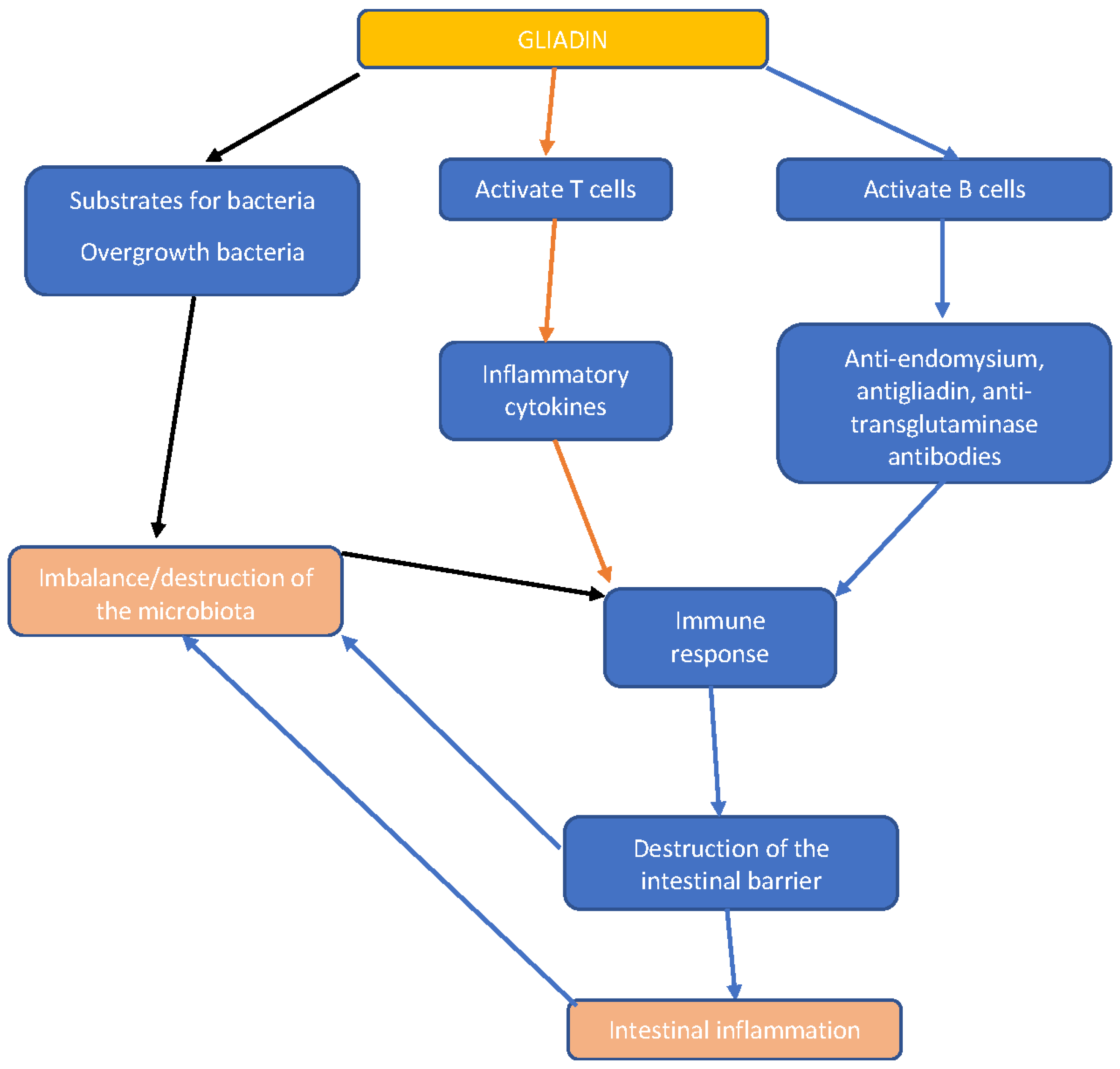
4.2. Pathophysiology
5. Intestinal Microbiota—General Aspects
6. Overview of CD Genetics and the Risk of Developing CD in Genetically Susceptible Individuals According to Their Microbiota Pattern
7. Implications of Intestinal Microbiota Alterations among Celiac Disease Patients
7.1. Possible Enviromental Causes of Intestinal Microbiota Alteration
7.1.1. Birth Gestational Age
7.1.2. Type of Delivery
7.1.3. Methods of Milk Feeding
7.1.4. Body Mass Index (BMI) Classes and Exercise Frequency
7.1.5. Antibiotics
7.1.6. Infections
7.1.7. Cause or Effect?
8. Intestinal Microbiome Modulation in Celiac Disease Patients—A New Therapeutic Perspective Besides Gluten-Free Diet
9. Conclusions
Author Contributions
Funding
Institutional Review Board Statement
Informed Consent Statement
Data Availability Statement
Conflicts of Interest
Abbreviations
| anti-tTg | Anti-transglutaminase antibodies |
| anti-tTG2 | Autoantibodies against tissue transglutaminase 2 |
| BMI | Body mass index |
| CD | Celiac disease |
| CDA | Celiac disease autoimmunity |
| CDGEMM | Celiac Disease Genomic, Environmental, Microbiome, and Metabolomic Study |
| DSS | Dextran sulfate sodium |
| EmA | Anti-endomysium antibodies |
| ETEC | Enterotoxigenic Escherichia coli |
| E. coli | Escherichia coli |
| GI | Gastrointestinal |
| GSRS | Gastrointestinal symptom rating scale |
| GFD | Gluten-free diet |
| IBD | Inflammatory bowel disease |
| IBS | Irritable bowel syndrome |
| IBS-SSS | Irritative bowel syndrome severity scoring system |
| IgA | Immunoglobulin A |
| IL-10 | Interleukin 10 |
| INF-γ | Interferon-γ |
| NLS | Natren life start |
| NCGS | Non-celiac gluten sensitivity |
| sIgA | Secretory IgA |
| SCFAs | Short-chain fatty acids |
| TGF | Transforming growth factor |
| TNF-α | Tumor necrosis factor-alpha |
| TLR | Toll-like receptor |
| tTG | Tissue transglutaminase |
| T1DM | Type 1 diabetes mellitus |
| WA | Wheat allergy |
References
- Catassi, C.; Bai, J.C.; Bonaz, B.; Bouma, G.; Calabrò, A.; Carroccio, A.; Castillejo, G.; Ciacci, C.; Cristofori, F.; Dolinsek, J.; et al. Non-Celiac Gluten Sensitivity: The New Frontier of Gluten Related Disorders. Nutrients 2013, 5, 3839. [Google Scholar] [CrossRef]
- Catassi, C.; Kryszak, D.; Louis-Jacques, O.; Duerksen, D.R.; Hill, I.; Crowe, S.E.; Brown, A.R.; Procaccini, N.J.; Wonderly, B.A.; Hartley, P.; et al. Detection of Celiac Disease in Primary Care: A Multicenter Case-Finding Study in North America. Am. J. Gastroenterol. 2007, 102, 1454–1460. [Google Scholar] [CrossRef] [PubMed]
- Rodríguez Del Río, P.; Díaz-Perales, A.; Sanchez-García, S.; Escudero, C.; do Santos, P.; Catarino, M.; Ibañez, M.D. Oral Immunotherapy in Children with IgE-Mediated Wheat Allergy: Outcome and Molecular Changes. J. Investig. Allergol. Clin. Immunol. 2014, 24, 240–248. [Google Scholar] [PubMed]
- Sapone, A.; Bai, J.C.; Ciacci, C.; Dolinsek, J.; Green, P.H.R.; Hadjivassiliou, M.; Kaukinen, K.; Rostami, K.; Sanders, D.S.; Schumann, M.; et al. Spectrum of Gluten-Related Disorders: Consensus on New Nomenclature and Classification. BMC Med. 2012, 10, 13. [Google Scholar] [CrossRef] [PubMed]
- West, J.; Logan, R.F.A.; Hill, P.G.; Khaw, K.T. The Iceberg of Celiac Disease: What Is Below the Waterline? Clin. Gastroenterol. Hepatol. 2007, 5, 59–62. [Google Scholar] [CrossRef]
- Caio, G.; Volta, U.; Sapone, A.; Leffler, D.A.; De Giorgio, R.; Catassi, C.; Fasano, A. Celiac Disease: A Comprehensive Current Review. BMC Med. 2019, 17, 142. [Google Scholar] [CrossRef]
- Chibbar, R.; Dieleman, L.A. The Gut Microbiota in Celiac Disease and Probiotics. Nutrients 2019, 11, 2375. [Google Scholar] [CrossRef]
- Marasco, G.; Cirota, G.G.; Rossini, B.; Lungaro, L.; Di Biase, A.R.; Colecchia, A.; Volta, U.; De Giorgio, R.; Festi, D.; Caio, G. Probiotics, Prebiotics and Other Dietary Supplements for Gut Microbiota Modulation in Celiac Disease Patients. Nutrients 2020, 12, 2674. [Google Scholar] [CrossRef]
- Corazza, G.R.; Andreani, M.L.; Biagi, F.; Corrao, G.; Pretolani, S.; Giulianelli, G.; Ghironzi, G.; Gasbarrini, G. The Smaller Size of the “coeliac Iceberg” in Adults. Scand. J. Gastroenterol. 1997, 32, 917–919. [Google Scholar] [CrossRef]
- Ivarsson, A.; Persson, L.Å.; Juto, P.; Peltonen, M.; Suhr, O.; Hernell, O. High Prevalence of Undiagnosed Coeliac Disease in Adults: A Swedish Population-Based Study. J. Intern. Med. 1999, 245, 63–68. [Google Scholar] [CrossRef]
- Riestra, S.; Fernández-Rodriguez, E.; Rodrigo, L.; Garcia, S.; Ocio, G. Prevalence of Coeliac Disease in the General Population of Northern Spain. Strategies of Serologic Screening. Scand. J. Gastroenterol. 2000, 35, 398–402. [Google Scholar] [CrossRef] [PubMed]
- Volta, U.; Bellentani, S.; Bianchi, F.B.; Brandi, G.; De Franceschi, L.; Miglioli, L.; Granito, A.; Balli, F.; Tiribelli, C. High Prevalence of Celiac Disease in Italian General Population. Dig. Dis. Sci. 2001, 46, 1500–1505. [Google Scholar] [CrossRef] [PubMed]
- Mustalahti, K.; Catassi, C.; Reunanen, A.; Fabiani, E.; Heier, M.; McMillan, S.; Murray, L.; Metzger, M.H.; Gasparin, M.; Bravi, E.; et al. The Prevalence of Celiac Disease in Europe: Results of a Centralized, International Mass Screening Project. Ann. Med. 2010, 42, 587–595. [Google Scholar] [CrossRef]
- Rubio-Tapia, A.; Ludvigsson, J.F.; Brantner, T.L.; Murray, J.A.; Everhart, J.E. The Prevalence of Celiac Disease in the United States. Am. J. Gastroenterol. 2012, 107, 1538–1544. [Google Scholar] [CrossRef]
- Singh, P.; Arora, S.; Singh, A.; Strand, T.A.; Makharia, G.K. Prevalence of Celiac Disease in Asia: A Systematic Review and Meta-Analysis. J. Gastroenterol. Hepatol. 2016, 31, 1095–1101. [Google Scholar] [CrossRef] [PubMed]
- Catassi, C.; Kryszak, D.; Bhatti, B.; Sturgeon, C.; Helzlsouer, K.; Clipp, S.L.; Gelfond, D.; Puppa, E.; Sferruzza, A.; Fasano, A. Natural History of Celiac Disease Autoimmunity in a USA Cohort Followed since 1974. Ann. Med. 2010, 42, 530–538. [Google Scholar] [CrossRef]
- Fasano, A.; Catassi, C. Clinical Practice. Celiac Disease. N. Engl. J. Med. 2012, 367, 2419–2426. [Google Scholar] [CrossRef]
- Sahin, Y. Celiac Disease in Children: A Review of the Literature. World J. Clin. Pediatr. 2021, 10, 53–71. [Google Scholar] [CrossRef]
- Lindfors, K.; Ciacci, C.; Kurppa, K.; Lundin, K.E.A.; Makharia, G.K.; Mearin, M.L.; Murray, J.A.; Verdu, E.F.; Kaukinen, K. Coeliac Disease. Nat. Rev. Dis. Primers 2019, 5, 3. [Google Scholar] [CrossRef]
- Cataldo, F.; Montalto, G. Celiac Disease in the Developing Countries: A New and Challenging Public Health Problem. World J. Gastroenterol. WJG 2007, 13, 2153. [Google Scholar] [CrossRef]
- Hill, I.D.; Fasano, A.; Guandalini, S.; Hoffenberg, E.; Levy, J.; Reilly, N.; Verma, R. NASPGHAN Clinical Report on the Diagnosis and Treatment of Gluten-Related Disorders. J. Pediatr. Gastroenterol. Nutr. 2016, 63, 156–165. [Google Scholar] [CrossRef]
- Dieterich, W.; Ehnis, T.; Bauer, M.; Donner, P.; Volta, U.; Riecken, E.O.; Schuppan, D. Identification of Tissue Transglutaminase as the Autoantigen of Celiac Disease. Nat. Med. 1997, 3, 797–801. [Google Scholar] [CrossRef]
- McGowan, K.E.; Castiglione, D.A.; Butzner, J.D. The Changing Face of Childhood Celiac Disease in North America: Impact of Serological Testing. Pediatrics 2009, 124, 1572–1578. [Google Scholar] [CrossRef] [PubMed]
- Lebwohl, B.; Rubio-Tapia, A.; Assiri, A.; Newland, C.; Guandalini, S. Diagnosis of Celiac Disease. Gastrointest. Endosc. Clin. N. Am. 2012, 22, 661–677. [Google Scholar] [CrossRef]
- Hujoel, I.A.; Van Dyke, C.T.; Brantner, T.; Larson, J.; King, K.S.; Sharma, A.; Murray, J.A.; Rubio-Tapia, A. Natural History and Clinical Detection of Undiagnosed Coeliac Disease in a North American Community. Aliment. Pharmacol. Ther. 2018, 47, 1358–1366. [Google Scholar] [CrossRef]
- Sanders, D.S.; Hurlstone, D.P.; Stokes, R.O.; Rashid, F.; Milford-Ward, A.; Hadjivassiliou, M.; Lobo, A.J. Changing Face of Adult Coeliac Disease: Experience of a Single University Hospital in South Yorkshire. Postgrad. Med. J. 2002, 78, 31–33. [Google Scholar] [CrossRef]
- Lo, W.; Sano, K.; Lebwohl, B.; Diamond, B.; Green, P.H.R. Changing Presentation of Adult Celiac Disease. Dig. Dis. Sci. 2003, 48, 395–398. [Google Scholar] [CrossRef] [PubMed]
- Singh, P.; Wadhwa, N.; Chaturvedi, M.K.; Bhatia, V.; Saini, S.; Tandon, N.; Makharia, G.K.; Maki, M.; Not, T.; Phillips, A.; et al. Validation of Point-of-Care Testing for Coeliac Disease in Children in a Tertiary Hospital in North India. Arch. Dis. Child 2014, 99, 1004–1008. [Google Scholar] [CrossRef] [PubMed]
- Nenna, R.; Tiberti, C.; Petrarca, L.; Lucantoni, F.; Mennini, M.; Luparia, R.P.L.; Panimolle, F.; Mastrogiorgio, G.; Pietropaoli, N.; Magliocca, F.M.; et al. The Celiac Iceberg: Characterization of the Disease in Primary Schoolchildren. J. Pediatr. Gastroenterol. Nutr. 2013, 56, 416–421. [Google Scholar] [CrossRef] [PubMed]
- Singh, P.; Arora, A.; Strand, T.A.; Leffler, D.A.; Catassi, C.; Green, P.H.; Kelly, C.P.; Ahuja, V.; Makharia, G.K. Global Prevalence of Celiac Disease: Systematic Review and Meta-Analysis. Clin. Gastroenterol. Hepatol. 2018, 16, 823–836.e2. [Google Scholar] [CrossRef] [PubMed]
- Tamai, T.; Ihara, K. Celiac Disease Genetics, Pathogenesis, and Standard Therapy for Japanese Patients. Int. J. Mol. Sci. 2023, 24, 2075. [Google Scholar] [CrossRef]
- Hall, E.J.; Batt, R.M. Dietary Modulation of Gluten Sensitivity in a Naturally Occurring Enteropathy of Irish Setter Dogs. Gut 1992, 33, 198. [Google Scholar] [CrossRef] [PubMed]
- Okada, H.; Kuhn, C.; Feillet, H.; Bach, J.F. The “hygiene Hypothesis” for Autoimmune and Allergic Diseases: An Update. Clin. Exp. Immunol. 2010, 160, 1–9. [Google Scholar] [CrossRef]
- Verdu, E.F.; Galipeau, H.J.; Jabri, B. Novel Players in Coeliac Disease Pathogenesis: Role of the Gut Microbiota. Nat. Rev. Gastroenterol. Hepatol. 2015, 12, 497–506. [Google Scholar] [CrossRef]
- Petersen, J.; Ciacchi, L.; Tran, M.T.; Loh, K.L.; Kooy-Winkelaar, Y.; Croft, N.P.; Hardy, M.Y.; Chen, Z.; McCluskey, J.; Anderson, R.P.; et al. T Cell Receptor Cross-Reactivity between Gliadin and Bacterial Peptides in Celiac Disease. Nat. Struct. Mol. Biol. 2020, 27, 49–61. [Google Scholar] [CrossRef] [PubMed]
- Caminero, A.; Galipeau, H.J.; McCarville, J.L.; Johnston, C.W.; Bernier, S.P.; Russell, A.K.; Jury, J.; Herran, A.R.; Casqueiro, J.; Tye-Din, J.A.; et al. Duodenal Bacteria From Patients With Celiac Disease and Healthy Subjects Distinctly Affect Gluten Breakdown and Immunogenicity. Gastroenterology 2016, 151, 670–683. [Google Scholar] [CrossRef] [PubMed]
- Araya, R.E.; Jury, J.; Bondar, C.; Verdu, E.F.; Chirdo, F.G. Intraluminal Administration of Poly I:C Causes an Enteropathy That Is Exacerbated by Administration of Oral Dietary Antigen. PLoS ONE 2014, 9, e99236. [Google Scholar] [CrossRef]
- Cristofori, F.; Indrio, F.; Miniello, V.L.; De Angelis, M.; Francavilla, R. Probiotics in Celiac Disease. Nutrients 2018, 10, 1824. [Google Scholar] [CrossRef] [PubMed]
- Bach, J.F. The Hygiene Hypothesis in Autoimmunity: The Role of Pathogens and Commensals. Nat. Rev. Immunol. 2018, 18, 105–120. [Google Scholar] [CrossRef]
- Talapko, J.; Včev, A.; Meštrović, T.; Pustijanac, E.; Jukić, M.; Škrlec, I. Homeostasis and Dysbiosis of the Intestinal Microbiota: Comparing Hallmarks of a Healthy State with Changes in Inflammatory Bowel Disease. Microorganisms 2022, 10, 2405. [Google Scholar] [CrossRef]
- Caio, G.; Lungaro, L.; Segata, N.; Guarino, M.; Zoli, G.; Volta, U.; De Giorgio, R. Effect of Gluten-Free Diet on Gut Microbiota Composition in Patients with Celiac Disease and Non-Celiac Gluten/Wheat Sensitivity. Nutrients 2020, 12, 1832. [Google Scholar] [CrossRef]
- Valitutti, F.; Cucchiara, S.; Fasano, A. Celiac Disease and the Microbiome. Nutrients 2019, 11, 2403. [Google Scholar] [CrossRef] [PubMed]
- Rinninella, E.; Raoul, P.; Cintoni, M.; Franceschi, F.; Miggiano, G.A.D.; Gasbarrini, A.; Mele, M.C. What Is the Healthy Gut Microbiota Composition? A Changing Ecosystem across Age, Environment, Diet, and Diseases. Microorganisms 2019, 7, 14. [Google Scholar] [CrossRef]
- Pecora, F.; Persico, F.; Gismondi, P.; Fornaroli, F.; Iuliano, S.; de’Angelis, G.L.; Esposito, S. Gut Microbiota in Celiac Disease: Is There Any Role for Probiotics? Front. Immunol. 2020, 11, 957. [Google Scholar] [CrossRef] [PubMed]
- Wu, X.; Qian, L.; Liu, K.; Wu, J.; Shan, Z. Gastrointestinal Microbiome and Gluten in Celiac Disease. Ann. Med. 2021, 53, 1797. [Google Scholar] [CrossRef]
- Abdukhakimova, D.; Dossybayeva, K.; Poddighe, D. Fecal and Duodenal Microbiota in Pediatric Celiac Disease. Front. Pediatr. 2021, 9, 158. [Google Scholar] [CrossRef]
- Losurdo, G.; Principi, M.; Iannone, A.; Ierardi, E.; Di Leo, A. The Interaction Between Celiac Disease and Intestinal Microbiota. J. Clin. Gastroenterol. 2016, 50, S145–S147. [Google Scholar] [CrossRef] [PubMed]
- Lundin, K.E.A.; Wijmenga, C. Coeliac Disease and Autoimmune Disease-Genetic Overlap and Screening. Nat. Rev. Gastroenterol. Hepatol. 2015, 12, 507–515. [Google Scholar] [CrossRef]
- Mazzilli, M.C.; Ferrante, P.; Mariani, P.; Martone, E.; Petronzelli, F.; Triglione, P.; Bonamico, M. A Study of Italian Pediatric Celiac Disease Patients Confirms That the Primary HLA Association Is to the DQ(Alpha 1*0501, Beta 1*0201) Heterodimer. Hum. Immunol. 1992, 33, 133–139. [Google Scholar] [CrossRef]
- Dieli-Crimi, R.; Cénit, M.C.; Núñez, C. The Genetics of Celiac Disease: A Comprehensive Review of Clinical Implications. J. Autoimmun. 2015, 64, 26–41. [Google Scholar] [CrossRef]
- Olivares, M.; Neef, A.; Castillejo, G.; De Palma, G.; Varea, V.; Capilla, A.; Palau, F.; Nova, E.; Marcos, A.; Polanco, I.; et al. The HLA-DQ2 Genotype Selects for Early Intestinal Microbiota Composition in Infants at High Risk of Developing Coeliac Disease. Gut 2015, 64, 406–417. [Google Scholar] [CrossRef]
- Vaahtovuo, J.; Munukka, E.; Korkeamäki, M.; Luukkainen, R.; Toivanen, P. Fecal Microbiota in Early Rheumatoid Arthritis. J. Rheumatol. 2008, 35, 1500–1505. [Google Scholar] [PubMed]
- Bamba, T.; Matsuda, H.; Endo, M.; Fujiyama, Y. The Pathogenic Role of Bacteroides Vulgatus in Patients with Ulcerative Colitis. J. Gastroenterol. 1995, 30 (Suppl. S8), 45–47. [Google Scholar] [PubMed]
- Sánchez, E.; De Palma, G.; Capilla, A.; Nova, E.; Pozo, T.; Castillejo, G.; Varea, V.; Marcos, A.; Garrote, J.A.; Polanco, I.; et al. Influence of Environmental and Genetic Factors Linked to Celiac Disease Risk on Infant Gut Colonization by Bacteroides Species. Appl. Environ. Microbiol. 2011, 77, 5316. [Google Scholar] [CrossRef]
- De Palma, G.; Capilla, A.; Nadal, I.; Nova, E.; Pozo, T.; Varea, V.; Polanco, I.; Castillejo, G.; López, A.; Garrote, J.A.; et al. Interplay between Human Leukocyte Antigen Genes and the Microbial Colonization Process of the Newborn Intestine. Curr. Issues Mol. Biol. 2010, 12, 1–10. [Google Scholar] [CrossRef] [PubMed]
- Olivares, M.; Benítez-Páez, A.; de Palma, G.; Capilla, A.; Nova, E.; Castillejo, G.; Varea, V.; Marcos, A.; Garrote, J.A.; Polanco, I.; et al. Increased Prevalence of Pathogenic Bacteria in the Gut Microbiota of Infants at Risk of Developing Celiac Disease: The PROFICEL Study. Gut Microbes 2018, 9, 551–558. [Google Scholar] [CrossRef] [PubMed]
- Sellitto, M.; Bai, G.; Serena, G.; Fricke, W.F.; Sturgeon, C.; Gajer, P.; White, J.R.; Koenig, S.S.K.; Sakamoto, J.; Boothe, D.; et al. Proof of Concept of Microbiome-Metabolome Analysis and Delayed Gluten Exposure on Celiac Disease Autoimmunity in Genetically at-Risk Infants. PLoS ONE 2012, 7, e33387. [Google Scholar] [CrossRef]
- Leonard, M.M.; Camhi, S.; Huedo-Medina, T.B.; Fasano, A. Celiac Disease Genomic, Environmental, Microbiome, and Metabolomic (CDGEMM) Study Design: Approach to the Future of Personalized Prevention of Celiac Disease. Nutrients 2015, 7, 9325–9336. [Google Scholar] [CrossRef]
- Leonard, M.M.; Fasano, A. The Microbiome as a Possible Target to Prevent Celiac Disease. Expert. Rev. Gastroenterol. Hepatol. 2016, 10, 555–556. [Google Scholar] [CrossRef]
- Leonard, M.M.; Karathia, H.; Pujolassos, M.; Troisi, J.; Valitutti, F.; Subramanian, P.; Camhi, S.; Kenyon, V.; Colucci, A.; Serena, G.; et al. Multi-Omics Analysis Reveals the Influence of Genetic and Environmental Risk Factors on Developing Gut Microbiota in Infants at Risk of Celiac Disease. Microbiome 2020, 8, 130. [Google Scholar] [CrossRef]
- Wei, Y.; Li, Y.; Yan, L.; Sun, C.; Miao, Q.; Wang, Q.; Xiao, X.; Lian, M.; Li, B.; Chen, Y.; et al. Alterations of Gut Microbiome in Autoimmune Hepatitis. Gut 2020, 69, 569–577. [Google Scholar] [CrossRef]
- Ye, Z.; Zhang, N.; Wu, C.; Zhang, X.; Wang, Q.; Huang, X.; Du, L.; Cao, Q.; Tang, J.; Zhou, C.; et al. A Metagenomic Study of the Gut Microbiome in Behcet’s Disease. Microbiome 2018, 6, 135. [Google Scholar] [CrossRef] [PubMed]
- Stricker, S.; Müller, M.; Zimmer, K.P.; Jacob, R. Altered Posttranslational Modification of Microtubules Contributes to Disturbed Enterocyte Morphology in Celiac Disease. Int. J. Mol. Sci. 2023, 24, 2635. [Google Scholar] [CrossRef]
- Aboulaghras, S.; Piancatelli, D.; Taghzouti, K.; Balahbib, A.; Alshahrani, M.M.; Al Awadh, A.A.; Goh, K.W.; Ming, L.C.; Bouyahya, A.; Oumhani, K. Meta-Analysis and Systematic Review of HLA DQ2/DQ8 in Adults with Celiac Disease. Int. J. Mol. Sci. 2023, 24, 1188. [Google Scholar] [CrossRef]
- Mårild, K.; Stephansson, O.; Montgomery, S.; Murray, J.A.; Ludvigsson, J.F. Pregnancy Outcome and Risk of Celiac Disease in Offspring: A Nationwide Case-Control Study. Gastroenterology 2012, 142, 39. [Google Scholar] [CrossRef]
- Cenit, M.C.; Olivares, M.; Codoñer-Franch, P.; Sanz, Y. Intestinal Microbiota and Celiac Disease: Cause, Consequence or Co-Evolution? Nutrients 2015, 7, 6900–6923. [Google Scholar] [CrossRef]
- Bresser, L.R.F.; de Goffau, M.C.; Levin, E.; Nieuwdorp, M. Gut Microbiota in Nutrition and Health with a Special Focus on Specific Bacterial Clusters. Cells 2022, 11, 3091. [Google Scholar] [CrossRef] [PubMed]
- Olshan, K.L.; Leonard, M.M.; Serena, G.; Zomorrodi, A.R.; Fasano, A. Gut Microbiota in Celiac Disease: Microbes, Metabolites, Pathways and Therapeutics. Expert. Rev. Clin. Immunol. 2020, 16, 1075–1092. [Google Scholar] [CrossRef]
- Trovato, C.M.; Montuori, M.; Anania, C.; Barbato, M.; Vestri, A.R.; Guida, S.; Oliva, S.; Mainiero, F.; Cucchiara, S.; Valitutti, F. Are ESPGHAN “Biopsy-Sparing” Guidelines for Celiac Disease Also Suitable for Asymptomatic Patients? Am. J. Gastroenterol. 2015, 110, 1485–1489. [Google Scholar] [CrossRef]
- Lionetti, E.; Castellaneta, S.; Francavilla, R.; Pulvirenti, A.; Tonutti, E.; Amarri, S.; Barbato, M.; Barbera, C.; Barera, G.; Bellantoni, A.; et al. Introduction of Gluten, HLA Status, and the Risk of Celiac Disease in Children. N. Engl. J. Med. 2014, 371, 1295–1303. [Google Scholar] [CrossRef] [PubMed]
- Vriezinga, S.L.; Auricchio, R.; Bravi, E.; Castillejo, G.; Chmielewska, A.; Crespo Escobar, P.; Kolaček, S.; Koletzko, S.; Korponay-Szabo, I.R.; Mummert, E.; et al. Randomized Feeding Intervention in Infants at High Risk for Celiac Disease. N. Engl. J. Med. 2014, 371, 1304–1315. [Google Scholar] [CrossRef] [PubMed]
- Ohkubo, T.; Tsuda, M.; Tamura, M.; Yamamura, M. Impaired Superoxide Production in Peripheral Blood Neutrophils of Germ-Free Rats. Scand. J. Immunol. 1990, 32, 727–729. [Google Scholar] [CrossRef]
- Mitsuyama, M.; Ohara, R.; Amako, K.; Nomoto, K.; Yokokura, T.; Nomoto, K. Ontogeny of Macrophage Function to Release Superoxide Anion in Conventional and Germfree Mice. Infect. Immun. 1986, 52, 236–239. [Google Scholar] [CrossRef]
- Mention, J.J.; Ben Ahmed, M.; Bègue, B.; Barbe, U.; Verkarre, V.; Asnafi, V.; Colombel, J.F.; Cugnenc, P.H.; Ruemmele, F.M.; McIntyre, E.; et al. Interleukin 15: A Key to Disrupted Intraepithelial Lymphocyte Homeostasis and Lymphomagenesis in Celiac Disease. Gastroenterology 2003, 125, 730–745. [Google Scholar] [CrossRef]
- Meresse, B.; Chen, Z.; Ciszewski, C.; Tretiakova, M.; Bhagat, G.; Krausz, T.N.; Raulet, D.H.; Lanier, L.L.; Groh, V.; Spies, T.; et al. Coordinated Induction by IL15 of a TCR-Independent NKG2D Signaling Pathway Converts CTL into Lymphokine-Activated Killer Cells in Celiac Disease. Immunity 2004, 21, 357–366. [Google Scholar] [CrossRef] [PubMed]
- Khosravi, A.; Mazmanian, S.K. Disruption of the Gut Microbiome as a Risk Factor for Microbial Infections. Curr. Opin. Microbiol. 2013, 16, 221. [Google Scholar] [CrossRef]
- Wacklin, P.; Kaukinen, K.; Tuovinen, E.; Collin, P.; Lindfors, K.; Partanen, J.; Mäki, M.; Mättuö, J. The Duodenal Microbiota Composition of Adult Celiac Disease Patients Is Associated with the Clinical Manifestation of the Disease. Inflamm. Bowel Dis. 2013, 19, 934–941. [Google Scholar] [CrossRef]
- O’Hara, A.M.; Shanahan, F. The Gut Flora as a Forgotten Organ. EMBO Rep. 2006, 7, 688. [Google Scholar] [CrossRef] [PubMed]
- Wijmenga, C.; Gutierrez-Achury, J. Celiac disease genetics: Past, present and future challenges. J. Pediatr. Gastroenterol. Nutr. 2014, 59 (Suppl. S1), S4–S7. [Google Scholar] [CrossRef] [PubMed]
- De Palma, G.; Capilla, A.; Nova, E.; Castillejo, G.; Varea, V.; Pozo, T.; Garrote, J.A.; Polanco, I.; López, A.; Ribes-Koninckx, C.; et al. Influence of Milk-Feeding Type and Genetic Risk of Developing Coeliac Disease on Intestinal Microbiota of Infants: The PROFICEL Study. PLoS ONE 2012, 7, e30791. [Google Scholar] [CrossRef]
- Asakuma, S.; Hatakeyama, E.; Urashima, T.; Yoshida, E.; Katayama, T.; Yamamoto, K.; Kumagai, H.; Ashida, H.; Hirose, J.; Kitaoka, M. Physiology of Consumption of Human Milk Oligosaccharides by Infant Gut-Associated Bifidobacteria. J. Biol. Chem. 2011, 286, 34583–34592. [Google Scholar] [CrossRef]
- Wang, C.; Zhang, M.; Guo, H.; Yan, J.; Liu, F.; Chen, J.; Li, Y.; Ren, F. Human Milk Oligosaccharides Protect against Necrotizing Enterocolitis by Inhibiting Intestinal Damage via Increasing the Proliferation of Crypt Cells. Mol. Nutr. Food Res. 2019, 63, 1900262. [Google Scholar] [CrossRef]
- Lammers, K.M.; Lu, R.; Brownley, J.; Lu, B.; Gerard, C.; Thomas, K.; Rallabhandi, P.; Shea-Donohue, T.; Tamiz, A.; Alkan, S.; et al. Gliadin Induces an Increase in Intestinal Permeability and Zonulin Release by Binding to the Chemokine Receptor CXCR3. Gastroenterology 2008, 135, 194. [Google Scholar] [CrossRef]
- Vorobjova, T.; Raikkerus, H.; Kadaja, L.; Talja, I.; Uibo, O.; Heilman, K.; Uibo, R. Circulating Zonulin Correlates with Density of Enteroviruses and Tolerogenic Dendritic Cells in the Small Bowel Mucosa of Celiac Disease Patients. Dig. Dis. Sci. 2017, 62, 358–371. [Google Scholar] [CrossRef]
- Nadal, I.; Donant, E.; Ribes-Koninckx, C.; Calabuig, M.; Sanz, Y. Imbalance in the Composition of the Duodenal Microbiota of Children with Coeliac Disease. J. Med. Microbiol. 2007, 56 Pt 12, 1669–1674. [Google Scholar] [CrossRef]
- De Palma, G.; Cinova, J.; Stepankova, R.; Tuckova, L.; Sanz, Y. Pivotal Advance: Bifidobacteria and Gram-Negative Bacteria Differentially Influence Immune Responses in the Proinflammatory Milieu of Celiac Disease. J. Leukoc. Biol. 2009, 87, 765–778. [Google Scholar] [CrossRef] [PubMed]
- Caminero, A.; Herrán, A.R.; Nistal, E.; Pérez-Andrés, J.; Vaquero, L.; Vivas, S.; Ruiz de Morales, J.M.G.; Albillos, S.M.; Casqueiro, J. Diversity of the Cultivable Human Gut Microbiome Involved in Gluten Metabolism: Isolation of Microorganisms with Potential Interest for Coeliac Disease. FEMS Microbiol. Ecol. 2014, 88, 309–319. [Google Scholar] [CrossRef]
- Olivares, M.; Laparra, M.; Sanz, Y. Influence of Bifidobacterium Longum CECT 7347 and Gliadin Peptides on Intestinal Epithelial Cell Proteome. J. Agric. Food Chem. 2011, 59, 7666–7671. [Google Scholar] [CrossRef]
- Duar, R.M.; Clark, K.J.; Patil, P.B.; Hernández, C.; Brüning, S.; Burkey, T.E.; Madayiputhiya, N.; Taylor, S.L.; Walter, J. Identification and Characterization of Intestinal Lactobacilli Strains Capable of Degrading Immunotoxic Peptides Present in Gluten. J. Appl. Microbiol. 2015, 118, 515–527. [Google Scholar] [CrossRef]
- Mandile, R.; Picascia, S.; Parrella, C.; Camarca, A.; Gobbetti, M.; Greco, L.; Troncone, R.; Gianfrani, C.; Auricchio, R. Lack of Immunogenicity of Hydrolysed Wheat Flour in Patients with Coeliac Disease after a Short-Term Oral Challenge. Aliment. Pharmacol. Ther. 2017, 46, 440–446. [Google Scholar] [CrossRef]
- Francavilla, R.; De Angelis, M.; Rizzello, C.G.; Cavallo, N.; Dal Bello, F.; Gobbetti, M. Selected Probiotic Lactobacilli Have the Capacity To Hydrolyze Gluten Peptides during Simulated Gastrointestinal Digestion. Appl. Environ. Microbiol. 2017, 83, e00376-17. [Google Scholar] [CrossRef] [PubMed]
- Serena, G.; Yan, S.; Camhi, S.; Patel, S.; Lima, R.S.; Sapone, A.; Leonard, M.M.; Mukherjee, R.; Nath, B.J.; Lammers, K.M.; et al. Proinflammatory Cytokine Interferon-γ and Microbiome-Derived Metabolites Dictate Epigenetic Switch between Forkhead Box Protein 3 Isoforms in Coeliac Disease. Clin. Exp. Immunol. 2017, 187, 490–506. [Google Scholar] [CrossRef]
- Lindfors, K.; Blomqvist, T.; Juuti-Uusitalo, K.; Stenman, S.; Venäläinen, J.; Mäki, M.; Kaukinen, K. Live Probiotic Bifidobacterium Lactis Bacteria Inhibit the Toxic Effects Induced by Wheat Gliadin in Epithelial Cell Culture. Clin. Exp. Immunol. 2008, 152, 552–558. [Google Scholar] [CrossRef]
- Medina, M.; De Palma, G.; Ribes-Koninckx, C.; Calabuig, M.; Sanz, Y. Bifidobacterium Strains Suppress in Vitro the Pro-Inflammatory Milieu Triggered by the Large Intestinal Microbiota of Coeliac Patients. J. Inflamm. 2008, 5, 19. [Google Scholar] [CrossRef]
- Laparra, J.M.; Olivares, M.; Gallina, O.; Sanz, Y. Bifidobacterium Longum CECT 7347 Modulates Immune Responses in a Gliadin-Induced Enteropathy Animal Model. PLoS ONE 2012, 7, e30744. [Google Scholar] [CrossRef]
- Zyrek, A.A.; Cichon, C.; Helms, S.; Enders, C.; Sonnenborn, U.; Schmidt, M.A. Molecular Mechanisms Underlying the Probiotic Effects of Escherichia Coli Nissle 1917 Involve ZO-2 and PKCzeta Redistribution Resulting in Tight Junction and Epithelial Barrier Repair. Cell Microbiol. 2007, 9, 804–816. [Google Scholar] [CrossRef]
- D’Arienzo, R.; Stefanile, R.; Maurano, F.; Mazzarella, G.; Ricca, E.; Troncone, R.; Auricchio, S.; Rossi, M. Immunomodulatory Effects of Lactobacillus Casei Administration in a Mouse Model of Gliadin-Sensitive Enteropathy. Scand. J. Immunol. 2011, 74, 335–341. [Google Scholar] [CrossRef] [PubMed]
- Cinova, J.; de Palma, G.; Stepankova, R.; Kofronova, O.; Kverka, M.; Sanz, Y.; Tuckova, L. Role of Intestinal Bacteria in Gliadin-Induced Changes in Intestinal Mucosa: Study in Germ-Free Rats. PLoS ONE 2011, 6, e16169. [Google Scholar] [CrossRef] [PubMed]
- Sánchez, E.; Laparra, J.M.; Sanz, Y. Discerning the Role of Bacteroides Fragilis in Celiac Disease Pathogenesis. Appl. Environ. Microbiol. 2012, 78, 6507. [Google Scholar] [CrossRef]
- Moreno, M.D.L.; Cebolla, Á.; Munõz-Suano, A.; Carrillo-Carrion, C.; Comino, I.; Pizarro, Á.; León, F.; Rodríguez-Herrera, A.; Sousa, C. Detection of Gluten Immunogenic Peptides in the Urine of Patients with Coeliac Disease Reveals Transgressions in the Gluten-Free Diet and Incomplete Mucosal Healing. Gut 2017, 66, 250–257. [Google Scholar] [CrossRef]
- West, J.; Logan, R.F.A.; Card, T.R.; Smith, C.; Hubbard, R. Risk of Vascular Disease in Adults with Diagnosed Coeliac Disease: A Population-Based Study. Aliment. Pharmacol. Ther. 2004, 20, 73–79. [Google Scholar] [CrossRef]
- Hallert, C.; Grant, C.; Grehn, S.; Grännö, C.; Hultén, S.; Midhagen, G.; Ström, M.; Svensson, H.; Valdimarsson, T. Evidence of Poor Vitamin Status in Coeliac Patients on a Gluten-Free Diet for 10 Years. Aliment. Pharmacol. Ther. 2002, 16, 1333–1339. [Google Scholar] [CrossRef]
- Midhagen, G.; Hallert, C. High Rate of Gastrointestinal Symptoms in Celiac Patients Living on a Gluten-Free Diet: Controlled Study. Am. J. Gastroenterol. 2003, 98, 2023–2026. [Google Scholar] [CrossRef]
- Roos, S.; Kärner, A.; Hallert, C. Psychological Well-Being of Adult Coeliac Patients Treated for 10 Years. Dig. Liver Dis. 2006, 38, 177–180. [Google Scholar] [CrossRef]
- Aziz, I.; Evans, K.E.; Papageorgiou, V.; Sanders, D.S. Are Patients with Coeliac Disease Seeking Alternative Therapies to a Gluten-Free Diet? J. Gastrointest. Liver Dis. 2011, 20, 27–31. [Google Scholar] [CrossRef]
- McCarville, J.L.; Caminero, A.; Verdu, E.F. Pharmacological Approaches in Celiac Disease. Curr. Opin. Pharmacol. 2015, 25, 7–12. [Google Scholar] [CrossRef] [PubMed]
- Jeon, S.G.; Kayama, H.; Ueda, Y.; Takahashi, T.; Asahara, T.; Tsuji, H.; Tsuji, N.M.; Kiyono, H.; Ma, J.S.; Kusu, T.; et al. Probiotic Bifidobacterium Breve Induces IL-10-Producing Tr1 Cells in the Colon. PLoS Pathog. 2012, 8, e1002714. [Google Scholar] [CrossRef] [PubMed]
- Zheng, B.; Van Bergenhenegouwen, J.; Overbeek, S.; Van De Kant, H.J.G.; Garssen, J.; Folkerts, G.; Vos, P.; Morgan, M.E.; Kraneveld, A.D. Bifidobacterium Breve Attenuates Murine Dextran Sodium Sulfate-Induced Colitis and Increases Regulatory T Cell Responses. PLoS ONE 2014, 9, e95441. [Google Scholar] [CrossRef] [PubMed]
- Orlando, A.; Linsalata, M.; Bianco, G.; Notarnicola, M.; D’attoma, B.; Scavo, M.P.; Tafaro, A.; Russo, F. Lactobacillus Rhamnosus GG Protects the Epithelial Barrier of Wistar Rats from the Pepsin-Trypsin-Digested Gliadin (PTG)-Induced Enteropathy. Nutrients 2018, 10, 1698. [Google Scholar] [CrossRef] [PubMed]
- Vanderpool, C.; Yan, F.; Polk, D.B. Mechanisms of Probiotic Action: Implications for Therapeutic Applications in Inflammatory Bowel Diseases. Inflamm. Bowel Dis. 2008, 14, 1585–1596. [Google Scholar] [CrossRef] [PubMed]
- De Angelis, M.; Rizzello, C.G.; Fasano, A.; Clemente, M.G.; De Simone, C.; Silano, M.; De Vincenzi, M.; Losito, I.; Gobbetti, M. VSL#3 Probiotic Preparation Has the Capacity to Hydrolyze Gliadin Polypeptides Responsible for Celiac Sprue. Biochim. Biophys. Acta 2006, 1762, 80–93. [Google Scholar] [CrossRef] [PubMed]
- Medina, M.; Izquierdo, E.; Ennahar, S.; Sanz, Y. Differential Immunomodulatory Properties of Bifidobacterium Logum Strains: Relevance to Probiotic Selection and Clinical Applications. Clin. Exp. Immunol. 2007, 150, 531–538. [Google Scholar] [CrossRef]
- Papista, C.; Gerakopoulos, V.; Kourelis, A.; Sounidaki, M.; Kontana, A.; Berthelot, L.; Moura, I.C.; Monteiro, R.C.; Yiangou, M. Gluten Induces Coeliac-like Disease in Sensitised Mice Involving IgA, CD71 and Transglutaminase 2 Interactions That Are Prevented by Probiotics. Lab. Investig. 2012, 92, 625–635. [Google Scholar] [CrossRef]
- Smecuol, E.; Hwang, H.J.; Sugai, E.; Corso, L.; Cherñavsky, A.C.; Bellavite, F.P.; González, A.; Vodánovich, F.; Moreno, M.L.; Vázquez, H.; et al. Exploratory, Randomized, Double-Blind, Placebo-Controlled Study on the Effects of Bifidobacterium Infantis Natren Life Start Strain Super Strain in Active Celiac Disease. J. Clin. Gastroenterol. 2013, 47, 139–147. [Google Scholar] [CrossRef] [PubMed]
- Golfetto, L.; de Senna, F.D.; Hermes, J.; Beserra, B.T.S.; da Silva França, F.; Martinello, F. Lower Bifidobacteria Counts in Adult Patients with Celiac Disease on a Gluten-Free Diet. Arq. Gastroenterol. 2014, 51, 139–143. [Google Scholar] [CrossRef]
- Pisarello, M.L.J.; Vintiñi, E.O.; González, S.N.; Pagani, F.; Medina, M.S. Decrease in Lactobacilli in the Intestinal Microbiota of Celiac Children with a Gluten-Free Diet, and Selection of Potentially Probiotic Strains. Can. J. Microbiol. 2015, 61, 32–37. [Google Scholar] [CrossRef] [PubMed]
- Olivares, M.; Castillejo, G.; Varea, V.; Sanz, Y. Double-Blind, Randomised, Placebo-Controlled Intervention Trial to Evaluate the Effects of Bifidobacterium Longum CECT 7347 in Children with Newly Diagnosed Coeliac Disease. Br. J. Nutr. 2014, 112, 30–40. [Google Scholar] [CrossRef]
- Klemenak, M.; Dolinšek, J.; Langerholc, T.; Di Gioia, D.; Mičetić-Turk, D. Administration of Bifidobacterium Breve Decreases the Production of TNF-α in Children with Celiac Disease. Dig. Dis. Sci. 2015, 60, 3386–3392. [Google Scholar] [CrossRef]
- Harnett, J.; Myers, S.P.; Rolfe, M. Probiotics and the Microbiome in Celiac Disease: A Randomised Controlled Trial. Evid. Based Complement. Alternat. Med. 2016, 2016, 9058574. [Google Scholar] [CrossRef]
- Quagliariello, A.; Aloisio, I.; Bozzi Cionci, N.; Luiselli, D.; D’Auria, G.; Martinez-Priego, L.; Pérez-Villarroya, D.; Langerholc, T.; Primec, M.; Mičetić-Turk, D.; et al. Effect of Bifidobacterium Breve on the Intestinal Microbiota of Coeliac Children on a Gluten Free Diet: A Pilot Study. Nutrients 2016, 8, 660. [Google Scholar] [CrossRef]
- Pinto-Sánchez, M.I.; Smecuol, E.C.; Temprano, M.P.; Sugai, E.; González, A.; Moreno, M.L.; Huang, X.; Bercik, P.; Cabanne, A.; Vázquez, H.; et al. Bifidobacterium Infantis NLS Super Strain Reduces the Expression of α-Defensin-5, a Marker of Innate Immunity, in the Mucosa of Active Celiac Disease Patients. J. Clin. Gastroenterol. 2017, 51, 814–817. [Google Scholar] [CrossRef] [PubMed]
- Martinello, F.; Roman, C.F.; de Souza, P.A. Effects of Probiotic Intake on Intestinal Bifidobacteria of Celiac Patients. Arq. Gastroenterol. 2017, 54, 85–90. [Google Scholar] [CrossRef] [PubMed]
- Primec, M.; Klemenak, M.; Di Gioia, D.; Aloisio, I.; Bozzi Cionci, N.; Quagliariello, A.; Gorenjak, M.; Mičetić-Turk, D.; Langerholc, T. Clinical Intervention Using Bifidobacterium Strains in Celiac Disease Children Reveals Novel Microbial Modulators of TNF-α and Short-Chain Fatty Acids. Clin. Nutr. 2019, 38, 1373–1381. [Google Scholar] [CrossRef] [PubMed]
- Francavilla, R.; Piccolo, M.; Francavilla, A.; Polimeno, L.; Semeraro, F.; Cristofori, F.; Castellaneta, S.; Barone, M.; Indrio, F.; Gobbetti, M.; et al. Clinical and Microbiological Effect of a Multispecies Probiotic Supplementation in Celiac Patients With Persistent IBS-Type Symptoms: A Randomized, Double-Blind, Placebo-Controlled, Multicenter Trial. J. Clin. Gastroenterol. 2019, 53, E117–E125. [Google Scholar] [CrossRef] [PubMed]
- Uusitalo, U.; Aronsson, C.A.; Liu, X.; Kurppa, K.; Yang, J.; Liu, E.; Skidmore, J.; Winkler, C.; Rewers, M.J.; Hagopian, W.A.; et al. Early Probiotic Supplementation and the Risk of Celiac Disease in Children at Genetic Risk. Nutrients 2019, 11, 1790. [Google Scholar] [CrossRef] [PubMed]
- Drabińska, N.; Krupa-Kozak, U.; Jarocka-Cyrta, E. Intestinal Permeability in Children with Celiac Disease after the Administration of Oligofructose-Enriched Inulin into a Gluten-Free Diet-Results of a Randomized, Placebo-Controlled, Pilot Trial. Nutrients 2020, 12, 1736. [Google Scholar] [CrossRef] [PubMed]
- Krupa-Kozak, U.; Drabińska, N.; Jarocka-Cyrta, E. The Effect of Oligofructose-Enriched Inulin Supplementation on Gut Microbiota, Nutritional Status and Gastrointestinal Symptoms in Paediatric Coeliac Disease Patients on a Gluten-Free Diet: Study Protocol for a Pilot Randomized Controlled Trial. Nutr. J. 2017, 16, 47. [Google Scholar] [CrossRef] [PubMed]
- Adebola, O.O.; Corcoran, O.; Morgan, W.A. Synbiotics: The Impact of Potential Prebiotics Inulin, Lactulose and Lactobionic Acid on the Survival and Growth of Lactobacilli Probiotics. J. Funct. Foods 2014, 10, 75–84. [Google Scholar] [CrossRef]
- Tuohy, K.M.; Finlay, R.K.; Wynne, A.G.; Gibson, G.R. A Human Volunteer Study on the Prebiotic Effects of HP-Inulin—Faecal Bacteria Enumerated Using Fluorescent In Situ Hybridisation (FISH). Anaerobe 2001, 7, 113–118. [Google Scholar] [CrossRef]

| Author | Country | Age, Years | First-Level Antibody Test | Prevalence of Celiac Disease |
|---|---|---|---|---|
| Corazza et al., 1997 [9] | Italy | 20–87 | EmA | 0.18% |
| Ivarsson et al., 1999 [10] | Sweden | 25–74 | EmA | 0.53% |
| Riestra et al., 2000 [11] | Spain | 14–89 | EmA | 0.26% |
| Volta et al., 2001 [12] | Italy | 14–65 | EmA | 0.57% |
| Mustalahti et al., 2010 [13] | Finland | 30–93 | Anti-tTG, EmA | 2.5% |
| Rubio-Tapia et al., 2012 [14] | USA | 23–66 | Anti-tTG, EmA | 0.71% |
| Singh et al., 2016 [15] | Asia | Not specified | Anti-tTG, EmA | 0.5% |
| Seroprevalence | Biopsy Prevalence | |
|---|---|---|
| Global | 1.4% (95% CI 1.1–1.7) | 0.7% (95% CI 0.5–0.9) |
| Africa | 1.1% (95% CI 0.4–2.2) | 0.5% (95% CI 0.2–0.9) |
| Asia | 1.8% (95% CI 1–2.9) | 0.6% (95% CI 0.4–0.8) |
| Europe | 1.3% (95% CI 1.1–1.5) | 0.8% (95% CI 0.6–1.1) |
| Oceania | 1.4% (95% CI 1.1–1.8) | 0.8% (95% CI 0.2–1.7) |
| North America | 1.4% (95% CI 0.7–2.2) | 0.5% |
| South America | 1.3% (95% CI 0.5–2.5) | 0.4% (95% CI 0.1–0.6) |
| Phylum | Class | Order | Family | Genus | Species |
|---|---|---|---|---|---|
| Actinobacteria | Actinobacteria | Actinomycetales | Corynebacteriaceae | Corynebacterium | |
| Bifidobacteriales | Bifidobacteriaceae | Bifidobacterium | B. longum | ||
| B. bifidum | |||||
| Coriobacteriia | Coriobacteriales | Coriobacteriaceae | Atoopobium | ||
| Firmicutes | Clostridia | Clostridiales | Clostridiaceae | Faecalibacterium | F. prausnitzii |
| Clostridium | Clostridium spp. | ||||
| Lachnospiraceae | Roseburia | R. intestinalis | |||
| Ruminococcaceae | Ruminococcus | R. faecis | |||
| Veillonellales | Veillonellaceae | Dialister | D. invisus | ||
| Negativicutes | Lactobacillales | Lactobacillaceae | Lactobacillus | L. reuteri | |
| Bacilli | Enterococcaceae | Enterococcus | E. faecium | ||
| Bacillales | Staphylocoecaceae | Staphylococcus | S. leei | ||
| Bacteroidetes | Sphingobacteriia | Sphingobacteriales | Sphingobacteriaceae | Sphingobacterium | |
| Bacteroidia | Bacteroidales | Bacteroidaceae | Bacteroides | B. vulgatus | |
| B. fragilis | |||||
| B. uniformis | |||||
| Tannerellaceae | Tannerella | ||||
| Parabacteroides | P. distasonis | ||||
| Rikenellaceae | Alistipes | A. finegoldi | |||
| Prevotellaceae | Prevotella | Prevotella spp. | |||
| Proteobacteria | Delta Proteobacteria | Enterobacterales | Enterobacter | Escherichia | E. coli |
| Shigella | S. flexneri | ||||
| Desulfovibrionales | Desulfovibrionaceae | Desulfovibrio | D. intestinalis | ||
| Bilophilia | B. wadsorthia | ||||
| Epsilon Proteobacteria | Campylobacterales | Helicobacteraceae | Helicobacter | H. pylori | |
| Fusobacteria | Fusobacteriia | Fusobacteriales | Fusobacteriaceae | Fusobacterium | F. nucleatum |
| Verrucomicrobia | Verrucomicrobiae | Verrucomicrobiaales | Akkermansiaceae | Akkermansia | A.muciniphila |
| Author | Year | Probiotic Variety | Objectives and Discoveries | Conclusions |
|---|---|---|---|---|
| De Angelis et al. [111] | 2006 | VSL#3 | VSL#3 exhibits a significant capacity for long-term colonization of the intestine. | Administration of VSL#3 would result in the complete eradication of toxic peptides in processed foods, thereby reducing long-term health risks and enhancing overall quality of life. |
| Medina M. et al. [112] | 2007 | Bifidobacterium longum | The genomic DNA of certain strains elicited a Th1 and proinflammatory cytokine response, characterized by the production of interferon-gamma and TNF-α, rather than IL-10. | The capacity of B. longum to modulate the immune system. |
| Lindfors K. et al. [93] | 2008 | Bifidobacterium lactis | Prevent the harmful effects on the mucous membrane of the small intestine induced by gluten/gliadin. | The inhibition is dose-dependent and results in increased permeability of epithelial cells induced by gliadin. Additionally, it stimulates the production of IL-10 by regulatory T cells. |
| D’Arienzo et al. [97] | 2011 | Lactobacillus casei ATCC 9595 | There was a full recovery of the blunting of villi, decreased weight loss, and the basal levels of TNF-α were restored. | The use of L. casei was successful in restoring the normal structure of the mucosal lining and maintaining homeostasis in the gut-associated lymphoid tissue. |
| Papista et al. [113] | 2012 | Saccharomyces boulardii KK1 strain, hydrolyzed the 28 kDa gliadin fraction | Administration of S. boulardii improved the development of enteropathy, reduced expression of CD71 in epithelial cells, and limited the production of cytokines in localized areas. | A novel mouse model has been developed for studying human CD that shares histopathological features and common biomarkers. The treatment of CD using S. boulardii was found to be effective in reversing the development of the disease. |
| Laparra et al. [95] | 2012 | Bifidobacterium longum CECT 7347 | In animals with gluten sensitivity, the administration of B. longum resulted in increased expression of NF-κB, IL-10, and CD8+ cells but reduced expression of TNF-α, CD4+ cells, and CD4+/Fox3+ cell populations. | In an animal model of gliadin-induced enteropathy, B. longum modulates the production of inflammatory cytokines and the immune response mediated by CD4+ T cells. |
| Smecuol et al. [114] | 2013 | Bifidobacterium natren life start | The impact of gluten on intestinal permeability, clinical symptomatology assessed through the GSRS questionnaire, and changes in immunological markers. | Supplementation of untreated CD patients with Bifidobacterium NLS did not alter protein abnormalities but demonstrated potential for symptom improvement and elicited immunological changes. |
| Golfetto et al. [115] | 2014 | Bifidobacteria spp. | The quantity of Bifidobacteria per gram of fecal matter was markedly greater in healthy controls (1.5 ± 0.63 × 10⁸ CFU/g) in comparison to celiac patients (2.5 ± 1.5 × 10⁷ CFU/g). | Diminished levels of Bifidobacteria may disrupt the intestinal microbiota equilibrium in individuals with CD, irrespective of pH and adherence to a GFD. |
| Pisarello et al. [116] | 2014 | Lactobacillus rhamnosus; Lactobacillus paracasei | The group of children with CD following a GFD exhibited markedly lower Lactobacillus counts compared to the healthy control group. | Probiotic therapy is not a substitute for a GFD, but it has the potential to mitigate the aberrant inflammatory parameters observed in individuals with CD, as well as modulate the composition of the intestinal microbiota. |
| Olivares et al. [117] | 2014 | Bifidobacterium longum CECT 7347 | Measures of immune phenotype in peripheral blood cells, serum cytokine levels, fecal secretory IgA, anthropometric parameters, and composition of intestinal microbiota at both baseline and following intervention. | Patients undergoing probiotic therapy exhibited an increase in height percentile, a reduction in peripheral CD3+ T lymphocytes, and a slight decrease in TNF-α concentration. Furthermore, decreased levels of B. fragilis and secretory IgA were observed in the stool. |
| Klemenak et al. [118] | 2015 | Bifidobacterium breve BRO3 and B. breve B632 | Results: serum levels of IL-10 and TNF- production. | Following 3 months of probiotic therapy, TNF-α levels exhibited a decrease; however, on subsequent follow-ups after another 3 months, levels showed an increase. The levels of IL-10 were below the detection threshold. |
| Harnett et al. [119] | 2016 | The De Simone formulation, previously known as VSL#3, is a blend of 9 strains of lyophilized bacteria, containing 450 billion viable microorganisms. | Quantitative analysis of microbial populations, with comparisons made between baseline and end-of-study measurements of dominant, pathogenic, and opportunistic bacteria. Evaluation of urinary metabolomics and fecal lactoferrin. | Over 12 weeks, no noteworthy alterations were observed in the gastrointestinal microbial populations of individuals with CD who exhibited persistent symptoms. |
| Quagliariello et al. [120] | 2016 | Bifidobacterium breve strains B632 and BRO3 | Assessment of the microbiome following the probiotic intervention. | A 3-month course of probiotic therapy can result in the recovery of the microbiota of children with CD to a level similar to that of healthy individuals. |
| Pinto-Sanchez et al. [121] | 2017 | B. infantis Natren Life Start super strain. | Assess the mucosal expression of innate immune markers through the evaluation of the number of macrophages, Paneth cells, and α-defensin-5 expression using immunohistochemistry in duodenal biopsies. | Analysis of duodenal biopsies showed that the administration of Bifidobacterium infantis NLS-SS resulted in a reduction in all three innate markers in patients with CD. However, the decrease in macrophage counts was more significant in patients who followed a GFD. |
| Martinello et al. [122] | 2017 | Yogurt containing probiotic strains from PIA, Nova Petropolis-RS, with an unspecified concentration of microorganisms. | Bifidobacterial levels in fecal samples following the ingestion of 100 g of yogurt in the morning. | The fecal count of Bifidobacteria was greater in healthy individuals than in those with CD. While the consumption of probiotic yogurt resulted in increased Bifidobacteria levels in CD patients, there was no such effect observed in healthy participants. |
| Primec et al. [123] | 2019 | Bifidobacterium breve strains B632 and BRO3. | Assess the impact of probiotics on the composition of the fecal microbiota, levels of SCFA, and the concentration of TNF-α in the serum. | There was a strong correlation between CD and the presence of Verrucomicrobia, Paracubacteria, and some unidentified phyla of bacteria and archaea. |
| Francavilla et al. [124] | 2019 | A probiotic product comprising five bacterial strains: L. casei, L. plantarum, B. animalis subsp. Lacti, B. breve Bbr8 LMG P-17501, and B. breve B110 LMG P-17500. | Assess the efficacy of probiotics in improving gastrointestinal symptoms using the IBS-SSS. | Probiotic treatment resulted in a significant reduction in IBS-SSS and GSRS scores and improvement in IBS symptoms compared to placebo. Additionally, in CD patients adhering to a strict GFD, probiotics were able to modify the gut microbiota by increasing the number of Bifidobacteria. |
| Uusitalo et al. [125] | 2019 | L. reuteri; L. rhamnosus, and some unidentified. | To investigate the potential link between probiotic intake via dietary supplements or infant formula starting from one year of age and the risk of developing CD or CDA. | In general, exposure to probiotics during the first year of life was not linked to the development of CDA or CD. Nonetheless, the consumption of probiotics through dietary supplements was related to a higher risk of CDA. |
Disclaimer/Publisher’s Note: The statements, opinions and data contained in all publications are solely those of the individual author(s) and contributor(s) and not of MDPI and/or the editor(s). MDPI and/or the editor(s) disclaim responsibility for any injury to people or property resulting from any ideas, methods, instructions or products referred to in the content. |
© 2023 by the authors. Licensee MDPI, Basel, Switzerland. This article is an open access article distributed under the terms and conditions of the Creative Commons Attribution (CC BY) license (https://creativecommons.org/licenses/by/4.0/).
Share and Cite
Belei, O.; Jugănaru, I.; Basaca, D.-G.; Munteanu, A.I.; Mărginean, O. The Role of Intestinal Microbiota in Celiac Disease and Further Therapeutic Perspectives. Life 2023, 13, 2039. https://doi.org/10.3390/life13102039
Belei O, Jugănaru I, Basaca D-G, Munteanu AI, Mărginean O. The Role of Intestinal Microbiota in Celiac Disease and Further Therapeutic Perspectives. Life. 2023; 13(10):2039. https://doi.org/10.3390/life13102039
Chicago/Turabian StyleBelei, Oana, Iulius Jugănaru, Diana-Georgiana Basaca, Andrei Ioan Munteanu, and Otilia Mărginean. 2023. "The Role of Intestinal Microbiota in Celiac Disease and Further Therapeutic Perspectives" Life 13, no. 10: 2039. https://doi.org/10.3390/life13102039
APA StyleBelei, O., Jugănaru, I., Basaca, D.-G., Munteanu, A. I., & Mărginean, O. (2023). The Role of Intestinal Microbiota in Celiac Disease and Further Therapeutic Perspectives. Life, 13(10), 2039. https://doi.org/10.3390/life13102039